Recent insights into the morphological diversity in the amniote primary and secondary palates
- PMID: 26293818
- PMCID: PMC4715671
- DOI: 10.1002/dvdy.24338
Recent insights into the morphological diversity in the amniote primary and secondary palates
Abstract
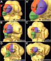
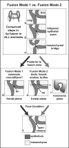
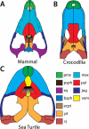
The assembly of the upper jaw is a pivotal moment in the embryonic development of amniotes. The upper jaw forms from the fusion of the maxillary, medial nasal, and lateral nasal prominences, resulting in an intact upper lip/beak and nasal cavities; together called the primary palate. This process of fusion requires a balance of proper facial prominence shape and positioning to avoid craniofacial clefting, whilst still accommodating the vast phenotypic diversity of adult amniotes. As such, variation in craniofacial ontogeny is not tolerated beyond certain bounds. For clarity, we discuss primary palatogenesis of amniotes into in two categories, according to whether the nasal and oral cavities remain connected throughout ontogeny or not. The transient separation of these cavities occurs in mammals and crocodilians, while remaining connected in birds, turtles and squamates. In the latter group, the craniofacial prominences fuse around a persistent choanal groove that connects the nasal and oral cavities. Subsequently, all lineages except for turtles, develop a secondary palate that ultimately completely or partially separates oral and nasal cavities. Here, we review the shared, early developmental events and highlight the points at which development diverges in both primary and secondary palate formation.
Keywords: choana; craniofacial; lip fusion; palatal shelves; primary palate; reptile.
© 2015 Wiley Periodicals, Inc.
Figures


Similar articles
-
Diversity in primary palate ontogeny of amniotes revealed with 3D imaging.J Anat. 2015 May;226(5):420-33. doi: 10.1111/joa.12291. Epub 2015 Apr 22. J Anat. 2015. PMID: 25904546 Free PMC article.
-
Divergent palate morphology in turtles and birds correlates with differences in proliferation and BMP2 expression during embryonic development.J Exp Zool B Mol Dev Evol. 2014 Feb;322(2):73-85. doi: 10.1002/jez.b.22547. Epub 2013 Dec 9. J Exp Zool B Mol Dev Evol. 2014. PMID: 24323766 Free PMC article.
-
Morphological observations in normal primary palate and cleft lip embryos in the Kyoto collection.Teratology. 1990 Jun;41(6):663-77. doi: 10.1002/tera.1420410603. Teratology. 1990. PMID: 2353315
-
Recent advances in primary palate and midface morphogenesis research.Crit Rev Oral Biol Med. 1992;4(1):111-30. doi: 10.1177/10454411920040010201. Crit Rev Oral Biol Med. 1992. PMID: 1457684 Review.
-
Palatogenesis: morphogenetic and molecular mechanisms of secondary palate development.Development. 2012 Jan;139(2):231-43. doi: 10.1242/dev.067082. Development. 2012. PMID: 22186724 Free PMC article. Review.
Cited by
-
Polarized Sonic Hedgehog Protein Localization and a Shift in the Expression of Region-Specific Molecules Is Associated With the Secondary Palate Development in the Veiled Chameleon.Front Cell Dev Biol. 2020 Jul 28;8:572. doi: 10.3389/fcell.2020.00572. eCollection 2020. Front Cell Dev Biol. 2020. PMID: 32850780 Free PMC article.
-
Development of the squamate naso-palatal complex: detailed 3D analysis of the vomeronasal organ and nasal cavity in the brown anole Anolis sagrei (Squamata: Iguania).Front Zool. 2020 Sep 22;17:28. doi: 10.1186/s12983-020-00369-7. eCollection 2020. Front Zool. 2020. PMID: 32983242 Free PMC article.
-
Developmental origin of the mammalian premaxilla.Dev Biol. 2023 Nov;503:1-9. doi: 10.1016/j.ydbio.2023.07.005. Epub 2023 Jul 29. Dev Biol. 2023. PMID: 37524195 Free PMC article.
-
Using frogs faces to dissect the mechanisms underlying human orofacial defects.Semin Cell Dev Biol. 2016 Mar;51:54-63. doi: 10.1016/j.semcdb.2016.01.016. Epub 2016 Jan 15. Semin Cell Dev Biol. 2016. PMID: 26778163 Free PMC article. Review.
-
Novel insights into the development of the avian nasal cavity.Anat Rec (Hoboken). 2021 Feb;304(2):247-257. doi: 10.1002/ar.24349. Epub 2020 Jan 1. Anat Rec (Hoboken). 2021. PMID: 31872940 Free PMC article.
References
-
- Abzhanov A, Protas M, Grant BR, Grant PR, Tabin CJ. Bmp4 and morphological variation of beaks in Darwin's finches. Science. 2004;305:1462–5. - PubMed
-
- Ashique AM, Fu K, Richman JM. Endogenous bone morphogenetic proteins regulate outgrowth and epithelial survival during avian lip fusion. Development. 2002;129:4647–60. - PubMed
Publication types
MeSH terms
Grants and funding
LinkOut - more resources
Full Text Sources
Other Literature Sources
Medical
Research Materials

