. 2015 Nov;5(11):1194-209.
doi: 10.1158/2159-8290.CD-15-0460. Epub 2015 Aug 20.

PtdIns(3,4,5)P3-Dependent Activation of the mTORC2 Kinase Complex

Affiliations

PtdIns(3,4,5)P3-Dependent Activation of the mTORC2 Kinase Complex

Pengda Liu et al. Cancer Discov. 2015 Nov.

Abstract

mTOR serves as a central regulator of cell growth and metabolism by forming two distinct complexes, mTORC1 and mTORC2. Although mechanisms of mTORC1 activation by growth factors and amino acids have been extensively studied, the upstream regulatory mechanisms leading to mTORC2 activation remain largely elusive. Here, we report that the pleckstrin homology (PH) domain of SIN1, an essential and unique component of mTORC2, interacts with the mTOR kinase domain to suppress mTOR activity. More importantly, PtdIns(3,4,5)P3, but not other PtdInsPn species, interacts with SIN1-PH to release its inhibition on the mTOR kinase domain, thereby triggering mTORC2 activation. Mutating critical SIN1 residues that mediate PtdIns(3,4,5)P3 interaction inactivates mTORC2, whereas mTORC2 activity is pathologically increased by patient-derived mutations in the SIN1-PH domain, promoting cell growth and tumor formation. Together, our study unravels a PI3K-dependent mechanism for mTORC2 activation, allowing mTORC2 to activate AKT in a manner that is regulated temporally and spatially by PtdIns(3,4,5)P3.

Significance: The SIN1-PH domain interacts with the mTOR kinase domain to suppress mTOR activity, and PtdIns(3,4,5)P3 binds the SIN1-PH domain to release its inhibition on the mTOR kinase domain, leading to mTORC2 activation. Cancer patient-derived SIN1-PH domain mutations gain oncogenicity by loss of suppressing mTOR activity as a means to facilitate tumorigenesis.


Conflict of interest statement

Conflict of interest: None

Figures

Fig. 1
Fig. 1
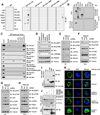
The PH domain of Sin1 binds the mTOR kinase domain and inhibits mTORC2 catalytic activity. A, A schematic illustration of the Sin1 domain structures. B, The Sin1-PH domain binds the mTOR kinase domain (KD, aa 2180–2431). Flag-mTOR-KD was transfected into HEK293 cells and affinity purified by Flag-M2 beads 48 hr post-transfection. Then Flag-mTOR-KD immunoprecipitates (IP) were used to pull down indicated Sin1 truncations expressed in HEK293 cells after serum starvation for 24 hr and subjected to immunoblot (IB) analyses. C, Sin1-PH suppresses mTOR to phosphorylate Akt in vitro. In vitro kinase assays of recombinant active mTOR kinases with GST-Akt1-tail (aa 409–480) as a substrate, in the presence of increasing doses of bacterially purified GST or GST-Sin1-PH proteins. D, Sin1-PH suppresses mTORC2 activity in cells. IB of whole cell lysates (WCL) derived from MDA-MB-231 cells transfected with increasing amounts of HA-Sin1-PH. E, Expression of full-length, but not PH domain-deleted Sin1, leads to reduced Akt-pS473 in cells. IB analysis of WCLs derived from HEK293T cells transfected with increasing doses of HA-Sin1. F, Binding of Sin1-PH to mTOR-KD does not disrupt mTORC2 complex integrity. IB of Flag-IP and WCLs derived from HEK293T cells transfected with HA-Sin1, Flag-mTOR and increasing doses of HA-Sin1-PH. G, Sin1-PH competes with full-length Sin1 for binding mTOR-KD. IB analysis of WCLs and Flag-IPs derived from HEK293T cells transfected with indicated constructs.
Fig. 2
Fig. 2
The PH domain of Sin1 largely replaces the PH domain of Akt1 for function in cells. A, A schematic illustration of the Akt1-WT (left) and the Sin1-PH-Akt1 chimera domain structures (right). B, Deleting the PH domain of Akt1 leads to attenuated Akt phosphorylation. Immunoblot (IB) analysis of whole cell lysates (WCLs) derived from DLD1-Akt1/2−/− cells transfected with indicated constructs. C–D, Sin1-PH in large functionally replaces Akt1-PH in cells. IB of WCLs derived from Akt1/2−/− MEFs (C) or HeLa cells (D) transfected with indicated constructs. Where indicated, cells were serum starved for 36 hr before adding insulin (100 nM) for 30 min or EGF (100 ng/ml) for 10 min (C) or IGF-1 (100 ng/ml) for the indicated time periods (D).
Fig. 3
Fig. 3
PI(3,4,5)P3 directly interacts with the Sin1-PH domain and recruits mTORC2 to plasma membrane proximity. A, PIPn overlay assays indicate that GST-Sin1-PH mainly interacts with PI(3,5)P2 and PI(3,4,5)P3 under 2-dimentional conditions. B, PI(3,4,5)P3, but not PI(3,5)P2-coupled beads pull down ectopically expressed Sin1, but not other mTORC2 components in cells. Immunoblot (IB) of indicated PIP beads pulldowns and whole cell lysates (WCLs) derived from HEK293T cells transfected with indicated constructs. C, PI(3,4,5)P3, but not other PIPs examined, pulled down ectopically expressed Sin1. IB of WCLs and PIP beads pulldowns derived from HEK293T cells transfected with indicated constructs. Please note that various Sin1 mutants were included here to screen for critical residues mediating the PI(3,4,5)P3 interaction with Sin1-PH. The detailed description of these mutants can be found in Fig. 4E and Fig. 5 or associated text. D, Inhibition of PI(3,4,5)P3, but not PI(3,5)P2 generation, leads to reduced Akt-S473 phosphorylation in cells. IB of WCLs derived from HeLa cells treated with indicated inhibitors for 2 hr. Drug doses used: wortmannin (100 nM), LY2940002 (1 µM), Plk90 (20 nM), BKM120 (100 nM) and YM201636 (500 nM). E–H, Depletion of endogenous PIK3CA, but not PIKFYVE, leads to attenuated mTORC2 activity towards phosphorylating Akt-S473 in cells. IB of WCLs derived from primary foreskin fibroblasts (E, G) or PC3 (F, H) cells infected with shPIK3CA (E, F) or shPIKFYVE (G, H) lenti-viruses. 72 hrs post-puromycin selection (1 µg/ml), cells were harvested for IB analysis. I, PI(3,4,5)P3 mainly binds the PH domain of Sin1. IB of PIP3 beads pulldowns and WCLs derived from HEK293T cells transfected with indicated constructs. J, PI(3,4,5)P3 pulls down intact mTORC2 complexes. IB of PIP3 pulldowns (in CHAPS buffers) and WCLs derived from HEK293T cells transfected with indicated constructs. K, Representative confocal images to illustrate that GFP-Sin1-PH enriches in plasma membrane proximity upon insulin (100 nM) stimulation for 15 min, which was abolished by inhibiting PIP3 generation via 20 nM Plk90 or 100 nM BKM120, but not by inhibiting PI(3,5)P2 generation with 500 nM YM201636.
Fig. 4
Fig. 4
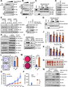
PI(3,4,5)P3 promotes mTORC2 activation to phosphorylate Akt-S473. A, In vitro kinase assays indicating that mTORC2 complexes immunoprecipitated (IP) by HA-Rictor are active upon insulin stimulation in phosphorylating Akt-S473. B, In vitro kinase assays showing that PI(3,4,5)P3-polysomes activate the purified inactive mTORC2 complexes in vitro. HA-Rictor containing mTORC2 complexes were immunoprecipitated from HEK293 cells and serum starved for 36 hr before harvest in CHAPS buffers. 25 µL of 1 mM polyPIPsomes containing 5% indicated PIP species were incubated with 10 µL of HA-Rictor precipitates in kinase assays. C, Insulin treatment attenuated Sin1-PH interaction with mTOR-KD-L. Immunoblot (IB) analysis of whole cell lysates (WCLs) and GST-pulldowns derived from 293 cells transfected with indicated constructs. Where indicated, cells were serum-starved for 24 hr and stimulated by 100 nM insulin for 30 min before harvesting. D, An illustration of solved Akt1-PH/IP4 co-crystal structure (PDB: IUNQ) by PyMOL. E, An illustration of possible Sin1-PH/IP4 complex structure by super-imposing IP4 into solved Sin1-PH structure (PDB: 3VOQ) by PyMOL.
Fig. 5
Fig. 5
PI(3,4,5)P3 mainly interacts with the Sin1-PH domain via R393, K428 and K464 residues to govern mTORC2 activation. A, The Sin1-R393C/K428A/K464A mutant is deficient in phosphorylating Akt-S473. Immunoblot (IB) analysis of whole cell lysates (WCLs) derived from Sin1−/− MEFs transfected with indicated constructs. Cells were serum-starved for 24 hr before 100 ng/ml IGF-1 was added for 30 min. B, Sin1-CAA is deficient in activating mTORC2 upon insulin stimulation. HAP1-Sin1−/− cells were infected with MSCV-Sin1-HA-WT or CAA retro-viruses, selected with 1 µg/ml puromycin for 3 days to eliminate non-infected cells and serum-starved for 24 hr and stimulated with 100 nM insulin for indicated periods before harvesting for IB analyses. C, PI(3,4,5)P3 loses its interaction with Sin1-CAA. IB of PIP3 beads pulldowns derived from HEK293T cells transfected with indicated constructs. D, In vitro kinase assays demonstrating that PI(3,4,5)P3 pulls down active mTORC2 complexes. IB of in vitro kinase assays derived from incubating PI(3,4,5)P3 beads pulldowns from HEK293T cells transfected with indicated constructs, using GST-Akt1-tail (aa 409–480) as a substrate. E, Sin1-PH-CAA is deficient in functionally replacing Akt1-PH. IB analyses of WCLs derived from Akt1/2−/− MEFs transfected with indicated constructs. Where indicated, cells were serum-starved for 36 hr before stimulated by insulin (100 nM) for 30 min or EGF (100 ng/ml) for 10 min. F, Sin1-CAA binds the mTOR kinase domain. IB analysis of HA-IPs and WCLs derived from HEK293T cells transfected with indicated constructs. G, Sin1-PH-CAA retains its ability to suppress mTOR kinase in vitro. In vitro kinase assays of recombinant active mTOR kinases with GST-Akt1-tail (aa 409–480) as a substrate, in the presence of increasing doses of bacterially purified GST-Sin1-PH-CAA recombinant proteins. H, Representative confocal images to illustrate that GFP-Sin1-PH-WT, but not GFP-Sin1-PH-CAA, enriches and co-localizes with Akt1-PH at plasma membrane proximity upon insulin (100 nM) stimulation. I, Sin1-CAA is deficient in activating Akt in cells. IB analysis of WCLs derived from Sin1-depleted OVCAR5 cells stably expressing MSCV-Sin1-WT-HA or MSCV-Sin1-CAA-HA. Where indicated, cells were serum-starved for 36 hr before stimulation by EGF (100 ng/ml) for 10 min. J–K, Soft agar assays using OVCAR5 cell lines generated in (I).
Fig. 6
Fig. 6
PI(3,4,5)P3 releases Sin1-PH-mediated inhibition on mTOR-KD, leading to mTORC2 activation. A–B, PI(3,4,5)P3 competes with mTOR-KD (A), but not full-length mTOR (B) in binding Sin1. Immunoblot (IB) analysis of PIP3 pulldowns, Flag-immunoprecipitates (IPs) and whole cell lysates (WCLs) derived from HEK293T cells transfected with indicated constructs. C, PI(3,4,5)P3-ploysomes attenuate mTOR-KD interaction with Sin1-PH-WT, but not Sin1-PH-CAA. IB analysis of GST pulldowns in the presence of increasing amounts of PIP-polysomes (0,10 or 20 µl) in CHAPS buffers. D, Compared to Sin1-WT, ectopic expression of Sin1-D412G leads to elevated Akt-pS473 in cells. IB analysis of WCLs derived from Sin1−/− MEFs transfected with indicated Sin1 constructs. E, Compared to Sin1-WT, ectopic expression of Sin1-D412G leads to elevated Akt-pS473 under indicated experimental conditions. IB analysis of WCLs derived from Sin1−/− MEFs transfected with indicated Sin1 constructs. Cells were serum-starved for 24 hr before stimulation by insulin (100 nM) for 30 min. F–G, Sin1-D412G loses interaction with mTOR-KD in cells (F) and in vitro (G). IB of HA-IPs and WLCs derived from HEK293T cells transfected with indicated constructs (F), or GST pulldowns in the presence of indicated PIP-polysomes (G). H, Compared to WT-Sin1, Sin1-D412G leads to an elevated Akt activation upon EGF stimulation. IB of WCLs derived from endogenous-Sin1-depleted OVCAR5 cells stably expressing indicated Sin1 constructs. Where indicated, cells were serum-starved for 36 hr before stimulation by EGF (100 ng/ml) for 10 min. I, Compared to WT-Sin1, Sin1-D412G results in reduced apoptosis. IB of WCLs derived from endogenous-Sin1-depleted OVCAR5 cells stably expressing indicated Sin1 constructs. Where marked, cells were treated with etoposide for 24 hr before collection. J–K, Compared to WT-Sin1, Sin1-D412G expressing cells display an elevated resistance to etoposide (J) or cisplatin (K) challenges. Various cell lines generated in (H) were cultured in 10% FBS-containing medium with the indicated concentrations of etoposide (J) or cisplatin (K) for 24 hr before performing cell viability assays. Data was shown as mean ± SD for three independent experiments. * indicates p<0.05 (student’s t-test). L–M, Compared to WT-Sin1, Sin1-D412G expressing cells display enhanced colony formation (L) and soft-agar growth abilities (M). Data was shown as mean ± SD for three independent experiments. * indicates p<0.05 (student’s t-test). N–P, Compared to WT-Sin1, Sin1-D412G expressing cells display enhanced tumor formation in a xenograft mouse model. 3×10⁶ of the generated cells in (H) were injected into nude mice (n=10 for each group) and monitored for tumor formation (O). Formed tumors were dissected (N) and weighed (P). As indicated, * represents p<0.05 calculated by student’s t-test. Q, Compared to WT-Sin1, elevated Akt-pS473 levels were observed in Sin1-D412G expressing xenograft tumors. IB analysis of WCLs derived from xenografted tumors obtained in (N).
Fig. 7
Fig. 7
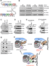
C-terminal tagging of the KRas-CAAX sequence to Sin1 partially rescues the deficiency of Sin1-CAA towards activating Akt. A, A schematic illustration of the Myr-Sin1 or Sin1-CAAX chimera structure. B, C-terminal addition of the KRas-CAAX sequence in part rescues Sin1-CAA in phosphorylating Akt-S473 in cells. Immunoblot (IB) analysis of whole cell lysates (WCLs) derived from Sin1−/− MEFs transfected with indicated constructs. Where indicated, cells were serum starved for 24 hr and stimulated by 100 nM insulin for indicated time periods. C–D, Addition of KRas-CAAX sequence does not affect mTORC2 complex integrity (C) or Sin1 interaction with Akt1 (D). IB of Flag-IPs and WCLs derived from 293T cells transfected with indicated constructs. E–F, The CAAX tag alleviates Sin1-PH interaction with mTOR-KD. IB of GST pulldowns and WCLs derived from 293T cells transfected with indicated constructs. G, Addition of a N-terminal-Myr tag, but not a C-terminal-CAAX tag largely rescues Akt phosphorylation in Akt1-R25C mutant. IB analysis of WCLs derived from DLD1-Akt1/2−/− cells transfected with indicated constructs. H, A proposed model for the PI(3,4,5)P3-mediated mTORC2 activation mechanism.

References

    1. Laplante M, Sabatini DM. mTOR signaling in growth control and disease. Cell. 2012;149:274–293. - PMC - PubMed
    1. Kim DH, Sarbassov DD, Ali SM, King JE, Latek RR, Erdjument-Bromage H, et al. mTOR interacts with raptor to form a nutrient-sensitive complex that signals to the cell growth machinery. Cell. 2002;110:163–175. - PubMed
    1. Sarbassov DD, Ali SM, Kim DH, Guertin DA, Latek RR, Erdjument-Bromage H, et al. Rictor, a novel binding partner of mTOR, defines a rapamycin-insensitive and raptor-independent pathway that regulates the cytoskeleton. Curr Biol. 2004;14:1296–1302. - PubMed
    1. Yang Q, Inoki K, Ikenoue T, Guan KL. Identification of Sin1 as an essential TORC2 component required for complex formation and kinase activity. Genes Dev. 2006;20:2820–2832. - PMC - PubMed
    1. Jacinto E, Facchinetti V, Liu D, Soto N, Wei S, Jung SY, et al. SIN1/MIP1 maintains rictor-mTOR complex integrity and regulates Akt phosphorylation and substrate specificity. Cell. 2006;127:125–137. - PubMed

Publication types

MeSH terms

Substances