. 2015 Oct 9;290(41):24657-68.
doi: 10.1074/jbc.M115.678086. Epub 2015 Aug 20.

Two-step Ligand Binding in a (βα)8 Barrel Enzyme: SUBSTRATE-BOUND STRUCTURES SHED NEW LIGHT ON THE CATALYTIC CYCLE OF HisA

Affiliations

Two-step Ligand Binding in a (βα)8 Barrel Enzyme: SUBSTRATE-BOUND STRUCTURES SHED NEW LIGHT ON THE CATALYTIC CYCLE OF HisA

Annika Söderholm et al. J Biol Chem. .

Abstract

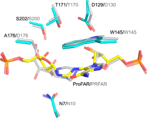
HisA is a (βα)8 barrel enzyme that catalyzes the Amadori rearrangement of N'-[(5'-phosphoribosyl)formimino]-5-aminoimidazole-4-carboxamide ribonucleotide (ProFAR) to N'-((5'-phosphoribulosyl) formimino)-5-aminoimidazole-4-carboxamide-ribonucleotide (PRFAR) in the histidine biosynthesis pathway, and it is a paradigm for the study of enzyme evolution. Still, its exact catalytic mechanism has remained unclear. Here, we present crystal structures of wild type Salmonella enterica HisA (SeHisA) in its apo-state and of mutants D7N and D7N/D176A in complex with two different conformations of the labile substrate ProFAR, which was structurally visualized for the first time. Site-directed mutagenesis and kinetics demonstrated that Asp-7 acts as the catalytic base, and Asp-176 acts as the catalytic acid. The SeHisA structures with ProFAR display two different states of the long loops on the catalytic face of the structure and demonstrate that initial binding of ProFAR to the active site is independent of loop interactions. When the long loops enclose the substrate, ProFAR adopts an extended conformation where its non-reacting half is in a product-like conformation. This change is associated with shifts in a hydrogen bond network including His-47, Asp-129, Thr-171, and Ser-202, all shown to be functionally important. The closed conformation structure is highly similar to the bifunctional HisA homologue PriA in complex with PRFAR, thus proving that structure and mechanism are conserved between HisA and PriA. This study clarifies the mechanistic cycle of HisA and provides a striking example of how an enzyme and its substrate can undergo coordinated conformational changes before catalysis.

Keywords: (beta/alpha)8 barrel; X-ray crystallography; conformational change; enzyme catalysis; enzyme mechanism; enzyme structure; histidine biosynthesis.


Figures

FIGURE 1.
FIGURE 1.
Mechanism for the isomerization of ProFAR to PRFAR catalyzed by HisA (7). AH/A, catalytic acid; B/HB, catalytic base. A, the ring oxygen of the reacting ribose in ProFAR is protonated by the general acid. The free electron pair of the neighboring amine nitrogen forms a double bond with the 1′ carbon. B, the resulting Schiff base intermediate acts as an electron sink allowing deprotonation of the 2′ carbon by a general base. C, the resulting enolamine form of PRFAR spontaneously tautomerizes to the corresponding keto-form (D).
FIGURE 2.
FIGURE 2.
Overall structure of SeHisA. The βα loops on the catalytic side of the (βα)8 barrel are colored by symmetry, and ligands are shown as sticks. A, apo-SeHisA with two phosphate ions. B, SeHisA(D7N) in complex with ProFAR. C, SeHisA(D7N/D176A) in complex with ProFAR. The reacting ribose is oriented to the left in all figures.
FIGURE 3.
FIGURE 3.
The ProFAR binding site in the SeHisA(D7N/D176A)-ProFAR structure. A, ProFAR (yellow) and interacting residues (core residues in cyan, loop residues colored as in Fig. 2) are shown as sticks, waters as red spheres, and hydrogen bonds as dotted lines. The unbiased FoFc simulated annealing omit map for ProFAR (green) is contoured at 3σ. B, two-dimensional projection showing the hydrogen-bond network between ProFAR and SeHisA(D7N/D176A). ProFAR is oriented with phosphate 1 to the left and phosphate 2 to the right.
FIGURE 4.
FIGURE 4.
The ProFAR binding site in the SeHisA(D7N)-ProFAR structure. A, ProFAR (yellow) and interacting residues (core residues in cyan, loop residues colored as in Fig. 2) are shown as sticks, waters as red spheres, and hydrogen bonds as dotted lines. The unbiased FoFc simulated annealing omit map for ProFAR (green) is contoured at 3σ. B, two-dimensional projection showing the hydrogen bonding network between ProFAR and SeHisA(D7N). ProFAR is oriented with phosphate 1 to the left and phosphate 2 to the right.
FIGURE 5.
FIGURE 5.
Comparison of ligand conformations and interactions in the active site of the open SeHisA(D7N) structure (cyan), the closed SeHisA(D7N/D176A) structure (yellow), and the apo-SeHisA structure (gray). A, superposition of ProFAR in the open SeHisA(D7N) structure and the closed SeHisA(D7N/D176A) structure. Right, side view showing the rotational movement of the aminoimidazole carboxamide moiety and the non-reacting ribose going from open to closed structure. B, hydrogen bond network stabilizing the central part of ProFAR in the open SeHisA(D7N) structure. C, the corresponding hydrogen bond network in the closed SeHisA(D7N/D176A) structure and the movement of these residues going from open to closed structure is shown. Trp-145 stacks with the aminoimidazole moiety of ProFAR in the closed structure. D, overlay showing interactions of SeHisA(D7N) and SeHisA(D7N/D176A) with the phosphates of ProFAR (left, phosphate 1; right, phosphate 2). E, overlay showing interactions of SeHisA(D7N/D176A) with the phosphates of ProFAR and SeHisA with free phosphates (left, phosphate 1; right, phosphate 2).
FIGURE 6.
FIGURE 6.
Superposition of MtPriA-PRFAR (PDB code 2Y88) (10) in gray, with the equivalent residues and ligand in the SeHisA(D7N/D176A)-ProFAR structure (cyan and yellow).
FIGURE 7.
FIGURE 7.
Structure-based multiple sequence alignment showing the secondary structure for SeHisA(D7N/D176A) (top) and MtPriA-PRFAR (PDB code 2Y88, bottom). The βα loops are colored as in Fig. 2. The boldface and boxed residues are conserved in >96% of sequences with >25% sequence identity to SeHisA according to our ConSurf analysis. SeHisA residues indicated by black asterisks were mutagenized in this study. The accession codes and amino acid sequence identities to SeHisA for the sequences are as follows: SeHisA (WP_000586409.1); EcHisA (WP_000586452.1, 93%); CjHisA (WP_002851391.1, 51%); TmHisA (WP_004080485.1, 25%); ScPriA (WP_003976766.1, 33%); and MtPriA (WP_003900374.1, 33%).
FIGURE 8.
FIGURE 8.
The catalytic cycle of SeHisA. A, in the uncharged enzyme, loops 1 and 6 are flexible, and loops 2 and 5 are distant from each other. B, ProFAR docks into the fully exposed active site. C, binding of the substrate induces conformational changes in the enzyme. Loops 1, 2, 5, and 6 form interactions with the substrate, resulting in loop closure and burying of the ligand. Coupled to loop ordering, the non-reacting half of ProFAR moves toward the edge of the barrel and adopts a product-like extended conformation stabilized by additional enzyme interactions. D, the conformational changes of substrate and enzyme prepare the complex for catalysis. After the reaction, the loops open, and PRFAR is released.

References

    1. Jürgens C., Strom A., Wegener D., Hettwer S., Wilmanns M., Sterner R. (2000) Directed evolution of a (βα)8-barrel enzyme to catalyze related reactions in two different metabolic pathways. Proc. Natl. Acad. Sci. U.S.A. 97, 9925–9930 - PMC - PubMed
    1. Näsvall J., Sun L., Roth J. R., Andersson D. I. (2012) Real-time evolution of new genes by innovation, amplification, and divergence. Science 338, 384–387 - PMC - PubMed
    1. Barona-Gómez F., Hodgson D. A. (2003) Occurrence of a putative ancient-like isomerase involved in histidine and tryptophan biosynthesis. EMBO Rep. 4, 296–300 - PMC - PubMed
    1. Noda-García L., Camacho-Zarco A. R., Medina-Ruíz S., Gaytán P., Carrillo-Tripp M., Fülöp V., Barona-Gómez F. (2013) Evolution of substrate specificity in a recipient's enzyme following horizontal gene transfer. Mol. Biol. Evol. 30, 2024–2034 - PubMed
    1. Lang D., Thoma R., Henn-Sax M., Sterner R., Wilmanns M. (2000) Structural evidence for evolution of the β/α barrel scaffold by gene duplication and fusion. Science 289, 1546–1550 - PubMed

Publication types

Substances