. 2015:2015:750752.
doi: 10.1155/2015/750752. Epub 2015 Jul 29.

Cocaine Causes Apoptotic Death in Rat Mesencephalon and Striatum Primary Cultures

Affiliations

Cocaine Causes Apoptotic Death in Rat Mesencephalon and Striatum Primary Cultures

Lucilia B Lepsch et al. Biomed Res Int. 2015.

Abstract

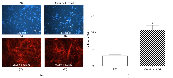
To study cocaine's toxic effects in vitro, we have used primary mesencephalic and striatal cultures from rat embryonic brain. Treatment with cocaine causes a dramatic increase in DNA fragmentation in both primary cultures. The toxicity induced by cocaine was paralleled with a concomitant decrease in the microtubule associated protein 2 (MAP2) and/or neuronal nucleus protein (NeuN) staining. We also observed in both cultures that the cell death caused by cocaine was induced by an apoptotic mechanism, confirmed by TUNEL assay. Therefore, the present paper shows that cocaine causes apoptotic cell death and inhibition of the neurite prolongation in striatal and mesencephalic cell culture. These data suggest that if similar neuronal damage could be produced in the developing human brain, it could account for the qualitative or quantitative defects in neuronal pathways that cause a major handicap in brain function following prenatal exposure to cocaine.


Figures

Figure 1
Figure 1
(a) Immunostaining of striatal primary cells treated with PBS (left panel) or treated with cocaine 1.0 mM (right panel) for 24 hours. Neurons were labeled with MAP2 and NeuN (red label). Hoechst 33342 (blue label) was added to monitor chromatin condensation. Arrows indicate dying neurons. Staining was observed under a fluorescent microscope. The treatment with cocaine caused a decrease in neuronal viability and an inhibition of neurite prolongation. (b) Percentage of cell death observed by immunostaining of striatal primary cultures treated with PBS and cocaine for 24 hours. Values are mean ± SEM from five independent experiments. The treatment with cocaine caused a decrease in the viability of the neurons. Significantly different from the control (PBS) value: P < 0.05 by Student's t-test.
Figure 2
Figure 2
(a) Immunostaining of mesencephalic primary cells treated with PBS (left panel) or treated with cocaine 1.0 mM (right panel) for 24 hours. The neurons were labeled with MAP2 and NeuN (red labels). Hoechst 33342 (blue label) was added to monitor chromatin condensation. Arrows indicate dying neurons. Staining was observed under a fluorescent microscope. Cocaine treatment caused a decrease in neuronal viability. (b) Percentage of cell death observed by immunostaining of the mesencephalic primary culture treated with PBS and cocaine for 24 hours. Values are mean ± SEM from four independent experiments. Cocaine treatment decreased neuronal viability. Significantly different from the control (PBS) value: P < 0.05 by Student's t-test.
Figure 3
Figure 3
(a) In situ histochemical evidence of DNA fragmentation after cocaine exposure. Striatal cultures were first established for 7 days and incubated with cocaine (1.0 mM) for 24 hours. After cells were fixed, the TUNEL method was performed. Cultures were photographed at the level of the neuronal layer. Note the labeling in the vast majority of treated cells, in contrast with the labeling of a few control cells. TUNEL positive cells were dUTP labeled (brown label). The neurons were labeled with MAP2 and NeuN (green label) and Hoechst 33342 (blue label) was added to monitor chromatin condensation. (b) Number of TUNEL positive cells observed by immunostaining of the striatal primary culture treated with PBS or cocaine for 24 hours. Values are mean ± SEM from five independent experiments. Cocaine treatment decreased neuronal viability. Significantly different from the control (PBS) value: P < 0.05 by Student's t-test.
Figure 4
Figure 4
(a) Mesencephalic cultures were first established for 7 days and incubated with cocaine (1.0 mM) for 24 hours. After cells were fixed, the TUNEL method was used. Cultures were photographed at the level of the neuronal layer. Note the labeling in the vast majority of treated cells, in contrast with the labeling of a few control cells. TUNEL positive cells were dUTP labeled (brown label). The neurons were labeled with MAP2 and NeuN (green label) and Hoechst 33342 (blue label) was added to monitor chromatin condensation. (b) Number of TUNEL positive cells observed by immunostaining of the mesencephalic primary culture treated with PBS or cocaine for 24 hours. Values are mean ± SEM from five independent experiments. Cocaine treatment decreased neuronal viability. Significantly different from the control (PBS) value: P < 0.05 by Student's t-test.

References

    1. Nestler E. J., Aghajanian G. K. Molecular and cellular basis of addiction. Science. 1997;278(5335):58–63. doi: 10.1126/science.278.5335.58. - DOI - PubMed
    1. Kennedy L. T., Hanbauer I. Sodium-sensitive cocaine binding to rat striatal membrane: possible relationship to dopamine uptake sites. Journal of Neurochemistry. 1983;41(1):172–178. doi: 10.1111/j.1471-4159.1983.tb13666.x. - DOI - PubMed
    1. Reith M. E. A., Meisler B. E., Sershen H., Lajtha A. Structural requirements for cocaine congeners to interact with dopamine and serotonin uptake sites in mouse brain and to induce stereotyped behavior. Biochemical Pharmacology. 1986;35(7):1123–1129. doi: 10.1016/0006-2952(86)90148-6. - DOI - PubMed
    1. Ritz M. C., Lamb R. J., Goldberg S. R., Kuhar M. J. Cocaine receptors on dopamine transporters are related to self-administration of cocaine. Science. 1987;237(4819):1219–1223. doi: 10.1126/science.2820058. - DOI - PubMed
    1. Wise R. A. Brain reward circuitry: Insights from unsensed incentives. Neuron. 2002;36(2):229–240. doi: 10.1016/S0896-6273(02)00965-0. - DOI - PubMed

Publication types

LinkOut - more resources