

Identification of Foot Pathologies Based on Plantar Pressure Asymmetry
- PMID: 26295239
- PMCID: PMC4570427
- DOI: 10.3390/s150820392
Identification of Foot Pathologies Based on Plantar Pressure Asymmetry
Abstract
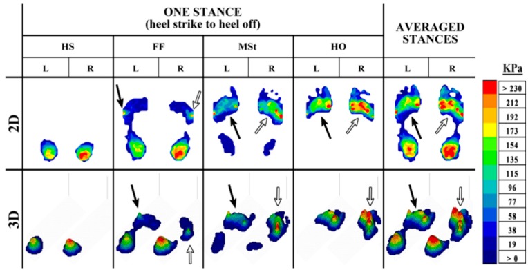
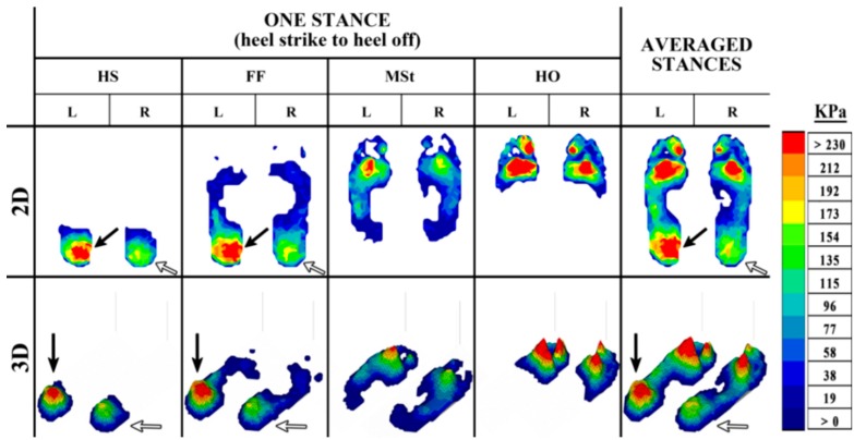
Foot pathologies can negatively influence foot function, consequently impairing gait during daily activity, and severely impacting an individual's quality of life. These pathologies are often painful and correspond with high or abnormal plantar pressure, which can result in asymmetry in the pressure distribution between the two feet. There is currently no general consensus on the presence of asymmetry in able-bodied gait, and plantar pressure analysis during gait is in dire need of a standardized method to quantify asymmetry. This paper investigates the use of plantar pressure asymmetry for pathological gait diagnosis. The results of this study involving plantar pressure analysis in fifty one participants (31 healthy and 20 with foot pathologies) support the presence of plantar pressure asymmetry in normal gait. A higher level of asymmetry was detected at the majority of the regions in the feet of the pathological population, including statistically significant differences in the plantar pressure asymmetry in two regions of the foot, metatarsophalangeal joint 3 (MPJ3) and the lateral heel. Quantification of plantar pressure asymmetry may prove to be useful for the identification and diagnosis of various foot pathologies.
Keywords: foot pathology; gait symmetry; plantar pressure.
Figures
Similar articles
-
Comparison of the asymmetries in foot posture, gait and plantar pressure between patients with unilateral and bilateral knee osteoarthritis based on a cross-sectional study.Sci Rep. 2024 Nov 5;14(1):26761. doi: 10.1038/s41598-024-78166-z. Sci Rep. 2024. PMID: 39501072 Free PMC article.
-
Plantar pressures during shod gait in diabetic neuropathic patients with and without a history of plantar ulceration.J Am Podiatr Med Assoc. 2009 Jul-Aug;99(4):285-94. doi: 10.7547/0980285. J Am Podiatr Med Assoc. 2009. PMID: 19605921
-
[Asymmetries in dynamic plantar pressure distribution measurement in able-bodied gait: application to the study of the gait asymmetries in children with hemiplegic cerebral palsy].Ann Readapt Med Phys. 2002 Mar;45(3):114-22. doi: 10.1016/s0168-6054(02)00186-1. Ann Readapt Med Phys. 2002. PMID: 11911930 French.
-
Gait-related strategies for the prevention of plantar ulcer development in the high risk foot.Curr Diabetes Rev. 2011 May;7(3):159-63. doi: 10.2174/157339911795843159. Curr Diabetes Rev. 2011. PMID: 21521160 Review.
-
A Review in Detection and Monitoring Gait Disorders Using In-Shoe Plantar Measurement Systems.IEEE Rev Biomed Eng. 2017;10:299-309. doi: 10.1109/RBME.2017.2747402. Epub 2017 Aug 30. IEEE Rev Biomed Eng. 2017. PMID: 28866600 Review.
Cited by
-
Influence of motor capacity of the lower extremity and mobility performance on foot plantar pressures in community-dwelling older women.Heliyon. 2024 Mar 19;10(6):e28114. doi: 10.1016/j.heliyon.2024.e28114. eCollection 2024 Mar 30. Heliyon. 2024. PMID: 38560666 Free PMC article.
-
Effect of Thoracic Kyphosis and Lumbar Lordosis on the Distribution of Ground Reaction Forces on the Feet.Orthop Res Rev. 2022 May 16;14:187-197. doi: 10.2147/ORR.S344972. eCollection 2022. Orthop Res Rev. 2022. PMID: 35601185 Free PMC article.
-
Flexible Smart Insole and Plantar Pressure Monitoring Using Screen-Printed Nanomaterials and Piezoresistive Sensors.ACS Appl Mater Interfaces. 2025 Aug 20;17(33):47153-47161. doi: 10.1021/acsami.5c08296. Epub 2025 Jul 29. ACS Appl Mater Interfaces. 2025. PMID: 40729702 Free PMC article.
-
Comparison of plantar pressure distribution patterns of patients with ankylosing spondylitis and asymptomatic healthy individuals: a cross-sectional study.Ir J Med Sci. 2025 Aug 16. doi: 10.1007/s11845-025-04064-6. Online ahead of print. Ir J Med Sci. 2025. PMID: 40817955
-
Optical-Based Foot Plantar Pressure Measurement System for Potential Application in Human Postural Control Measurement and Person Identification.Sensors (Basel). 2021 Jun 28;21(13):4437. doi: 10.3390/s21134437. Sensors (Basel). 2021. PMID: 34203534 Free PMC article.
References
-
- Winter D.A. The Biomechanics and Motor Control of Human Gait: Normal, Elderly and Pathological. Waterloo Press; Waterloo, ON, Canada: 1991.
-
- Perry J. Gait Analysis: Normal and Pathological Function. Slack; Thorofare, NJ, USA: 1992.
-
- Rai D., Aggarwal L. The study of plantar pressure distribution in normal and pathological foot. Polish J. Med. Phys. Eng. 2006;12:25–34.
MeSH terms
LinkOut - more resources
Full Text Sources
Other Literature Sources
Medical
