Review
. 2015 Sep;16(9):554-67.
doi: 10.1038/nrm4041.

Microfluidics: reframing biological enquiry

Affiliations
Review

Microfluidics: reframing biological enquiry

Todd A Duncombe et al. Nat Rev Mol Cell Biol. 2015 Sep.

Abstract

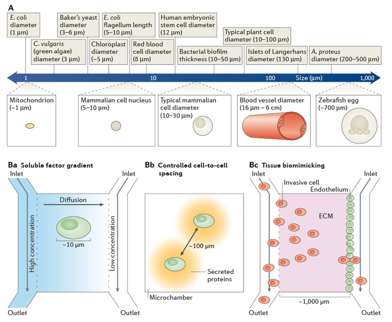
The underlying physical properties of microfluidic tools have led to new biological insights through the development of microsystems that can manipulate, mimic and measure biology at a resolution that has not been possible with macroscale tools. Microsystems readily handle sub-microlitre volumes, precisely route predictable laminar fluid flows and match both perturbations and measurements to the length scales and timescales of biological systems. The advent of fabrication techniques that do not require highly specialized engineering facilities is fuelling the broad dissemination of microfluidic systems and their adaptation to specific biological questions. We describe how our understanding of molecular and cell biology is being and will continue to be advanced by precision microfluidic approaches and posit that microfluidic tools - in conjunction with advanced imaging, bioinformatics and molecular biology approaches - will transform biology into a precision science.


Conflict of interest statement

Competing interests statement

The authors declare competing interests: see Web version for details.

Figures

Figure 1 |
Figure 1 |. A flow diagram highlighting crucial developments in microfluidics, from fabrication approaches and functional developments in the evolution of the technology platform to some of the applications discussed in this Review.
As early as the 1960s, seminal microfluidic technologies were increasing the speed and resolution of bioanalysis for a small number of powerful analytical tools. Crucial fabrication developments, which began in 1990, allowed researchers to transition from using pulled glass capillaries to microfabricated glass or polymer microchannels,. Microfluidic tools have precisely defined features and contain highly complex channel networks and fluidic components that can execute specific functions,,,–. In the past decade, the increasing availability of fluidic components and number of experienced microfluidic researchers have opened the door to new biological applications; these include new single-cell measurements,,, high-throughput monoclonal antibody screening, analysis of dynamic phenomena,, and organ-on-a-chip platforms. PDMS, polydimethylsiloxane.
Figure 2 |
Figure 2 |. Physics at the microscale.
a | Bioanalysis in small, confined microfluidic volumes can enhance reaction speeds for detecting low-abundance molecules, compared with reactions conducted in a conventional chamber. In the case of detecting proteins from a single HeLa cell (median protein abundance ~170,000 molecules per cell), a sufficient protein concentration is maintained in the microfluidic system for an antibody (assuming kon = 105 M−1 s−1 and a 60 kDa protein) to reach reaction equilibrium within ~1 hour, whereas it would take ~106 hours to reach equilibrium in the conventional system, owing to the relative concentration differences. The minimal diffusive losses that are associated with microfluidic volumes are essential for measuring proteins from single cells and other analytes that cannot be amplified, b | The predictable laminar flow that occurs in a microfluidic device is one of its most important features. The hydrodynamic flow focusing of cells is shown for different flow regimes, which vary according to the flow velocity, channel diameter or viscosity (these parameters are reflected in the Reynolds number (Re)). At near Stokes flow (Re <<1) and low Re flow (Re <10), which are the conditions in most microfluidic devices, the effects of inertial forces are minimal and for most applications can be ignored. In inertial microfluidics (typically 10 < Re <1,000), high pressures are used to create relatively high flow velocities, so that particles (such as cells) have inertial interactions with objects or flows. Inertial microfluidics can be used for several different applications, including size-based sorting, streamline focusing and mechanical measurements. Above a Re of 2,000 in pipe flow, which is not practical to achieve in microfluidic devices, a transition to turbulent flow occurs.
Figure 3 |
Figure 3 |. Dynamic process analysis.
a | The schematic depicts a microfluidic device designed for the generation of pulses of a soluble factor by alternating the pressures at two separate inlets. The reproducible behaviour of fluids at the microscale enables the input to be described mathematically. Microfluidic devices easily interface with real-time microscopy (owing to their planar form factor), which enables the output of cells cultured in situ to be directly monitored as they respond to the pulses of soluble factor. Rapid pulses (< 10 seconds between pulses) have been used to study rapid biological processes such as the nuclear localization of transcription factors and their interactions with promoters. b | The logarithmic ruler shows the timescales of dynamic biological processes and the time resolutions of the corresponding microfluidic techniques that can be used to study them (as indicated by corresponding colours). EGFR, epidermal growth factor receptor.
Figure 4 |
Figure 4 |. High-throughput microfluidics.
a | Integrating sample processing and analysis steps within microfluidic technologies has increased the throughput and data output of a range of analytical tools to orders of magnitude higher than those obtained using conventional approaches. For example, the mechanical properties of a cell can be measured using conventional methods (such as atomic force microscopy) at a throughput of ~10 cells per hour; by contrast, inertial microfluidics has been used to mechanically assess ~65,000 cells per second. b | Water-in-oil droplet microfluidic technologies are at the forefront of the increase in analytical through put. Thousands of droplets are produced per second, each of which is a precisely defined experimental reaction chamber. Downstream fluidic components carryout serial high-throughput sample processing, which facilitates the analysis of hundreds of millions of samples per day. In one study, the directed evolution of horseradish peroxidase (HRP) from a mutant yeast library was carried out using droplet microfluidics. The researchers screened 108 individual enzyme reactions in only 10 hours, which is an ~1,000-fold increase in speed and an ~10 million-fold decrease in cost compared with conventional tools. ELISA; enzyme-linked immunosorbent assay.
Figure 5 |
Figure 5 |. Biological length scales.
A | The logarithmic ruler shows the length scales of important biological features. The feature sizes of microfluidic devices facilitate experiments that are relevant at the micrometre scale and that are not possible with conventional techniques. Ba–c | Examples of how microfluidic length scales can match the relevant biology. Cells respond to differences in concentrations of soluble factors across their surfaces; therefore, experimental gradients of soluble factors must be generated at the same order of magnitude as the cell diameter (part Ba). By having reservoirs that are set at different concentrations of soluble factors at either end of a microchannel, linear concentration gradients can be formed by diffusion. Physiologically relevant cell-to-cell spacing can be realized by confining pairs of cells in microchambers (part Bb). Micrometre-scale tissue morphology can be replicated in microfluidic devices (part Bc). For example, events in cancer metastasis can be studied by establishing an endothelium adjacent to an extracellular matrix (ECM) in vitro and directly observing intravasation or extravasation with invasive cells. A. proteus, Amoeba proteus; C. vulgaris, Chlorella vulgaris; E. coli, Escherichia coli.

References

    1. Lanier LL Just the FACS. J. Immunol 193, 2043–2044 (2014). - PubMed
    1. Dovichi NJ & Zhang JZ How capillary electrophoresis sequenced the human genome. Angew. Chem. Int. Ed. Engl 39, 4463–4468 (2000). - PubMed
    1. Duffy DC, McDonald JC, Schueller OJ & Whitesides GM Rapid prototyping of microfluidic systems in poly(dimethylsiloxane). Anal. Chem 70, 4974–4984 (1998). - PubMed
    1. Agresti JJ et al. Ultrahigh-throughput screening in drop-based microfluidics for directed evolution. Proc. Natl Acad. Sci. USA 107, 4004–4009 (2010). - PMC - PubMed
    1. Shi Q et al. Single-cell proteomic chip for profiling intracellular signaling pathways in single tumor cells. Proc. Natl Acad. Sci. USA 109, 419–424 (2012). - PMC - PubMed
    2. Multiplexed single-cell resolution immunoassays were used to directly correlate protein phosphorylation in a signalling pathway within a single cell, for thousands of cells in parallel.

Publication types