. 2015 Aug 22;6(1):148.
doi: 10.1186/s13287-015-0142-x.

Comparison of adipose tissue- and bone marrow- derived mesenchymal stem cells for alleviating doxorubicin-induced cardiac dysfunction in diabetic rats

Affiliations

Comparison of adipose tissue- and bone marrow- derived mesenchymal stem cells for alleviating doxorubicin-induced cardiac dysfunction in diabetic rats

Hania Ibrahim Ammar et al. Stem Cell Res Ther. .

Abstract

Introduction: Doxorubicin (DOX) is a well-known anticancer drug. However its clinical use has been limited due to cardiotoxic effects. One of the major concerns with DOX therapy is its toxicity in patients who are frail, particularly diabetics. Several studies suggest that mesenchymal stem cells (MSCs) have the potential to restore cardiac function after DOX-induced injury. However, limited data are available on the effects of MSC therapy on DOX-induced cardiac dysfunction in diabetics. Our objective was to test the efficacy of bone marrow-derived (BM-MSCs) and adipose-derived MSCs (AT-MSCs) from age-matched humans in a non-immune compromised rat model.

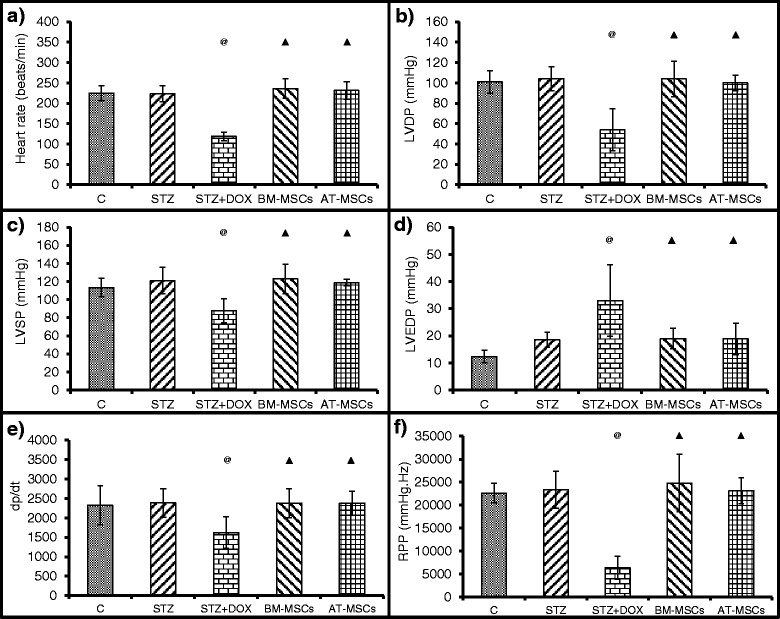
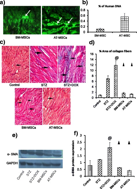
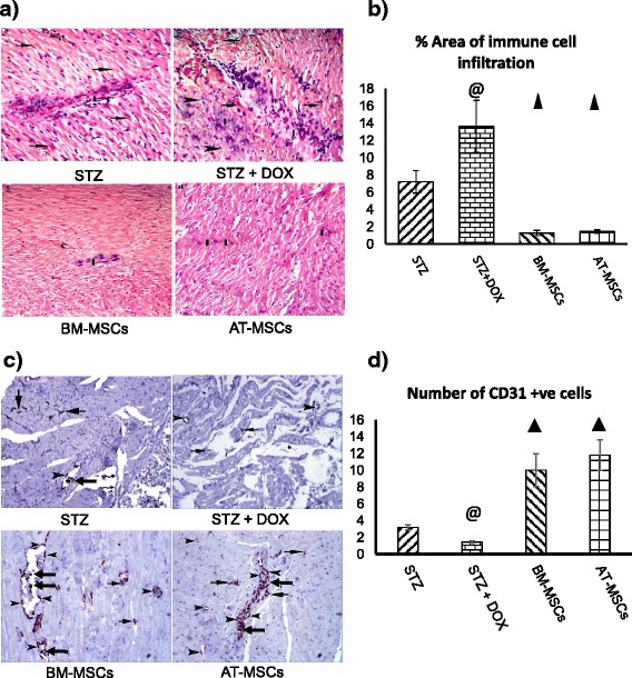
Methods: Diabetes mellitus was induced in rats by streptozotocin injection (STZ, 65 mg/kg b.w, i.p.). Diabetic rats were treated with DOX (doxorubicin hydrochloride, 2.5 mg/kg b.w, i.p) 3 times/wk for 2 weeks (DOX group); or with DOX+ GFP labelled BM-MSCs (2x10⁶cells, i.v.) or with DOX + GFP labelled AT-MSCs (2x10⁶cells, i.v.). Echocardiography and Langendorff perfusion analyses were carried out to determine the heart function. Immunostaining and western blot analysis of the heart tissue was carried out for CD31 and to assess inflammation and fibrosis. Statistical analysis was carried out using SPSS and data are expressed as mean ± SD.

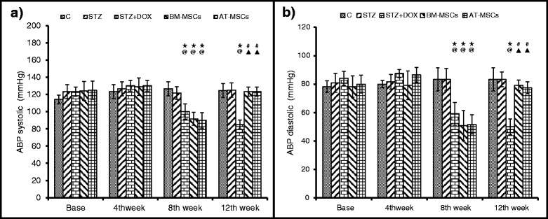
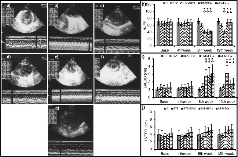
Results: Glucose levels in the STZ treated groups were significantly greater than control group. After 4 weeks of intravenous injection, the presence of injected MSCs in the heart was confirmed through fluorescent microscopy and real time PCR for ALU transcripts. Both BM-MSCs and AT-MSCs injection prevented DOX-induced deterioration of %FS, LVDP, dp/dt max and rate pressure product. Staining for CD31 showed a significant increase in the number of capillaries in BM-MSCs and AT-MSCs treated animals in comparison to DOX treated group. Assessment of the inflammation and fibrosis revealed a marked reduction in the DOX-induced increase in immune cell infiltration, collagen deposition and αSMA in the BM-MSCs and AT-MSCs groups.

Conclusions: In conclusion BM-MSCs and AT-MSCs were equally effective in mitigating DOX-induced cardiac damage by promoting angiogenesis, decreasing the infiltration of immune cells and collagen deposition.


Figures

Fig. 1
Fig. 1
Immunophenotyping of bone marrow-derived mesenchymal stem cells (BM-MSCs) by flow cytometry. a Flow gate; b CD45; c CD34; d CD44; e CD90 and f CD105. All the cells were negative for CD45 and CD34 and >90 % of the cells were positive for CD44, CD90 and CD105. FS fractional shortening, FITC fluorescein isothiocyanate
Fig. 2
Fig. 2
Blood glucose, body weight, serum insulin and fibrinogen levels were measured in different groups. Data are mean ± SD. a Blood glucose levels increased after streptozotocin (STZ) treatment, and both bone marrow-derived mesenchymal stem cell (BM-MSC) and adipose tissue derived mesenchymal stem cell (AT-MSC) treatment normalized glucose levels. b Body weight increased normally in the control group (C) after 4,8 and 12 weeks; in the STZ and the STZ + doxorubicin (STZ + DOX) groups body weight decreased at these time points, which was rescued after treatment with BM-MSCs and AT-MSCs. c Insulin and d fibrinogen levels deviated in the STZ and STZ + DOX groups; treatment with BM-MSCs and AT-MSCs normalized these parameters. *P <0.05 compared to respective baselines, @ P <0.05 compared to respective controls at same time points, # P <0.05 compared to respective pretreatment (8 weeks), P <0.05 compared to the untreated STZ + DOX group
Fig. 3
Fig. 3
Arterial blood pressure (ABP) was measured in different groups. Data are mean ± SD. a Systolic ABP and b diastolic ABP significantly decreased after 4 weeks of doxorubicin (DOX) treatment (8 weeks after streptozotocin (STZ) treatment). Both BM-MSC and AT-MSC treatment rescued ABP levels. *P <0.05 compared to respective baselines, @ P <0.05 compared to respective controls (C) at time points, # P <0.05 compared to respective pretreatment (8 weeks), P <0.05 compared to the untreated STZ + DOX group
Fig. 4
Fig. 4
Heart function was assessed in different groups by echocardiography. Data are mean ± SD. a-g M mode pictures in different treatment groups. a Control, b streptozotocin (STZ), c STZ + doxorubicin (STZ + DOX, d bone marrow-derived mesenchymal stem cells (BM-MSCs) before injection, e BM-MSCs post-treatment, f adipose tissue-derived mesenchymal stem cells (AT-MSCs) before injection, g AT-MSCs post-treatment, h percent fractional shortening (% FS), i LVESD, and j left ventricular end diastolic dimension (LVEDD). After 4 weeks of DOX treatment (8 weeks after STZ) heart function deteriorated; both BM-MSC and AT-MSC implantation improved heart function. *P <0.05 compared to respective baselines, @ P <0.05 compared to respective controls at time points, # P <0.05 compared to respective pretreatment (8 weeks), P <0.05 compared to the untreated STZ + DOX group
Fig. 5
Fig. 5
Retrograde Langendorff perfusion was performed on isolated hearts at the end of the study (12 weeks after streptozotocin (STZ) injection) to measure a heart rate, b left ventricular developed pressure (LVDP), c LVSP, d left ventricular end diastolic pressure (LVEDP), e maximum rate of pressure rise (dp/dt max) and f rate pressure product (RPP) in different groups. Data are mean ± SD. @ P <0.05 compared to respective controls at time points, P <0.05 compared to the untreated STZ + doxorubicin (STZ + DOX) group. BM-MSCs bone marrow-derived mesenchymal stem cells, AT-MSCs adipose tissue-derived mesenchymal stem cells
Fig. 6
Fig. 6
a Bone marrow-derived mesenchymal stem cells (BM-MSCs) and adipose tissue-derived mesenchymal stem cells (AT-MSCs) were labeled with green fluorescent protein (GFP) before injection. Implanted cells were detected in the myocardium 4 weeks after intravenous injection through the tail vein (magnification × 10). b Real-time PCR was performed to quantify transplanted MSCs in the heart. Genomic DNA was isolated from the heart samples, and ALU PCR was performed using TaqMan probes to detect transplanted human MSCs in the rat myocardium. Histograms show percentage of human DNA found in total DNA extracted from rat heart tissues from the BM-MSC and AT-MSC groups. c and d Myocardial sections were stained with Masson’s trichrome, (magnification × 200) to detect fibrosis in the heart in the different groups (control, streptozotocin (STZ), STZ + doxorubicin (STZ + DOX), BM-MSCs and AT-MSCs; d histograms show percent area of collagen deposition. Black arrows indicate collagen fibers. Quantification of the cells in various groups was performed using the Leica Qwin 500 LTD computer-assisted image analysis system (Cambridge, UK). Measurements were done in 10 high power fields (HPF) in all the experimental groups. e and f α-smooth muscle actin (α-SMA) expression was assessed by western blot in different groups. f Histograms show α-SMA levels, values were normalized with glyceraldehyde-3-phosphate dehydrogenase (GAPDH). @ P <0.05 compared to STZ, P <0.05 compared to STZ and STZ + DOX
Fig. 7
Fig. 7
a H & E staining was performed to detect infiltration of immune cells in the heart. Photomicrographs of heart sections (×200) from different groups: control, streptozotocin (STZ), STZ + doxorubicin (STZ + DOX), bone marrow-derived mesenchymal stem cells (BM-MSCs) and adipose tissue-derived mesenchymal stem cells (AT-MSCs) . Black arrows indicate infiltrating immune cells. b Histograms show percent area of immune cell infiltration. c CD31 expression in the myocardium was examined by immunohistochemistry (magnification × 200) in different groups. Black arrows indicate CD31+ cells; wavy arrows show damaged muscle tissue. d Histograms show percentage of CD31+ cells. Quantification of the cells in various groups was performed using the Leica Qwin 500 LTD computer-assisted image analysis system (Cambridge, UK). The measurements were done in 10 high power fields in all the experimental groups. @ P <0.05 compared to STZ, P <0.05 compared to STZ and STZ + DOX

References

    1. International Diabetes Federation. IDF Diabetes Atlas. In: Guariguata L, Nolan T, Beagley J, Linnenkamp U, Jacqmain O, editors. International Diabetes Federation. 6th ed. 2013. Chapter 2: 32-49.
    1. Onitilo AA, Engel JM, Glurich I, Stankowski RV, Williams GM, Doi SA. Diabetes and cancer I: risk, survival, and implications for screening. Cancer Causes Control. 2012;23:967–81. doi: 10.1007/s10552-012-9972-3. - DOI - PMC - PubMed
    1. Xu CX, Zhu HH, Zhu YM. Diabetes and cancer: Associations, mechanisms, and implications for medical practice. World J Diabetes. 2014;5:372–80. doi: 10.4239/wjd.v5.i3.372. - DOI - PMC - PubMed
    1. Tacar O, Sriamornsak P, Dass CR. Doxorubicin: an update on anticancer molecular action, toxicity and novel drug delivery systems. J Pharm Pharmacol. 2013;65:157–70. doi: 10.1111/j.2042-7158.2012.01567.x. - DOI - PubMed
    1. Al-Shabanah OA, El-Kashef HA, Badary OA, Al-Bekairi AM, Elmazar MM. Effect of streptozotocin-induced hyperglycaemia on intravenous pharmacokinetics and acute cardiotoxicity of doxorubicin in rats. Pharmacol Res. 2000;41:31–7. doi: 10.1006/phrs.1999.0568. - DOI - PubMed

Publication types

LinkOut - more resources