. 2015 Sep 22;6(28):26437-56.
doi: 10.18632/oncotarget.4664.

Protein kinase C-related kinase 1 and 2 play an essential role in thromboxane-mediated neoplastic responses in prostate cancer

Affiliations

Protein kinase C-related kinase 1 and 2 play an essential role in thromboxane-mediated neoplastic responses in prostate cancer

Aine G O'Sullivan et al. Oncotarget. .

Abstract

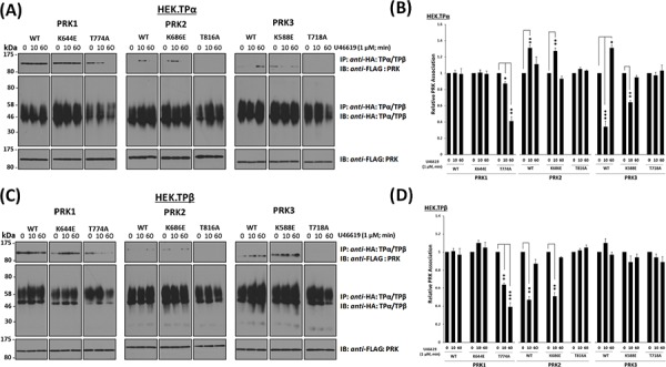
The prostanoid thromboxane (TX) A2 is increasingly implicated in neoplastic progression, including prostate cancer (PCa). Mechanistically, we recently identified protein kinase C-related kinase (PRK) 1 as a functional interactant of both the TPα and TPβ isoforms of the human T prostanoid receptor (TP). The interaction with PRK1 was not only essential for TPα/TPβ-induced PCa cell migration but also enabled the TXA2-TP axis to induce phosphorylation of histone H3 at Thr11 (H3Thr11), an epigenetic marker both essential for and previously exclusively associated with androgen-induced chromatin remodelling and transcriptional activation. PRK1 is a member of a subfamily of three structurally related kinases comprising PRK1/PKNα, PRK2/PKNγ and PRK3/PKNβ that are widely yet differentially implicated in various cancers. Hence, focusing on the setting of prostate cancer, this study investigated whether TPα and/or TPβ might also complex with PRK2 and PRK3 to regulate their activity and neoplastic responses. While TPα and TPβ were found in immune complexes with PRK1, PRK2 and PRK3 to regulate their activation and signalling, they do so differentially and in a TP agonist-regulated manner dependent on the T-loop activation status of the PRKs but independent of their kinase activity. Furthermore, TXA2-mediated neoplastic responses in prostate adenocarcinoma PC-3 cells, including histone H3Thr11 phosphorylation, was found to occur through a PRK1- and PRK2-, but not PRK3-, dependent mechanism. Collectively, these data suggest that TXA2 acts as both a neoplastic and epigenetic regulator and provides a mechanistic explanation, at least in part, for the prophylactic benefits of Aspirin in reducing the risk of certain cancers.

Keywords: cancer; prostate; protein kinase C-related kinase; receptor; thromboxane.


Conflict of interest statement

CONFLICTS OF INTEREST

The authors declare that they have no competing interests.

Figures

Figure 1
Figure 1. Association of PRK1 and PRK2 with TPα and TPβ in prostate PC-3 cells
Panel A. PC-3 cells were immunoprecipitated with anti-TPα, anti-TPβ or, as controls, with the pre-immune (IgG) sera. Thereafter, immunoprecipitates (upper panels) or equivalent aliquots of whole cell lysates (20 μg/lane, lower panels) were immunoblotted (IB) with anti-PRK1, anti-PRK2 or anti-PRK3 antisera. The relative positions of the molecular size markers (kDa) are indicated to the left of the panels. Data shown are representative of at least three independent experiments (n ≥ 3). Panel B. PC-3 cells were incubated with U46619 (1 μM; 0–60 min) prior to immunoprecipitation with anti-TPα, anti-TPβ or, as controls, with the pre-immune (IgG) sera. Thereafter, immunoprecipitates (upper panels) or equivalent aliquots of whole cell lysates (20 μg/lane, lower panels) were IB with anti-PRK1, anti-PRK2 or anti-PRK3 antisera. Data n ≥ 3. Panel C. Bar charts show the mean relative levels of PRK1 or PRK2 associated with the anti-TPα or anti-TPβ immunoprecipitates, as determined by quantitative densitometry (± SEM), where levels associated with the respective immunoprecipitates in the absence of agonist are expressed as 1. The asterisks indicate where U46619 stimulation resulted in significant changes in complex-associated PRK1 or PRK2, where * and ** indicate p < 0.05 and p < 0.01, respectively.
Figure 2
Figure 2. Association of PRK1, PRK2 and PRK3 with TPα and TPβ in HEK 293 cells
Panels A & B. HEK 293 cells stably over-expressing HA-tagged TPα (Panel A) or TPβ (Panel B) and co-transfected with FLAG-tagged PRK1, PRK2 or PRK3 were incubated with U46619 (1 μM; 0–60 min) prior to immunoprecipitation with anti-HA antiserum. Immunoprecipitates (IP) were immunoblotted (IB) with anti-FLAG or anti-HA (upper and middle panels, respectively). To verify uniform expression of the PRKs, aliquots of the whole cell lysates (20 μg/lane) were IB with anti-FLAG antiserum (lower panels). Data n ≥ 4. Panel C. Bar charts show the mean relative levels of PRK1, PRK2 or PRK3 associated with the anti-HA immunoprecipitates, as determined by quantitative densitometry (± SEM), where levels in the absence of agonist are expressed as 1. The asterisks indicate where U46619 stimulation resulted in significant changes in complex-associated PRK1, PRK2 or PRK3, where *, ** and *** indicate p < 0.05, p < 0.01 and p < 0.001, respectively. Panels D & E. HEK 293 cells stably over-expressing HA-tagged TPα (Panel D) or TPβ (Panel E) and co-transfected with FLAG-tagged PRK1, PRK2 and PRK3 (FL, RBD, RBD+C2, kinase domain/KD) were incubated with U46619 (1 μM; 0–10 min) prior to immunoprecipitation with anti-HA antiserum and then immunoblotted (IB) with anti-FLAG or anti-HA (upper and middle panels, respectively). To verify uniform expression of the PRKs, aliquots of the whole cell lysates (20 μg/lane) were IB with anti-FLAG antiserum (lower panels). The inset panels show long duration exposures of the anti-FLAG-PRK3 immunoblots of the immunoprecipitates from HEK.TPα and HEK.TPβ cells. Data n ≥ 3.
Figure 3
Figure 3. Influence of TP Agonist on the Activation of PRK1, PRK2 and PRK3
Panel A. PC-3 cells, serum-starved (0% FBS, 16 hr), were pre-incubated for 30 min with the listed protein kinase inhibitors or, as controls, with drug vehicle (0.001% DMSO) prior to stimulation with U46619 (1 μM) or vehicle (0.01% EtOH) for 0, 10 or 60 min. Cells were harvested and aliquots (20 μg/lane) were immunoblotted (IB) with anti-phospho-PRK1Thr774/PRK2Thr816/PRK3Thr718 (T-loop phosphorylation) and thereafter with anti-HDJ2 antisera to verify equal protein loading. Data n ≥ 3. Panel B. Bar charts show the mean relative activation levels of the PRK doublet species corresponding to PRK1 and PRK2, as determined by quantitative densitometry (± SEM), where levels in the absence of agonist are expressed as 1. The asterisks indicate where U46619 stimulation resulted in significant changes in activation, where * and ** indicate p < 0.05 and p < 0.01, respectively. Panel C. In order to identify the species of PRK subject to U46619-induced T-loop phosphorylation, PC-3 cells were initially transfected for 72 hr with 30 nM siRNAPRK1, siRNAPRK2, siRNAPRK3 or, as controls, with a scrambled siRNAControl. Thereafter, cells were serum starved and stimulated with U46619 for 60 min or with vehicle and then immunoblotted (20 μg/lane) with anti-phospho-PRK1Thr774/PRK2Thr816/PRK3Thr718 and with anti-PRK1, anti-PRK2, anti-PRK3 or, as loading controls, HDJ2 antisera, as indicated. Panel D. HEK.TPα or HEK.TPβ cells, transiently co-transfected with FLAG-tagged PRK1, PRK2 or PRK3 and serum-starved (0% FBS, 16 hr), were stimulated with U46619 (1 μM) or vehicle (0.01% EtOH) for 0, 10 or 60 min. Cells were harvested and aliquots (20 μg/lane) immunoblotted (IB) with anti-phospho-PRK1Thr774/PRK2Thr816/PRK3Thr718 and thereafter, to verify equal protein loading, with anti-HDJ2 antisera. Data n ≥ 3. Panel E. Bar charts show the mean relative activation levels of PRK1, PRK2 and PRK3 in HEK.TPα and HEK.TPβ cells, as determined by quantitative densitometry (± SEM), where levels in the absence of agonist are expressed as 1. The asterisks indicate where U46619 stimulation resulted in significant changes in activation, where ** and *** indicate p < 0.01 and p < 0.001, respectively.
Figure 4
Figure 4. Influence of kinase activity and T-loop activation of PRK1, PRK2 and PRK3 on their agonist-regulated interaction with TPα and TPβ
Panels A & C. HEK 293 cells stably over-expressing HA-tagged TPα (Panel A) or TPβ (Panel C) and co-transfected with FLAG-tagged wild type (WT), mutated kinase dead/dominant negative (K → E) or T-loop activation loop defective (T → A) forms of PRK1, PRK2 or PRK3 were incubated with U46619 (1 μM; 0–60 min) prior to immunoprecipitation with anti-HA antibody. Immunoprecipitates (IP) were immunoblotted (IB) with anti-FLAG or anti-HA (upper and middle panels, respectively). To verify uniform expression of the PRKs, aliquots of the whole cell lysates (20 μg/lane) were IB with anti-FLAG antibody (lower panels). Data n ≥ 3. Panels B & D. Bar charts show the mean relative levels of the PRKs associated with the anti-HA immunoprecipitates, as determined by quantitative densitometry (± SEM), where levels in the absence of agonist are expressed as 1. The asterisks indicate where U46619 stimulation resulted in significant changes in complex-associated PRK1, PRK2 or PRK3, where *, ** and *** indicate p < 0.05, p < 0.01 and p < 0.001, respectively.
Figure 5
Figure 5. Effect of siRNA-mediated disruption of PRK1, PRK2 and PRK3 expression on TP agonist-induced proliferation and migration of PC-3 cells
Panels A-D. PC-3 cells were transfected for 48 hr with 30 nM siRNAPRK1, siRNAPRK2, siRNAPRK3 or, as controls, with a scrambled siRNA (siRNAControl) prior to stimulation with U46619 (10 nM) or vehicle (0.0001% EtOH) for the indicated time specific to the assay, where non-transfected cells served as a reference. Panel A: For analysis of proliferation, 72 hr post-siRNA transfection PC-3 cells were stimulated for 48 hr with U46619 (10 nM) or vehicle (0.0001% EtOH). Panel B: For analysis of colony formation, 48 hr post-siRNA transfection PC-3 cells were stimulated with U46619 (10 nM) or vehicle (0.0001% EtOH) in soft agar and assessed 10 day post-treatment for colony formation. Panel C: For analysis of migration, 72 hr post-siRNA transfection PC-3 cells were stimulated for 8 hr with U46619 (10 nM) or vehicle (0.0001% EtOH). In Panels A-C, the bar charts show mean relative levels of PC-3 cell proliferation, colony formation and migration (± SEM, n ≥ 3), where levels in the vehicle-treated cells are assigned a value of 100%. The asterisks indicate where U46619-stimulation resulted in significant increases in proliferation, colony formation or migration by PC-3 cells compared to vehicle-treated cells, where ** and *** indicates p < 0.01 and p < 0.001, respectively. Panel D: Validation of the specificity and sustained siRNA-mediated disruption of PRK1, PRK2 and PRK3 expression was confirmed by immunoblot analysis of whole cell lysates (20 μg/lane) with the respective anti-PRK1/2/3 or, to validate protein loading, with anti-HDJ2 antisera where analysis was carried out 3 and 6 day post-transfection. Data n ≥ 3.
Figure 6
Figure 6. Effect of TP agonist stimulation on PRK-mediated H3Thr11 phosphorylation in PC-3 cells
PC-3 cells were transfected for 72 hr with 30 nM siRNAPRK1, siRNAPRK2, siRNAPRK3 or, as controls, with a scrambled siRNA (siRNAControl) prior to stimulation with U46619 (50 nM; 0–240 min) or vehicle (0.0005% EtOH), where non-transfected cells served as a reference. As a positive reference for H3Thr11 phosphorylation, cells were growth-arrested by treatment with colcemid (50 ng/ml) for 24 hr. In all cases, extracted histones were immunoblotted (IB) with anti-phospho-H3Thr11 (upper panels) and back-blotted with anti-histone H3 (lower panels) antisera. The bar charts show the mean relative levels of H3Thr11 phosphorylation relative to total histone H3 levels, as determined by densitometry (± SEM, n ≥ 3), where levels in the vehicle-treated cells were assigned a value of 1. The asterisks indicate where U46619 stimulation resulted in significant changes in the levels of H3Thr11 phosphorylation, where * and ** indicate p < 0.05 and p < 0.01, respectively.
Figure 7
Figure 7. Effect of PRK1 on the Association of PRK2 with TPα and TPβ in PC-3 cells
PC-3 cells were transfected for 72 hr with 30 nM siRNAPRK1, siRNAPRK2 or, as controls, with a scrambled siRNA (siRNAControl) prior to stimulation with U46619 (1 μM; 10 min) or vehicle (0.01% EtOH), and then immunoprecipitated with anti-TPα, anti-TPβ or, as controls, with the pre-immune (IgG) sera. Thereafter, immunoprecipitates (upper panels) or equivalent aliquots of whole cell lysates (20 μg/lane, lower panels) were IB with anti-PRK1 or anti-PRK2 antisera. Data n ≥ 3.
Figure 8
Figure 8. Proposed model of the association of TPα/TPβ with PRK1-3, and the implications for prostate tumour progression
Panel A. In the prostate carcinoma PC-3 cell line, under constitutive/basal conditions PRK1 is recruited into a complex with both the TPα (i) and TPβ (ii) isoforms while PRK2 distinctly forms a complex only with that of TPβ. Following short-term agonist (U46619)-stimulation of TPα/TPβ (10 min), whereas there is no measurable alteration in the level of PRK1 associated with either TP isoform, a change occurs whereby PRK2 dissociates from TPβ (ii), while transiently associating into a complex with TPα (i). Following prolonged agonist-activation of TPα/TPβ (60 min), PRK2 dissociates from TPα (i) and is recruited once more into a complex with TPβ (ii). Hence, the PRK:TPα (i) and PRK:TPβ (ii) complexes observed at 60 min post-agonist mimic those found basally, in the absence of agonist. Although not found to occur in PC-3 cells, PRK3 also has the ability to associate with TPα (i) and TPβ (ii), such-as when co-expressed in HEK293 cells. In response to receptor stimulation, while there is no measurable alteration in the level of PRK3 associated with TPβ (ii), PRK3 transiently dissociates from TPα (i) at 10 min post-agonist before re-associating following prolonged agonist stimulation (60 min). Agonist-stimulation of both TPα and TPβ leads to T-loop phosphorylation-dependent activation of PRK1, PRK2 and PRK3, where each of the kinases remain in the activated state for at least 60 min post-agonist. Thus, while PRK2 and PRK3, but not PRK1, transiently associate/disassociate from the given TPα/TPβ complex in response to agonist stimulation, the agonist-induced phosphorylation & activation of each of the PRKs is more sustained. Panel B. In PC-3 cells, TPα/TPβ is found associated in a complex with both PRK1 and PRK2. Agonist-activation of TPα/TPβ leads to activation of phosphatidylinositol 3′kinase (PI3′K) and, in turn, to activation of 3-phosphoinositide-dependent protein kinase-1 (PDK-1), the master-regulator AGC kinase that leads to subsequent activation of the PRKs through phosphorylation of the essential Thr within their activation loop (T-loop phosphorylation). Critically, in response to TPα/TPβ stimulation, activation of both PRK1 and PRK2 leads to chromatin remodelling (H3Thr11 phosphorylation) in prostate cells and to enhanced cell proliferation and migration, thereby exacerbating the pathological state in prostate cancer. Note; TPα/TPβ-regulation of Gq/PLCβ, G12/Rho or of the ERK signalling cascades are not shown.

References

    1. Choi J, Kermode JC. New therapeutic approaches to combat arterial thrombosis: better drugs for old targets, novel targets, and future prospects. Mol Interv. 2011;11:111–123. - PubMed
    1. Davi G, Santilli F, Vazzana N. Thromboxane receptors antagonists and/or synthase inhibitors. Handb Exp Pharmacol. 2012:261–286. - PubMed
    1. Zha S, Yegnasubramanian V, Nelson WG, Isaacs WB, De Marzo AM. Cyclooxygenases in cancer: progress and perspective. Cancer Lett. 2004;215:1–20. - PubMed
    1. Cuzick J, Thorat MA, Bosetti C, Brown PH, Cook NR, Ford LG, Jacobs EJ, KJankowski JA, La Vecchia C, Law M, Meyskens F, Rothwell PM, Senn HJ, Uman A. Estimates of benefits and harms of prophylactic use of aspirin in the general population. Annals of Oncology. 2015;26:47–57. - PMC - PubMed
    1. Rothwell PM, Wilson M, Elwin CE, Norrving B, Algra A, Warlow CP, Meade TW. Long-term effect of aspirin on colorectal cancer incidence and mortality: 20-year follow-up of five randomised trials. Lancet. 2010;376:1741–1750. - PubMed

Publication types

MeSH terms

LinkOut - more resources