. 2015 Dec:6:278-286.
doi: 10.1016/j.redox.2015.07.016. Epub 2015 Jul 31.

Complex I dysfunction underlies the glycolytic switch in pulmonary hypertensive smooth muscle cells

Affiliations

Complex I dysfunction underlies the glycolytic switch in pulmonary hypertensive smooth muscle cells

Ruslan Rafikov et al. Redox Biol. 2015 Dec.

Abstract

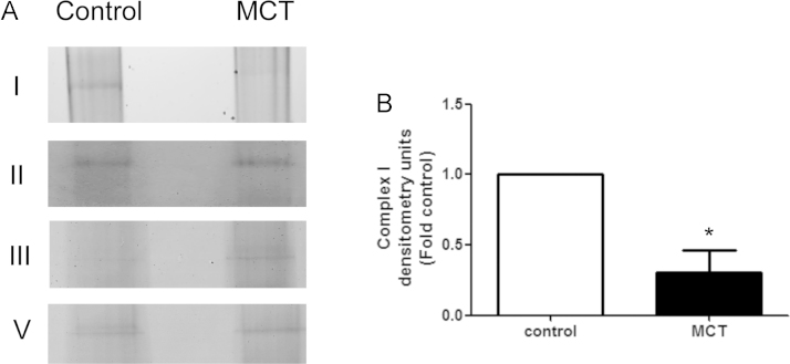
ATP is essential for cellular function and is usually produced through oxidative phosphorylation. However, mitochondrial dysfunction is now being recognized as an important contributing factor in the development cardiovascular diseases, such as pulmonary hypertension (PH). In PH there is a metabolic change from oxidative phosphorylation to mainly glycolysis for energy production. However, the mechanisms underlying this glycolytic switch are only poorly understood. In particular the role of the respiratory Complexes in the mitochondrial dysfunction associated with PH is unresolved and was the focus of our investigations. We report that smooth muscle cells isolated from the pulmonary vessels of rats with PH (PH-PASMC), induced by a single injection of monocrotaline, have attenuated mitochondrial function and enhanced glycolysis. Further, utilizing a novel live cell assay, we were able to demonstrate that the mitochondrial dysfunction in PH-PASMC correlates with deficiencies in the activities of Complexes I-III. Further, we observed that there was an increase in mitochondrial reactive oxygen species generation and mitochondrial membrane potential in the PASMC isolated from rats with PH. We further found that the defect in Complex I activity was due to a loss of Complex I assembly, although the assembly of Complexes II and III were both maintained. Thus, we conclude that loss of Complex I assembly may be involved in the switch of energy metabolism in smooth muscle cells to glycolysis and that maintaining Complex I activity may be a potential therapeutic target for the treatment of PH.

Keywords: Electron transport chain; Mitochondria; Pulmonary hypertension; Warburg effect.


Figures

None
Graphical abstract
Fig. 1
Fig. 1
Mitochondrial respiration is attenuated in pulmonary arterial smooth muscle cells isolated from rats with pulmonary hypertension. The Seahorse XF24 analyzer was used to take measurements in PASMC (40,000/0.32 cm2) isolated from control (red) and PH (blue) rats. Oligomycin (1 µM), FCCP (1 µM), and Rotenone and Antimycin A (1 µM each) were added at the indicated points (A). Basal mitochondrial respiration (B) is unchanged between control and PH-PASMC as estimated by measuring the rate of oxygen consumption rate (OCR, pmols/min). Both the maximal respiratory capacity (C) and the reserve respiratory capacity (D) are significantly attenuated in PH-PASMC. Values are means±SEM. *(p<0.05 vs. Control PH-PASMC, N=10 measurements from 3 independent SMC isolations). (For interpretation of the references to color in this figure legend,the reader is referred to the web version of this article.)
Fig. 2
Fig. 2
Glycolysis is increased in pulmonary arterial smooth muscle cells isolated from rats with pulmonary hypertension. The Seahorse XF24 analyzer was used to take extracellular acidification rate (ECAR) measurements in PASMC (20,000/0.32 cm2) isolated from control (red, N=10) and PH (blue, N=10) rats. D-glucose (2 mg/ml), oligomycin (1 µM) and 2-deoxy-D-glucose (100 mM) were added at the indicated points (A). There is no difference in the glycolytic rate between control and PH-PASMC in the absence of substrate (B). However, the addition of D-glucose caused a significant increase in ECAR in PH- compared to Control-PASMC (C). The addition of oligomycin increased ECAR significantly more in PH- compared to control-PASMC (D). Values are means±SEM. *(p<0.05 vs. Control PH-PASMC, N=10 measurements from 3 independent SMC isolations). (For interpretation of the references to color in this figure legend,the reader is referred to the web version of this article.)
Fig. 3
Fig. 3
Complex I and Complex II dependent respiration is attenuated in pulmonary arterial smooth muscle cells isolated from rats with pulmonary hypertension. The Seahorse XF24 analyzer was used to take OCR measurements in PASMC (40,000/0.32 cm2) isolated from control (red) and PH (blue) rats to determine Complex I and II respiratory activity (A). Both Complex I (B) and Complex II (C) activities are attenuated in PH- compared to Control-PASMC. Values are means±SEM. *(p<0.05 vs. Control PH-PASMC, N=5–15 measurements from 3 independent SMC isolations). (For interpretation of the references to color in this figure legend,the reader is referred to the web version of this article.)
Fig. 4
Fig. 4
Complex III dependent respiration is attenuated in pulmonary arterial smooth muscle cells isolated from rats with pulmonary hypertension. The Seahorse XF24 analyzer was used to take OCR measurements in PASMC (40,000/0.32 cm2) isolated from control (red) and PH (blue) rats to determine Complex I and III respiratory activity (A). Both Complex I (B) and Ccomplex III (C) activities are attenuated in PH- compared to Control-PASMC. Values are means±SEM. *(p<0.05 vs. Control PH-PASMC, N=10 measurements from 3 independent SMC isolations). (For interpretation of the references to color in this figure legend,the reader is referred to the web version of this article.)
Fig. 5
Fig. 5
Mitochondrial ROS production is increased in pulmonary arterial smooth muscle cells isolated from rats with pulmonary hypertension. PASMC were pre-incubated with the mitoSOX red fluorescent dye. Digital images of mitoSOX fluorescence were obtained by fluorescent microscopy. Representative digital images from PASMC isolated from control and PH rats SMC are shown. Quantification of the fluorescent signal indicates that ROS production is increased in PH-PASMC. Data are plotted as mean fluorescence intensity (±SEM). *(p<0.05, N=10).
Fig. 6
Fig. 6
Mitochondrial membrane potential is increased in pulmonary arterial smooth muscle cells isolated from rats with pulmonary hypertension. PASMC were pre-incubated with TMRM to polarized mitochondria. Digital images of TMRM fluorescence were obtained using fluorescent microscopy. Representative digital images from PASMC isolated from control and PH rats SMC are shown. Quantification of the fluorescent signal indicates that the mitochondrial membrane potential is increased in PH-PASMC. Data are plotted as mean fluorescence intensity (±SEM). *(p<0.05, N=10).
Fig. 7
Fig. 7
Complex I assembly is attenuated in pulmonary arterial smooth muscle cells isolated from rats with pulmonary hypertension. Mitochondria, obtained from PASMC isolated from control and PH rats SMC, were subjected to Native Blue electrophoresis to preserve the respiratory Complexes (A). Complex I levels are significantly reduced in PH-PASMC (B). Complex II and V levels are unchanged and Complex III levels are increased in PH-PASMC. The image shown is representative of three independent SMC isolations experiments. Data are plotted as mean±SEM, *p<0.05.

References

    1. Owens G.K., Kumar M.S., Wamhoff B.R. Molecular regulation of vascular smooth muscle cell differentiation in development and disease. Physiol. Rev. 2004;84:767–801. - PubMed
    1. Hao H., Gabbiani G., Bochaton-Piallat M.L. Arterial smooth muscle cell heterogeneity: implications for atherosclerosis and restenosis development. Arterioscler. Thromb. Vasc. Biol. 2003;23:1510–1520. - PubMed
    1. Lincoln T.M., Dey N.B., Boerth N.J., Cornwell T.L., Soff G.A. Nitric oxide-cyclic GMP pathway regulates vascular smooth muscle cell phenotypic modulation: implications in vascular diseases. Acta Physiol. Scand. 1998;164:507–515. - PubMed
    1. Negash S., Narasimhan S.R., Zhou W., Liu J., Wei F.L., Tian J., Raj J.U. Role of cGMP-dependent protein kinase in regulation of pulmonary vascular smooth muscle cell adhesion and migration: effect of hypoxia. Am. J. Physiol.: Heart Circ. Physiol. 2009;297:H304–H312. - PMC - PubMed
    1. Zhou W., Negash S., Liu J., Raj J.U. Modulation of pulmonary vascular smooth muscle cell phenotype in hypoxia: role of cGMP-dependent protein kinase and myocardin. Am. J. Physiol. Lung Cell. Mol. Physiol. 2009;296:L780–L789. - PMC - PubMed

Publication types

MeSH terms

Substances