. 2015 Oct 1;25(19):4219-24.
doi: 10.1016/j.bmcl.2015.07.095. Epub 2015 Aug 6.

Discovery, synthesis and biochemical profiling of purine-2,6-dione derivatives as inhibitors of the human poly(A)-selective ribonuclease Caf1

Affiliations

Discovery, synthesis and biochemical profiling of purine-2,6-dione derivatives as inhibitors of the human poly(A)-selective ribonuclease Caf1

Gopal P Jadhav et al. Bioorg Med Chem Lett. .

Abstract

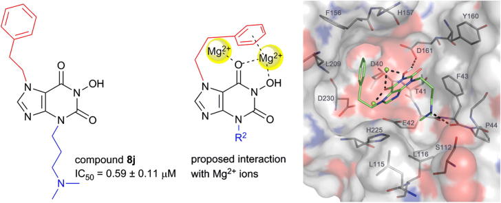
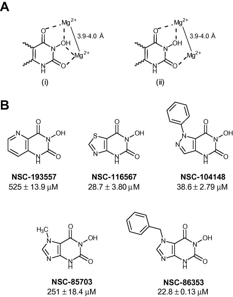
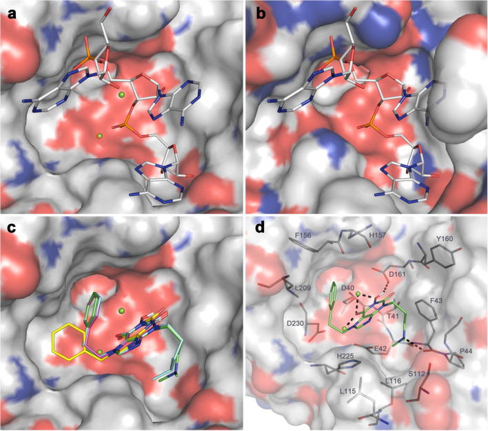
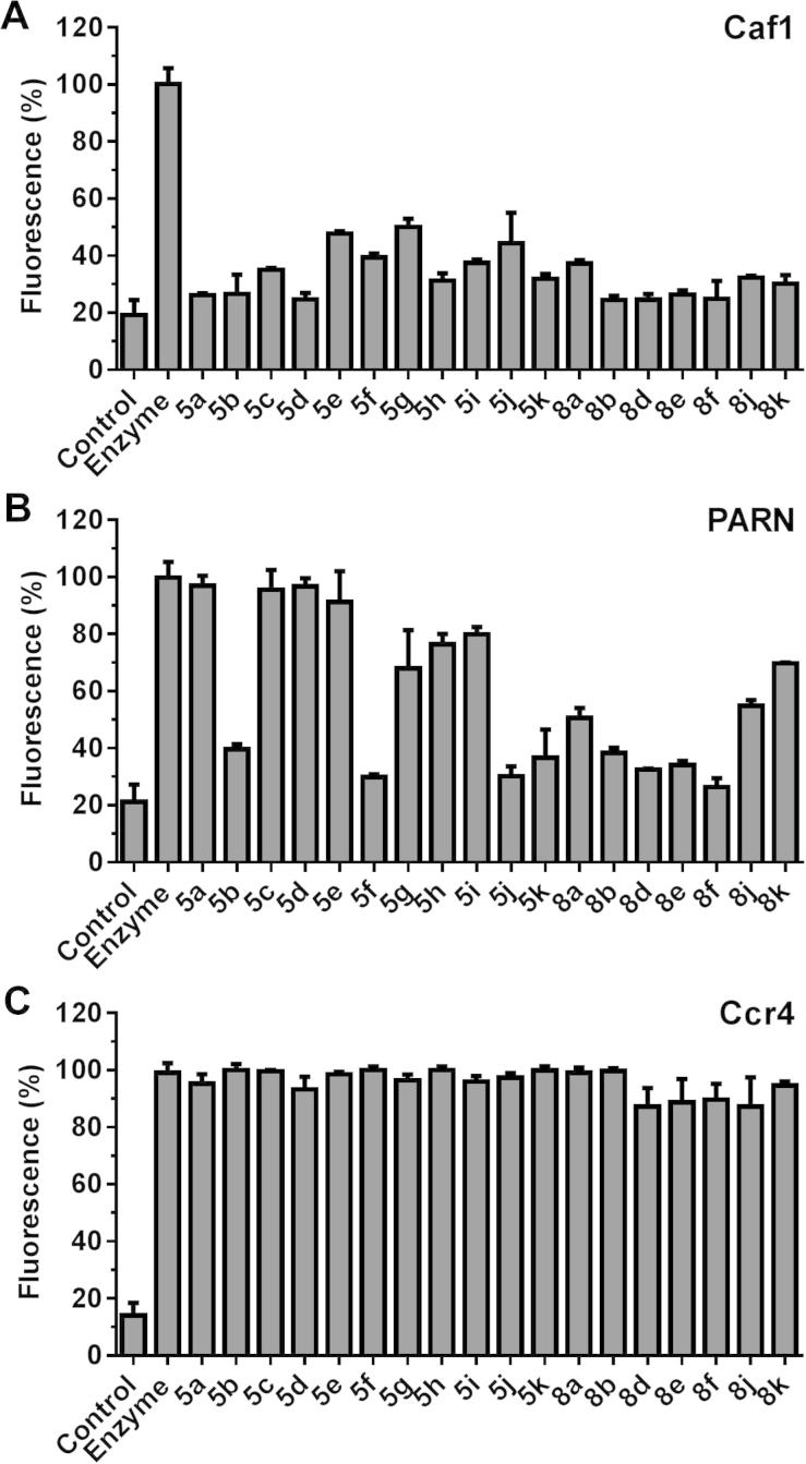
Eukaryotic mRNA contains a 3' poly(A) tail, which plays important roles in the regulation of mRNA stability and translation. Well-characterized enzymes involved in the shortening of the poly(A) tail include the multi-subunit Ccr4-Not deadenylase, which contains the Caf1 (Pop2) and Ccr4 catalytic components, and poly(A)-specific ribonuclease (PARN). Two Mg(2+) ions present in the active sites of these ribonucleases are required for RNA cleavage. Here, we report the discovery, synthesis and biochemical profiling of purine-2,6-dione derivatives as (sub)micromolar inhibitors of Caf1.

Keywords: Caf1/CNOT7; Deadenylase; Mg(2+) dependent nuclease; PARN; Ribonuclease.


Figures

None
Graphical abstract
Figure 1
Figure 1
Discovery of 3-hydroxy-pyrimidine-2,4-dione compounds as inhibitors of the Caf1 ribonuclease. (A) Possible interaction modes of 3-hydroxy-pyrimidine-2,4-dione compounds with two divalent metal ions in the active site of Caf1. (B) Structure and activity of compounds selected from the Open Chemical Repository Collection (NCI, Bethesda). IC50 values refer to inhibition of Caf1. Also indicated is the standard error of the mean (n = 3).
Figure 2
Figure 2
Molecular modelling of inhibitors in the active site of Caf1. Complex crystal structures of human Caf1 (a; PDB 4GMJ.B) and PARN (b; PDB 2A1R) show that the cognate poly(A) ligand (stick models) likely binds in a similar manner to the active site (protein surface with Mg2+ ions as green spheres), despite the fact that the active sites in Caf1 and PARN have somewhat different shapes. In (c) it can be seen that the top-ranking poses of the most potent compounds 5j (magenta), 8e (cyan), and 8j (green) adopt very similar conformations upon docking to the 4GMJ.B model, whereas several poses with similar scores were recorded for lead compound 5a (yellow), whereof only some (one shown) were similar to the bound conformations of the more potent compounds. Probable polar binding contacts of the most potent compound 8j with the Caf1 active site include a cation–π interaction between one of the Mg2+ ions and the phenyl ring of the inhibitor, as well as H-bonds and polar interactions involving the 3° amine and one of the carbonyl O and the OH of the N-hydroxyimide group as indicated (d; dashed lines). Furthermore, 8j makes numerous van-der-Waals contacts with residues (labelled in d) of the active site cavity.
Figure 3
Figure 3
Inhibition of the ribonuclease activities of (A) Caf1, (B) PARN and (C) Ccr4 by 3,7-N-substituted-1-hydroxy-3,7-dihydro-1H-purine-2,6-diones. Reactions contained no enzyme (control), complete reactions (enzyme) or complete reactions in the presence of the indicated compound. Compounds were used at 30 μM (5ae, 8ad, 8f), 10 μM (5hk), or 3 μM (8e, 8j). Error bars represent the standard deviation.
Scheme 1
Scheme 1
Synthesis of 7-substituted 1-hydroxy-3,7-dihydro-1H-purine-2,6-diones. Reagents and conditions: (a) ethyl 2-bromoacetate, CHCl3, rt, 2 h (37–79%); (b) (ethoxymethylene)cyanamide, THF, Δ, 16 h, then KOBut, EtOH, Δ, 2 h (46–80% over 2 steps); (c) CDI, PhMe, Δ, 2 h, then O-allylhydroxylamine, aq NaOH, EtOH, Δ, 2 h (21–61% over 2 steps); (d) Pd(OAc)2, PPh3, HCOOH, EtOH–H2O (8:2), 80 °C, 2 h (23–55%); (e) CDI, PhMe, Δ, 2 h, then O-benzylhydroxylamine, aq NaOH, EtOH, Δ, 2 h (74% over 2 steps); (f) R2X (X = Br or I), K2CO3, DMF, 80 °C, 2–12 h, (67–98%); (g) H2, 10% (w/w) Pd(C), CH2Cl2–MeOH (1:9) (33–53%); (h) urea, 2-methoxyethanol, 190 °C, 24 h, then aq NaOH, Δ, 3 h (33%). For definitions of R1 and R2 refer to Tables 1–3.
Scheme 2
Scheme 2
Synthesis of 6-hydroxy-1-phenethyl-1,4-dihydro-5H-imidazo[4,5-b]pyridin-5-one 13. Reagents and conditions: (a) CHCl3, KOBut, THF–DMF (2:1), −100 °C, 15 min, then CaCO3, H2O, 75 °C, 48 h, then H2, 10% (w/w) Pd(C), EtOAc–MeOH–AcOH (18:1:1), 55 psi, 18 h (60% over 3 steps); (b) ethyl allyloxyacetate, (Me3Si)2NLi, THF, −78 °C to rt over 16 h (36%); (c) Pd(OAc)2, PPh3, HCOOH, EtOH–H2O (8:2), 80 °C, 2 h (33%).

References

    1. Chang H., Lim J., Ha M., Kim V.N. Mol Cell. 2014;53:1044. - PubMed
    1. Subtelny A.O., Eichhorn S.W., Chen G.R., Sive H., Bartel D.P. Nature. 2014;508:66. - PMC - PubMed
    1. Goldstrohm A.C., Wickens M. Nat Rev Mol Cell Biol. 2008;9:337. - PubMed
    1. Collart M.A., Panasenko O.O. Gene. 2012;492:42. - PubMed
    1. Wahle E., Winkler G.S. Biochim Biophys Acta. 2013;1829:561. - PubMed

Publication types

LinkOut - more resources