Review
. 2016 Aug;92(Pt A):18-45.
doi: 10.1016/j.nbd.2015.08.003. Epub 2015 Aug 20.

Neuronal migration disorders: Focus on the cytoskeleton and epilepsy

Affiliations
Review

Neuronal migration disorders: Focus on the cytoskeleton and epilepsy

Melissa A Stouffer et al. Neurobiol Dis. 2016 Aug.

Abstract

A wide spectrum of focal, regional, or diffuse structural brain abnormalities, collectively known as malformations of cortical development (MCDs), frequently manifest with intellectual disability (ID), epilepsy, and/or autistic spectrum disorder (ASD). As the acronym suggests, MCDs are perturbations of the normal architecture of the cerebral cortex and hippocampus. The pathogenesis of these disorders remains incompletely understood; however, one area that has provided important insights has been the study of neuronal migration. The amalgamation of human genetics and experimental studies in animal models has led to the recognition that common genetic causes of neurodevelopmental disorders, including many severe epilepsy syndromes, are due to mutations in genes regulating the migration of newly born post-mitotic neurons. Neuronal migration genes often, though not exclusively, code for proteins involved in the function of the cytoskeleton. Other cellular processes, such as cell division and axon/dendrite formation, which similarly depend on cytoskeletal functions, may also be affected. We focus here on how the susceptibility of the highly organized neocortex and hippocampus may be due to their laminar organization, which involves the tight regulation, both temporally and spatially, of gene expression, specialized progenitor cells, the migration of neurons over large distances and a birthdate-specific layering of neurons. Perturbations in neuronal migration result in abnormal lamination, neuronal differentiation defects, abnormal cellular morphology and circuit formation. Ultimately this results in disorganized excitatory and inhibitory activity leading to the symptoms observed in individuals with these disorders.

Keywords: Cortical malformation; Molecular and cellular mechanisms; Mouse model; Neurodevelopment.


Conflict of interest statement

Conflict of interest statement

The authors declare no conflicts of interest.

Figures

Figure 1.
Figure 1.. Rodent neocortical development
a. Schematic representation of the developing rodent neocortex. Glutamatergic PNs produced in the dorsal telencephalon (pallium) migrate radially to generate the neocortex and hippocampus. Cortical inhibitory INs generated in the ventral telencephalon (subpallium) migrate tangentially. b. Early in development (E12), RG cells located in the VZ divide symmetrically (curved black arrow) to generate two more RGs, or asymmetrically to produce neurogenic progenitors (IPs/SNPs) or neurons that migrate via somal translocation toward the MZ. c-d. Later in development (E14–18), IPs located in the SVZ are the primary source of neurogenic divisions. SNPs remain in the VZ, and are possibly precursors for IPs. At this stage, neurons first migrate via RG cell guided locomotion followed by somal translocation. INs arriving from the subpallium migrate tangentially to the cortex, then radially to reach their final destinations. RG cells also give rise to OPCs, which produce oligodendrocytes, and astrocytes. Some RG processes in the hippocampus bend (c), causing clonally related neurons to be distributed horizontally. Clonally related neurons in the neocortex (d) form radial functional columns with layer specific input and output connectivities. Abbreviations: NCx= neocortex; LV= lateral ventricle; H= hippocampus; LGE= lateral geniculate nucleus; MGE=medial geniculate nucleus; Str= striatum; MZ= marginal zone; CP= cortical plate; SVZ= subventricular zone; IZ= intermediate zone; VZ= ventricular zone; RG= radial glial cell; SNP= short neural precursor; IP= intermediate progenitor; PN= projection neuron; IN= interneuron; OPC= oligodendrocyte precursor cell; A= astrocyte; CR= Cajal-Retzius cell
Figure 2.
Figure 2.. MCD genes and cytoskeletal regulatory pathways
Schematic representation of proteins regulating MTs and actin in a migrating post-mitotic neuron. In general, MT regulating proteins are shown in green, and actin regulating proteins are shown in orange. Disruption of cytoskeletal proteins and their regulatory pathways leads to various consequences. Mutations in MAPs (Dcx, Dclk, Tau, Map2, Map1b), which stabilize MTs, can impair polarization, formation of the leading process, neurite outgrowth and/or lead to excessive branching that hinders migration. Lis1/Ndel1/dynein are required for nuclear movement during migration. Molecular motors, such as dynein and kinesins, serve important roles in migration by transporting cargo in opposite directions along MTs, or by transporting MTs themselves. Mutations in components of the reelin pathway lead to impaired migration, interactions with RG cells and final positioning of neurons via impaired regulation of cytoskeletal dynamics. Actin plays complex roles in the growth cone and trailing processes of migrating neurons, with mutations in regulatory pathways leading to overmigration or migration arrest, which may be consequential to progenitor defects observed in various rodent models.
Figure 3.
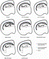
Figure 3.. Schematic diagrams of selected MCD rodent model neocortical and hippocampal phenotypes.
Schematic representations of morphological characteristics shown on coronal hemi-sections for adult animals. Hippocampal subregions are indicated in control. Location of early-born (purple, E12–13) and late-born (blue, E14–16) pyramidal neurons are depicted by dots in neocortex and SBH, and layer 1 by a grey line. INs are not displayed. In general, early-born and late-born neurons are considered to be L5–6 and L2–4, respectively, except in the case of the reeler mouse, which may show more complicated layer distributions in certain subregions (see Boyle et al., 2011). Discrete lamination defects or general dispersion of pyramidal cells or DG granule cells in the hippocampus are represented by multiple or thick lines, respectively. Changes in the corpus callosum (CC) are indicated by changes in line thickness or dispersion of line into SBH (Rapgef2 cKO, p35 KO). An abnormal layer 1 is depicted in the reeler mouse. Wavy L2–4 are depicted in Tuba1a mouse. Abbreviations: DG = dentate gyrus, LV = lateral ventricle, CC = corpus callosum

References

    1. Ackman JB, Aniksztejn L, Crepel V, Becq H, Pellegrino C, Cardoso C, Ben-Ari Y, Represa A, 2009. Abnormal network activity in a targeted genetic model of human double cortex. The Journal of neuroscience : the official journal of the Society for Neuroscience 29, 313–327. - PMC - PubMed
    1. Alkuraya FS, Cai X, Emery C, Mochida GH, Al-Dosari MS, Felie JM, Hill RS, Barry BJ, Partlow JN, Gascon GG, Kentab A, Jan M, Shaheen R, Feng Y, Walsh CA, 2011. Human mutations in NDE1 cause extreme microcephaly with lissencephaly [corrected]. American journal of human genetics 88, 536–547. - PMC - PubMed
    1. Anderson SA, Eisenstat DD, Shi L, Rubenstein JL, 1997. Interneuron migration from basal forebrain to neocortex: dependence on Dlx genes. Science (New York, N.Y.) 278, 474–476. - PubMed
    1. Anton ES, Marchionni MA, Lee KF, Rakic P, 1997. Role of GGF/neuregulin signaling in interactions between migrating neurons and radial glia in the developing cerebral cortex. Development 124, 3501–3510. - PubMed
    1. Arimura N, Kaibuchi K, 2007. Neuronal polarity: from extracellular signals to intracellular mechanisms. Nature reviews. Neuroscience 8, 194–205. - PubMed

Publication types

MeSH terms