. 2015 Aug 24:5:13429.
doi: 10.1038/srep13429.

Curcumin as therapeutics for the treatment of head and neck squamous cell carcinoma by activating SIRT1

Affiliations

Curcumin as therapeutics for the treatment of head and neck squamous cell carcinoma by activating SIRT1

An Hu et al. Sci Rep. .

Abstract

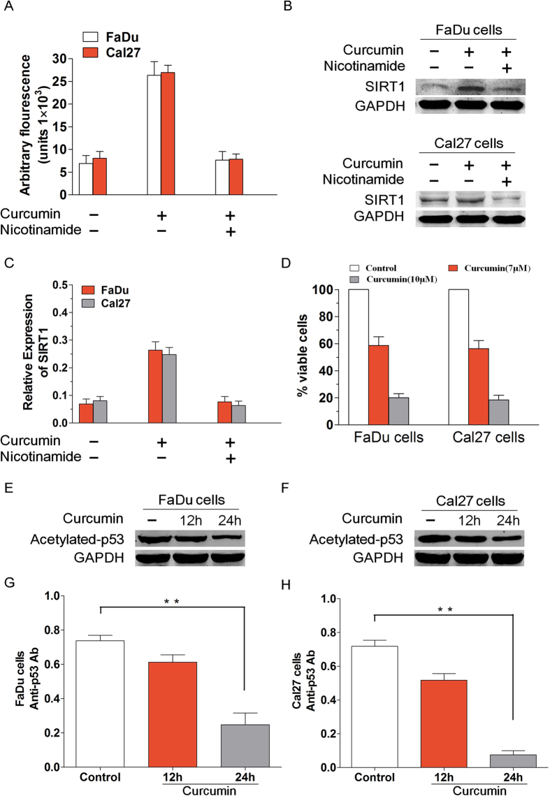
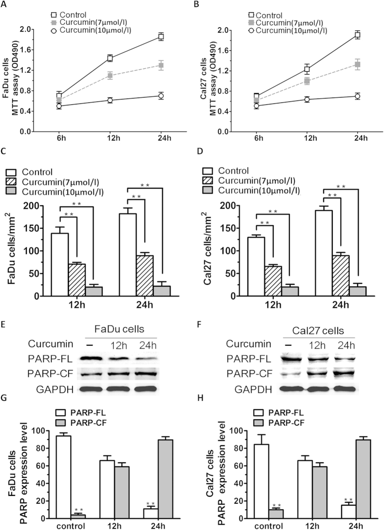
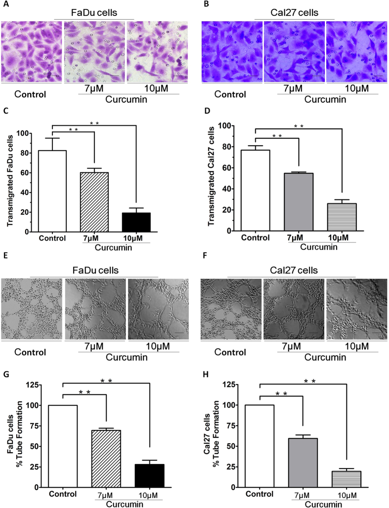
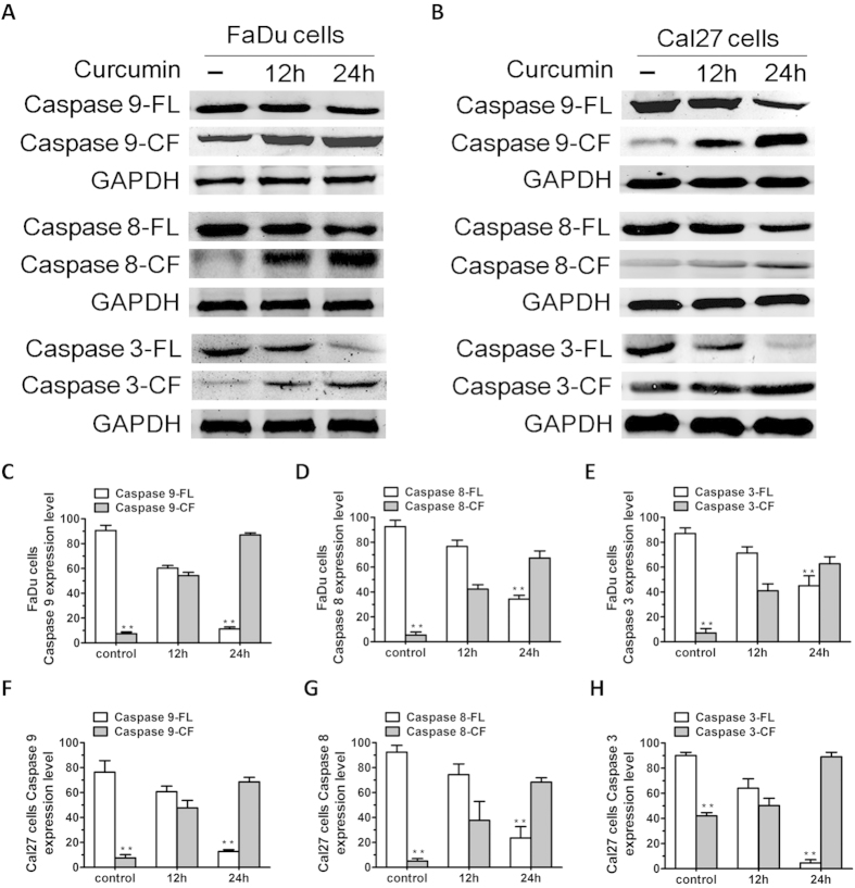
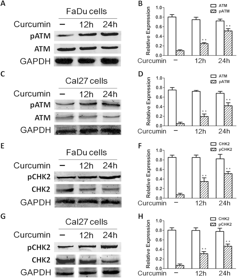
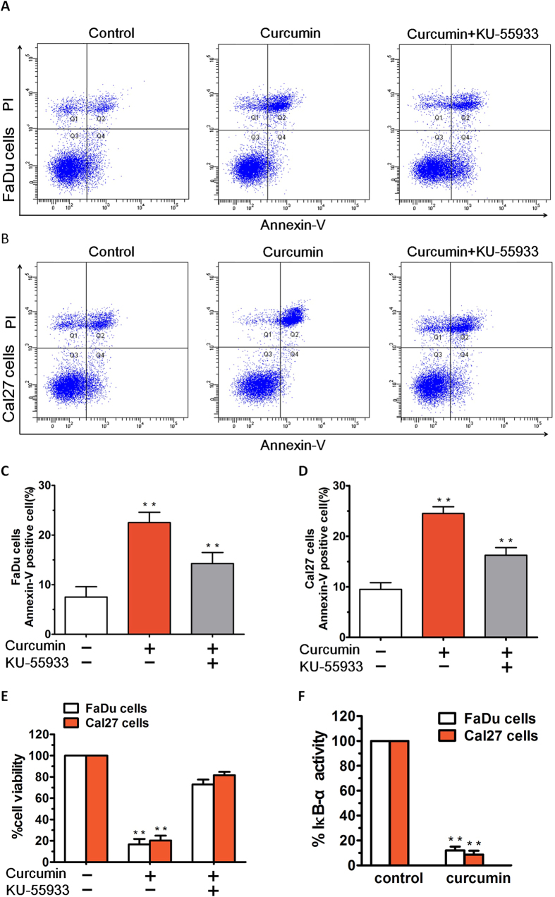
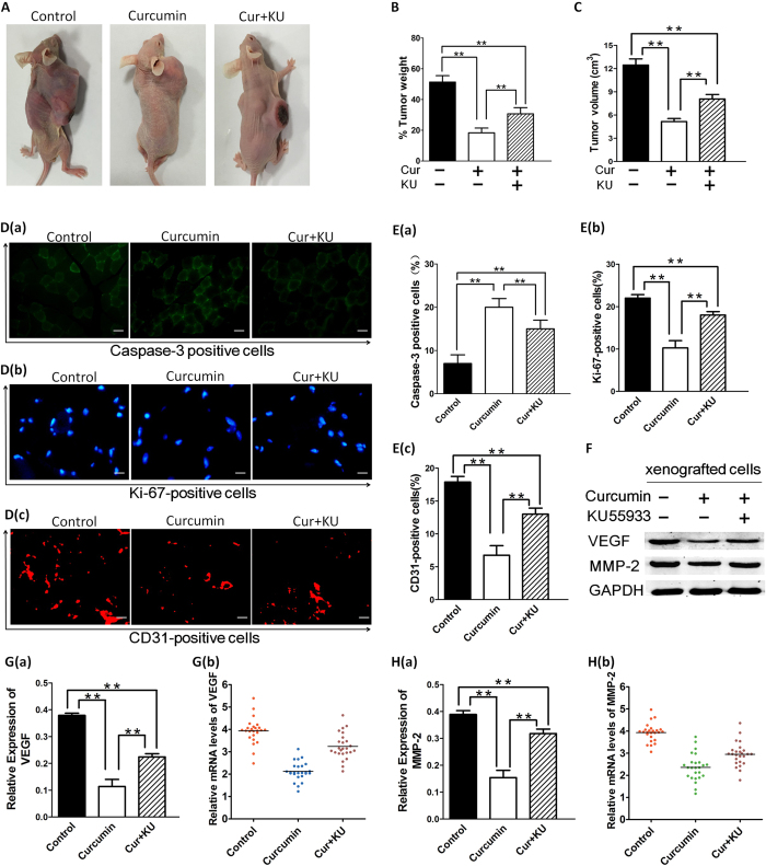
SIRT1 is one of seven mammalian homologs of Sir2 that catalyzes NAD(+)-dependent protein deacetylation. The aim of the present study is to explore the effect of SIRT1 small molecule activator on the anticancer activity and the underlying mechanism. We examined the anticancer activity of a novel oral agent, curcumin, which is the principal active ingredient of the traditional Chinese herb Curcuma Longa. Treatment of FaDu and Cal27 cells with curcumin inhibited growth and induced apoptosis. Mechanistic studies showed that anticancer activity of curcumin is associated with decrease in migration of HNSCC and associated angiogenesis through activating of intrinsic apoptotic pathway (caspase-9) and extrinsic apoptotic pathway (caspase-8). Our data demonstrating that anticancer activity of curcumin is linked to the activation of the ATM/CHK2 pathway and the inhibition of nuclear factor-κB. Finally, increasing SIRT1 through small molecule activator curcumin has shown beneficial effects in xenograft mouse model, indicating that SIRT1 may represent an attractive therapeutic target. Our studies provide the preclinical rationale for novel therapeutics targeting SIRT1 in HNSCC.


Figures

Figure 1
Figure 1. Curcumin induces SIRT1 enzyme activity in vitro.
(A) FaDu and Cal27 cells were treated with curcumin (7 μmol/l) in the presence or absence of SIRT1 inhibitor nicotinamide for 6 h and harvested. Then the extracts were analysed for SIRT1 enzyme activity. Treatment of FaDu and Cal27 cells with curcumin markedly increased the deacetylating activity; conversely, pre-treatment of cells with nicotinamide significantly blocked curcumin-triggered deacetylating activity. (B–C) To further confirm curcumin activity on SIRT1, we explored SIRT1 expression by Western blotting in FaDu and Cal27 cells treated or not with curcumin. Treatment of carcinoma cells with curcumin markedly increased the SIRT1 expression. However, pre-treatment of FaDu and Cal27 cells with nicotinamide significantly blocked curcumin-triggered SIRT1 activity. (D) We determined a decrease in cell viability using MTT assay and this was due to anti-proliferative activity of curcumin. FaDu as well as Cal27 cells were treated with or without curcumin at the indicated concentrations for 24 h, followed by assessment for cell viability using MTT assays. Raw data from MTT assays were normalized to the % of viable cells in control versus curcumin-treated carcinoma cells. (E–F) FaDu and Cal27 cells were treated with curcumin and then the protein lysates were subjected to Western blot analysis with antibodies specifically against acetylated p53 and GAPDH. (G) Curcumin pretreatment of FaDu cells diluted acetylated p53 expression level at 12 h, with increasing effect at 24 h. (P < 0.01, n = 3) (H) When the effect of curcumin on acetylated p53 expression level of Cal27 cells was tested, a significant reduction was observed at 24 h. (P < 0.01, n = 3) Blots shown are representative of three independent experiments. Data were expressed as mean ± SD. **p < 0.01
Figure 2
Figure 2. Curcumin inhibits HNSCC cells growth and proliferation in vitro.
(A,B) Cell viability assays measuring growth and proliferation were performed in FaDu and Cal27 cells. (C,D) Human HNSCC cell lines (FaDu and Cal27 cells) were treated with various concentrations (7 μmol/l and 10 μmol/l) of curcumin for 24 h. A concentration-dependent decrease in viability of all cell lines was observed in response to treatment with curcumin. (P < 0.01, n = 3) (E,F) FaDu and Cal27 cells were treated with curcumin and harvested. Whole cell lysates were subjected to Western Blot analysis with anti-PARP, or anti-GAPDH (FL, full length; CF, cleaved fragment). (G,H) Treatment of both FaDu and Cal27 cells with curcumin triggered a marked increasing in proteolytic cleavage of PARP (P < 0.01; n = 3). Data were expressed as mean ± SD. **p < 0.01, versus control group.
Figure 3
Figure 3. Curcumin inhibited HNSCCs migration.
(A,B) Curcumin significantly inhibited FaDu and Cal27 cells migration, as evidenced by a decrease in the number of crystal violet-stained cells. (C,D) Pretreatment of FaDu and Cal27 cells with curcumin resulted in inhibition of cell transmigration. 10 μM curcumin pretreatment of FaDu and Cal27 cells inhibited transmigration, which was statistically significant difference to the effect of control group. (E,F)Vasculogenic mimicry was measured in vitro using 3D-Matrigel capillary-like tubule structure formation assays: FaDu and Cal27 cells plated onto Matrigel form capillary-like tubule structures similar to in vivo neovascularization. FaDu and Cal27 cells were seeded in 96-well culture plates precoated with Matrigel; and then examined for tubule formation using an inverted microscope. (G,H)Tubule formation was markedly decreased in a dose-dependent manner in curcumin-treated cells. Cells remained >95% viable before and after performing the migration assay, excluding the possibility that drug-induced inhibition of migration was due to cell death. Data were expressed as mean ± SD. **p < 0.01.
Figure 4
Figure 4. Curcumin triggers activation of both intrinsic (caspase-8) and extrinsic (caspase-9) apoptotic signalling.
(A,B) FaDu and Cal27 cells were treated with curcumin (7 μM) for the indicated times and harvested. Total proteins were subjected to Western blot analysis (WB) with anti-caspase-9, caspase-8, caspase-3, or GAPDH Abs. (C–H) Bar graph represents quantification of the percentage of caspase/ GAPDH. Blots shown are representative of three independent experiments. Results are mean ± SD of three independent experiments (n = 3; P < 0.005).FL, full length; CF, cleaved fragment.
Figure 5
Figure 5. Curcumin-induced apoptosis is associated with activation of ATM/CHK2 signal pathway.
(A–D) FaDu and Cal27 cells were treated with curcumin (7 μM) and harvested; total proteins were subjected to Western blot analysis with anti-phosphorylated ATM (anti-pATM) or anti-ATM Abs. Blots shown are representative of three independent experiments. (E–H) FaDu and Cal27 cells were treated with curcumin (7 μM) and harvested; total proteins were subjected to Western blot analysis with anti-phosphorylated CHK2 (anti-pCHK2) or anti-CHK2 Abs. Blots shown are representative of three independent experiments.
Figure 6
Figure 6. Annexin-V/PI staining showed that curcumin treatment induced apparent early and late apoptosis in HNSCCs.
(A,B) After treatment with curcumin (7 μM), the FaDu cells and Cal27 cells groups showed early and late apoptosis at rates of 17.20% and 21.60%, respectively. (C,D) After treatment with 10 μM KU55933, the apoptosis rate was significantly reduced, not only in FaDu cells group, but also in Cal27 cells group. (E) FaDu and Cal27 cells were treated with curcumin in the presence or absence of the ATM inhibitor KU55933 for 24 h, followed by assessment for cell viability using MTT assays. (F) FaDu and Cal27 cells were treated with curcumin (7 μM) for 24 h, and extracts were then analysed for NF-κB activity by measuring phosphorylated IκB-α. Data presented are means ± SD (n = 3; P < 0.05).
Figure 7
Figure 7. Curcumin inhibits growth of xenografted FaDu cells in mice.
(A–C) FaDu cells alone (5 × 106 cells/mouse) were implanted in the right rear flank of female null mice (5–7 weeks of age at the time of tumor challenge). On Day 27–30, mice were randomized to treatment groups and treated with vehicle or curcumin (200 mg/kg). Mice were treated for seven consecutive days, repeated weekly for 4 weeks. Data are presented as mean tumor volume ± standard error of the mean (SEM) (n = 6; P < 0.05). A representative experiment is shown. (D–E) Tumors were removed from control untreated or curcumin-treated and/not KU-55933-treated mice, followed by immunofluorescence analysis with antibodies against Caspase-3 and Ki-67 and CD31. Micrographs are representative of tumor sections from two different mice in each group. (F–H) We further investigated the effect of curcumin on MMP-2 expression and VEGF production in curcumin-treated and/not KU-55933-treated mice. Whole-tissue lysates were subjected to western-blot analysis for determining levels of total MMP-2 and VEGF proteins. Pretreatment of cancer cells with either curcumin alone, or combination of curcumin and KU55933 significantly inhibited MMP-2 and VEGF protein expression. Quantitative reverse transcription–PCR of MMP-2 and VEGF in cancer cells demonstrates that MMP-2 and VEGF mRNA was down-regulated expression. GAPDH was used as an internal loading control. The data are expressed as mean ± SD of three independent experiments, and significant differences from the control are indicated by **p < 0.01.

References

    1. Hideshima T. et al. Small-molecule inhibition of proteasome and aggresome function induces synergistic antitumor activity in multiple myeloma. Proc Natl Acad Sci USA 102, 8567–8572, 10.1073/pnas.0503221102 (2005). - DOI - PMC - PubMed
    1. Catley L. et al. Aggresome induction by proteasome inhibitor bortezomib and alpha-tubulin hyperacetylation by tubulin deacetylase (TDAC) inhibitor LBH589 are synergistic in myeloma cells. Blood 108, 3441–3449, 10.1182/blood-2006-04-016055 (2006). - DOI - PMC - PubMed
    1. Dasmahapatra G. et al. The pan-HDAC inhibitor vorinostat potentiates the activity of the proteasome inhibitor carfilzomib in human DLBCL cells in vitro and in vivo. Blood 115, 4478–4487, 10.1182/blood-2009-12-257261 (2010). - DOI - PMC - PubMed
    1. He L. et al. Mcl-1 and FBW7 control a dominant survival pathway underlying HDAC and Bcl-2 inhibitor synergy in squamous cell carcinoma. Cancer Discov 3, 324–337, 10.1158/2159-8290.cd-12-0417 (2013). - DOI - PMC - PubMed
    1. Kurundkar D. et al. Vorinostat, an HDAC inhibitor attenuates epidermoid squamous cell carcinoma growth by dampening mTOR signaling pathway in a human xenograft murine model. Toxicol Appl Pharmacol 266, 233–244, 10.1016/j.taap.2012.11.002 (2013). - DOI - PMC - PubMed

Publication types

MeSH terms