Comparative Study
. 2015 Oct 6;6(30):29254-67.
doi: 10.18632/oncotarget.4201.

The metastatic infiltration at the metastasis/brain parenchyma-interface is very heterogeneous and has a significant impact on survival in a prospective study

Affiliations
Comparative Study

The metastatic infiltration at the metastasis/brain parenchyma-interface is very heterogeneous and has a significant impact on survival in a prospective study

Laila Siam et al. Oncotarget. .

Abstract

The current approach to brain metastases resection is macroscopic removal of metastasis until reaching the glial pseudo-capsule (gross total resection (GTR)). However, autopsy studies demonstrated infiltrating metastatic cells into the parenchyma at the metastasis/brain parenchyma (M/BP)-interface.

Aims/methods: To analyze the astrocyte reaction and metastatic infiltration pattern at the M/BP-interface with an organotypic brain slice coculture system. Secondly, to evaluate the significance of infiltrating metastatic tumor cells in a prospective biopsy study. Therefore, after GTR, biopsies were obtained from the brain parenchyma beyond the glial pseudo-capsule and analyzed histomorphologically.

Results: The coculture revealed three types of cancer cell infiltration. Interestingly, the astrocyte reaction was significantly different in the coculture with a benign, neuroectodermal-derived cell line. In the prospective biopsy study 58/167 (34.7%) samples revealed infiltrating metastatic cells. Altogether, 25/39 patients (64.1%) had proven to exhibit infiltration in at least one biopsy specimen with significant impact on survival (OS) (3.4 HR; p = 0.009; 2-year OS was 6.6% versus 43.5%). Exceptionally, in the non-infiltrating cohort three patients were long-term survivors.

Conclusions: Metastatic infiltration has a significant impact on prognosis. Secondly, the astrocyte reaction at the M/BP-interface is heterogeneous and supports our previous concept of the organ-specific defense against metastatic (organ-foreign) cells.

Keywords: astrocytes; brain metastasis; glial-pseudo capsule; metastatic infiltration; organ-specific defense.


Conflict of interest statement

CONFLICTS OF INTEREST

The authors do not report any conflicts of interest.

Figures

Figure 1
Figure 1. Coculture system with different cancer cells
A. Macroscopic picture of the coculture system: An organotypic living whole brain slice is cocultured with a 3D tumor plug of MDA-MB231 cells embedded in matrigel (white arrows mark the area of M/BP-interface). B. Characterization of the epithelial cells and their mesenchymal characteristics by N-Cadherin (N-Cad), E-Cadherin (E-Cad), Pan-Cytokeratin and Vimentin expression, respectively (see also supplementary Figure 1). C. The RPE cell line demonstrates no infiltration (type 0). Interestingly, the RPE also does not cause astrogliosis and astrocyte activation within the otherwise observed defense-reaction. D. As already shown, the luminal A breast cancer cell line MCF-7 infiltrates with tumor strands and cohorts (type I) with a significant astrocytic activation. E. In contrast, the basal-like breast cancer cell line infiltrates with single cells and mini-spheres (type II) also with a significant astrocytic activation.
Figure 2
Figure 2. Histological distribution of the investigated samples and impact of the metastatic infiltration on overall survival (OS)
A. White bars represent the total patient numbers, black bars are the patients with infiltration detected in the cavity wall after GTR. B. The gray line represents patients with infiltrated metastatic cells in at least one biopsy specimen, the dashed line those without infiltration. C. This diagram shows patients with a KPS ≥ 70% and < 70%. In contrast to the infiltration there is no significant difference.
Figure 3
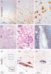
Figure 3. Types of NSCLC carcinoma infiltration
A. Type I cohort infiltration: Pan-cytokeratin IHC of the infiltrating NSCLC carcinoma cells in the cerebellum with glandular formation (100x and 400x magnification). B. Type II single cell and mini-sphere infiltration: HE staining of the infiltrating NSCLC carcinoma cells in the cortex, the arrow marks deeply infiltrated carcinoma cells. C. Micrometastasis of a completely resected NSCLC in almost 2 mm distance of the resection cavity measured from the infiltrating edge of the biopsy (on the right: higher magnification of the rectangle). Infiltration type of breast cancer: D. Infiltrating tumor strands combined with type I cohort infiltration: HE staining of the breast cancer biopsy tissue (100x and 400x magnification). E. Pan-cytokeratin IHC of the infiltrating breast cancer carcinoma cells. The higher magnification demonstrates a cluster of breast cancer cells closely aligned to the external surface of a blood vessel in the Virchow space far beyond the resection margin and pseudo-capsule. (100x and 400x magnification).
Figure 4
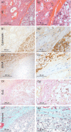
Figure 4. Renal cancer: Type 0 non-infiltration
A. HE staining of the harvested brain metastasis tissue of renal cancer (100x and 400x magnification). B. Pan-cytokeratin IHC of the non-infiltrating renal metastatic cancer mass with a smooth interface to the capsule (100x and 400x magnification). C. The GFAP IHC reveals that astrogliosis is not directly adjacent to the metastasis tissue, as is usually the case. D–E. Unexpectedly, the Elastica van Gieson (EvG) and the Masson Goldner trichrome staining demonstrated collagen fibers directly adjacent to the tumor cells (red/blue). This indicates that the renal cell cancer forms its own fibrous capsule in the distant organs. (100x and 400x magnification)
Figure 5
Figure 5. Infiltration of a neuroendocrine CUP
A. The carcinoma cells demonstrate a type II infiltration (single cell and mini-sphere) (Chromogranin IHC). The entire biopsy tissue is infiltrated with carcinoma cells: thus, the infiltration area is at least 2 mm (100x and 400x magnification). Infiltration of melanoma: B–D. The majority of the infiltrating melanoma cells tightly surround blood vessels in these three different patients as sheets/layers. Analogous to the previous in vivo description, we named this type III angio-cooptive infiltration. (40x, 100x and 400x magnification).
Figure 6
Figure 6. Schemas of different types of infiltration
Type 0 = displacing growth without infiltration. Non-infiltrating cancer cells with a significant glial-reaction. Type 1 = cluster-/cohort infiltration: Strands invade into the adjacent brain parenchyma with detached infiltrating cohorts and cluster sometimes found in the Virchow-Robin space, but also without contact to blood vessels. Type 2 = diffuse infiltration: Single cells or mini-spheres (few cells formed together a sphere) infiltrating the brain parenchyma. Type 3 = angio-cooptive: Typically, the infiltration into the adjacent brain parenchyma takes place along pre-existing blood vessels. The table summarizes our results of the biopsy study of NSCLC, SCLC, breast cancer, malignant melanoma and renal cell cancer (RCC) (* with extensive collagen capsule) and visualize the differences in the infiltration. The predominant infiltration pattern is highlighted in bold.

References

    1. Raore B, Schniederjan M, Prabhu R, Brat DJ, Shu HK, Olson JJ. Metastasis infiltration: an investigation of the postoperative brain-tumor interface. International journal of radiation oncology, biology, physics. 2011;81:1075–1080. - PubMed
    1. Baumert BG, Rutten I, Dehing-Oberije C, Twijnstra A, Dirx MJ, Debougnoux-Huppertz RM, Lambin P, Kubat B. A pathology-based substrate for target definition in radiosurgery of brain metastases. IntJRadiatOncolBiolPhys. 2006;66:187–194. - PubMed
    1. Neves S, Mazal PR, Wanschitz J, Rudnay AC, Drlicek M, Czech T, Wustinger C, Budka H. Pseudogliomatous growth pattern of anaplastic small cell carcinomas metastatic to the brain. Clinical neuropathology. 2001;20:38–42. - PubMed
    1. Berghoff AS, Rajky O, Winkler F, Bartsch R, Furtner J, Hainfellner JA, Goodman SL, Weller M, Schittenhelm J, Preusser M. Invasion patterns in brain metastases of solid cancers. Neuro-oncology. 2013;15:1664–1672. - PMC - PubMed
    1. Chuang HN, van Rossum D, Sieger D, Siam L, Klemm F, Bleckmann A, Bayerlova M, Farhat K, Scheffel J, Schulz M, Dehghani F, Stadelmann C, Hanisch UK, Binder C, Pukrop T. Carcinoma cells misuse the host tissue damage response to invade the brain. Glia. 2013;61:1331–1346. - PMC - PubMed

Publication types

MeSH terms

Substances