Review
. 2016 Apr:334:49-57.
doi: 10.1016/j.heares.2015.08.005. Epub 2015 Aug 20.

Auditory-limbic interactions in chronic tinnitus: Challenges for neuroimaging research

Affiliations
Review

Auditory-limbic interactions in chronic tinnitus: Challenges for neuroimaging research

Amber M Leaver et al. Hear Res. 2016 Apr.

Abstract

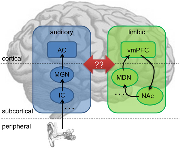
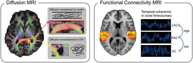
Tinnitus is a widespread auditory disorder affecting approximately 10-15% of the population, often with debilitating consequences. Although tinnitus commonly begins with damage to the auditory system due to loud-noise exposure, aging, or other etiologies, the exact neurophysiological basis of chronic tinnitus remains unknown. Many researchers point to a central auditory origin of tinnitus; however, a growing body of evidence also implicates other brain regions, including the limbic system. Correspondingly, we and others have proposed models of tinnitus in which the limbic and auditory systems both play critical roles and interact with one another. Specifically, we argue that damage to the auditory system generates an initial tinnitus signal, consistent with previous research. In our model, this "transient" tinnitus is suppressed when a limbic frontostriatal network, comprised of ventromedial prefrontal cortex and ventral striatum, successfully modulates thalamocortical transmission in the auditory system. Thus, in chronic tinnitus, limbic-system damage and resulting inefficiency of auditory-limbic interactions prevents proper compensation of the tinnitus signal. Neuroimaging studies utilizing connectivity methods like resting-state fMRI and diffusion MRI continue to uncover tinnitus-related anomalies throughout auditory, limbic, and other brain systems. However, directly assessing interactions between these brain regions and networks has proved to be more challenging. Here, we review existing empirical support for models of tinnitus stressing a critical role for involvement of "non-auditory" structures in tinnitus pathophysiology, and discuss the possible impact of newly refined connectivity techniques from neuroimaging on tinnitus research.

Keywords: Auditory; Connectivity; Frontostriatal; Limbic; MRI; Tinnitus.


Figures

Figure 1.
Figure 1.. A schematic model of auditory-limbic interactions in tinnitus.
In our model of tinnitus, dysregulation of the auditory system by specific structures of the limbic system is what causes subjective tinnitus to become chronic (see Rauschecker et al., 2010; Leaver et al., 2011). Specifically, peripheral deafferentation of the central auditory pathway (shown in blue) causes increased activity leading to tinnitus via lesion-induced plasticity (Rauschecker, 1999). Typically, transient tinnitus can be assessed by limbic frontostriatal networks (green) as an unwanted and/or irrelevant stimulus (Leaver et al., 2011), and thus suppressed. In patients with chronic tinnitus, this regulatory mechanism does not function properly (Rauschecker et al., 2010): a volume loss is consistently found in the ventromedial prefrontal cortex (vmPFC; Mühlau et al., 2006; Leaver et al., 2011, 2012), and hyperactivity is found in the nucleus accumbens (NAc; Leaver et al., 2011). However, as indicated by the red arrows, exactly how and whether the auditory and limbic networks interact in the context of tinnitus remains to be determined. The initial tinnitus signal could enter limbic networks via projections from the auditory thalamus (MGN, medial geniculate nucleus) and/or auditory cortex (AC) to the amygdala and NAc, which is a part of the ventral striatum (LeDoux et al., 1991)], but may also enter through projections between AC and vmPFC (Romanski et al., 1999)]. Similarly, limbic structures could suppress auditory activity via projections between the vmPFC and MGN [via the thalamic reticular nucleus, (Zikopoulos and Barbas, 2006)]; however, suppression may also occur via the medial dorsal nucleus [MDN; (Pandya et al., 1994; Tanibuchi and Goldman-Rakic, 2003)]. Studies are sorely needed to test this and other models of tinnitus pathophysiology. Note that the placement of brain regions on this schematic is approximate and not intended to be anatomically accurate. Left hemisphere is shown; posterior is on the left; anterior on the right.
Figure 2.
Figure 2.. Methodological approaches to connectivity MRI.
Diffusion MRI (left) measures the strength and directionality of water diffusion. Color overlaid on the brain at left indicates the strongest direction of diffusion. For example, red marks strong diffusion in the left-right direction through major white matter tracts of the corpus callosum where axons are oriented in the same direction (top inset). Regions that do not have a color indicate instances of relatively unconstrained diffusion, for example through cerebrospinal fluid (CSF) in ventricles (bottom inset). Functional connectivity MRI (right) identifies regions with temporally coherent (i.e., correlated) fMRI activity. For example, activity in left and right auditory cortex (LAC and RAC, respectively) is typically highly coherent, as indicated by the orange color overlaid on the brain image. By contrast, fMRI activity in auditory cortex and visual cortex (VC) will have lower temporal coherence. To illustrate this relationship, example voxel time-courses are shown at right, where LAC and RAC time-courses are more correlated with each other than with the VC time-course.

References

    1. Adjamian P, Hall DA, Palmer AR, Allan TW, Langers DRM, 2014. Neuroanatomical abnormalities in chronic tinnitus in the human brain. Neurosci. Biobehav. Rev 45, 119–33. doi:10.1016/j.neubiorev.2014.05.013 - DOI - PMC - PubMed
    1. Alain C, Reinke K, McDonald KL, Chau W, Tam F, Pacurar A, Graham S, 2005. Left thalamo-cortical network implicated in successful speech separation and identification. Neuroimage 26, 592–599. - PubMed
    1. Bar M, 2009. A cognitive neuroscience hypothesis of mood and depression. Trends Cogn Sci 13, 456–463. - PMC - PubMed
    1. Blood AJ, Zatorre RJ, Bermudez P, Evans AC, 1999. Emotional responses to pleasant and unpleasant music correlate with activity in paralimbic brain regions. Nat Neurosci 2, 382–387. - PubMed
    1. Boyen K, de Kleine E, van Dijk P, Langers DRM, 2014. Tinnitus-related dissociation between cortical and subcortical neural activity in humans with mild to moderate sensorineural hearing loss. Hear. Res 312, 48–59. doi:10.1016/j.heares.2014.03.001 - DOI - PubMed

Publication types