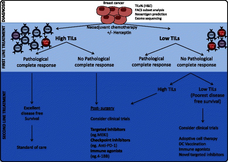
Review
. 2015 Aug 24:13:202.
doi: 10.1186/s12916-015-0431-3.

Relevance of tumor-infiltrating lymphocytes in breast cancer

Affiliations
Review

Relevance of tumor-infiltrating lymphocytes in breast cancer

Sathana Dushyanthen et al. BMC Med. .

Abstract

While breast cancer has not been considered a cancer amenable to immunotherapeutic approaches, recent studies have demonstrated evidence of significant immune cell infiltration via tumor-infiltrating lymphocytes in a subset of patient tumors. In this review we present the current evidence highlighting the clinical relevance and utility of tumor-infiltrating lymphocytes in breast cancer. Retrospective and prospective studies have shown that the presence of tumor-infiltrating lymphocytes is a prognostic marker for higher responses to neoadjuvant chemotherapy and better survival, particularly in triple negative and HER2-positive early breast cancer. Further work is required to determine the immune subsets important in this response and to discover ways of encouraging immune infiltrate in tumor-infiltrating lymphocytes-negative patients.


Figures

Fig. 1
Fig. 1
Interactions between the immune microenvironment and tumor cells in breast cancer. The antitumor immune response is dependent upon CD4+ (Th1) IFNγ production, which in turn mediates the expansion, differentiation, and activation of tumor-specific CD8+. CD8+ cytotoxic T cells induce cell lysis via recognition of specific TAAs such as MHC, FAS, and TRAILR on the surface of cancer cells/APCs. Similarly, CD4+ T cells are able to recognize MHC II on APCs. As a result of this complex formation (TCR-MHC/Peptide), high levels of granzymes, IFNγ, and perforin are released from CTLs, resulting in granule exocytosis and tumor cell death via apoptosis. NK and NKT cells with the help of APCs (DCs/M1) and CD4+Th1 are able to recognize and eliminate tumor cells. In the pro-tumor environment, CTLA-4, TIM-3, and PD-1 deliver inhibitory signals as a result of T-cell exhaustion/anergy caused by prolonged activation. CTLA-4 negatively regulates T-cell activation during the ‘priming’ phase of T-cell response. PD-1 expressed on T cells in the effector phase of T-cell response binds to its ligand PD-L1, expressed within the tumor microenvironment. This results in inhibition of T-cell activity (apoptosis). FOXP3+ Treg cells play a critical role during the selection of high-avidity CD8+ T cells, reducing their functionality. Tregs also have inhibitory action on APCs, CD8+ T cells, NKs, and CD4+ Th1 T cells. Both Tregs and tumor cells produce adenosine, which has inhibitory effects on T cells. Tumor cells can secrete cytokines and chemokines (e.g., TGF-β, CCL2) that recruit and stimulate suppressive cells such as Tregs, MDSCs, and M2 macrophages. M2 macrophages and MDSCs inhibit T-cell responses through nutrient sequestration via arginase, ROS, and NOS generation, as well as interference with trafficking into the tumor site. The upregulation of immunosuppressive enzymes such as IDO and arginase catabolizes essential nutrients required for effector cell activation. Furthermore, tumor cells downregulate MHC molecules, lose expression of antigenic molecules, and upregulate inhibitory molecules such as PD-L1, causing immune recognition to be inhibited and thus allowing immune escape and cancer progression. This figure was made exclusively for this manuscript. A2aR A2A adenosine receptor, ADP adenosine diphosphate, AMP adenosine monophosphate, APC antigen-presenting cell, ATP adenosine triphosphate, CCl-2 chemokine ligand-2, CTL cytotoxic T lymphocyte, CTLA-4 cytotoxic t lymphocyte-associated protein, DC dendritic cell, FAS fatty-acid synthase, GAL-9 galectin-9, IDO indolamine 2,3-dioxygenase, IFNγ interferon gamma, IL interleukin, M1/M1 TAM tumor-associated macrophage, MDSC myeloid-derived suppressor cell, MHC major histocompatibility complex, NK natural killer, NKT natural killer T cell, NOS nitric oxide synthase, PD-1 programmed death, ROS reactive oxygen species, TAA tumor-associated antigen, TCR T-cell receptor, TGF-β transforming growth factor beta, TNFα tumor necrosis factor alpha, TRAIL TNF-related apoptosis-inducing ligand, Treg T regulatory cell
Fig. 2
Fig. 2
Using the TIL infiltrate and response to frontline treatments to guide patient management decisions. The presence of tumor-infiltrating lymphocytes (TILs) and response to neoadjuvant chemotherapy (NAC) may be used to guide decisions on second line treatments. Patients with high TILs and exhibiting pathological complete responses to NAC (far left) have an excellent prognosis and may not require further intervention other than standard of care. Patients with high TILs at diagnosis but no pathological complete response, or patients with low TILs at diagnosis but high TILs post-NAC, may benefit from immunotherapies, such as checkpoint inhibition (PD-1 blockade), or immune agonists (e.g., 4-1BB). However, patients with little TIL infiltrate either pre-NAC or post-NAC (far right) require additional or different treatment strategies to induce an immune response, such as adoptive cellular therapy or vaccination strategies. Targeted inhibitors (e.g., MEK inhibitors) should be considered for all patient groups where appropriate, but the impact of targeted inhibitors on the immune response should be a therapeutic consideration. This figure was made exclusively for this manuscript. DC dendritic cells, FACS fluorescence-activated cell sorting, H&E hematoxylin and eosin staining

References

    1. Schreiber RD, Old LJ, Smyth MJ. Cancer immunoediting: integrating immunity’s roles in cancer suppression and promotion. Science. 2011;331:1565–70. doi: 10.1126/science.1203486. - DOI - PubMed
    1. Mittal D, Gubin MM, Schreiber RD, Smyth MJ. New insights into cancer immunoediting and its three component phases--elimination, equilibrium and escape. Curr Opin Immunol. 2014;27:16–25. doi: 10.1016/j.coi.2014.01.004. - DOI - PMC - PubMed
    1. Andre F, Dieci MV, Dubsky P, Sotiriou C, Curigliano G, Denkert C, et al. Molecular pathways: involvement of immune pathways in the therapeutic response and outcome in breast cancer. Clin Cancer Res. 2013;19:28–33. doi: 10.1158/1078-0432.CCR-11-2701. - DOI - PubMed
    1. Caras I, Grigorescu A, Stavaru C, Radu DL, Mogos I, Szegli G, et al. Evidence for immune defects in breast and lung cancer patients. Cancer Immunol Immunother. 2004;53:1146–52. doi: 10.1007/s00262-004-0556-2. - DOI - PMC - PubMed
    1. Monjazeb AM, Zamora AE, Grossenbacher SK, Mirsoian A, Sckisel GD, Murphy WJ. Immunoediting and antigen loss: overcoming the Achilles heel of immunotherapy with antigen non-specific therapies. Front Oncol. 2013;3:197. doi: 10.3389/fonc.2013.00197. - DOI - PMC - PubMed