. 2015 Sep 17;59(6):891-903.
doi: 10.1016/j.molcel.2015.07.022. Epub 2015 Aug 20.

Ancient Origin of cGAS-STING Reveals Mechanism of Universal 2',3' cGAMP Signaling

Affiliations

Ancient Origin of cGAS-STING Reveals Mechanism of Universal 2',3' cGAMP Signaling

Philip J Kranzusch et al. Mol Cell. .

Abstract

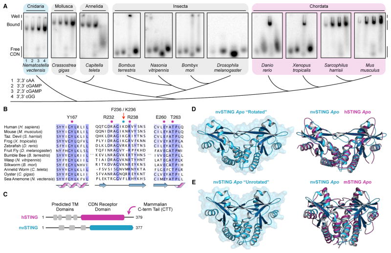
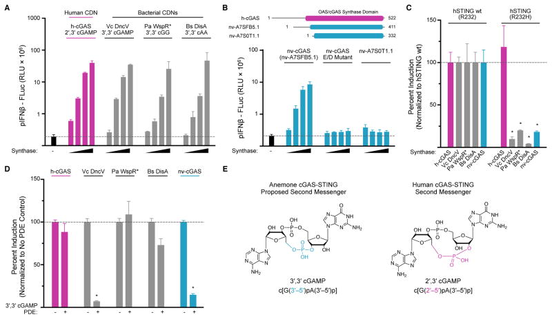
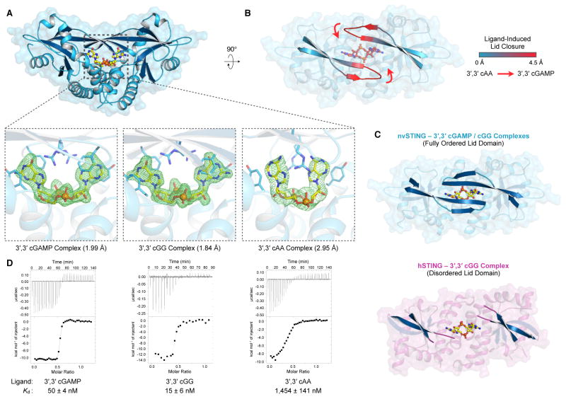
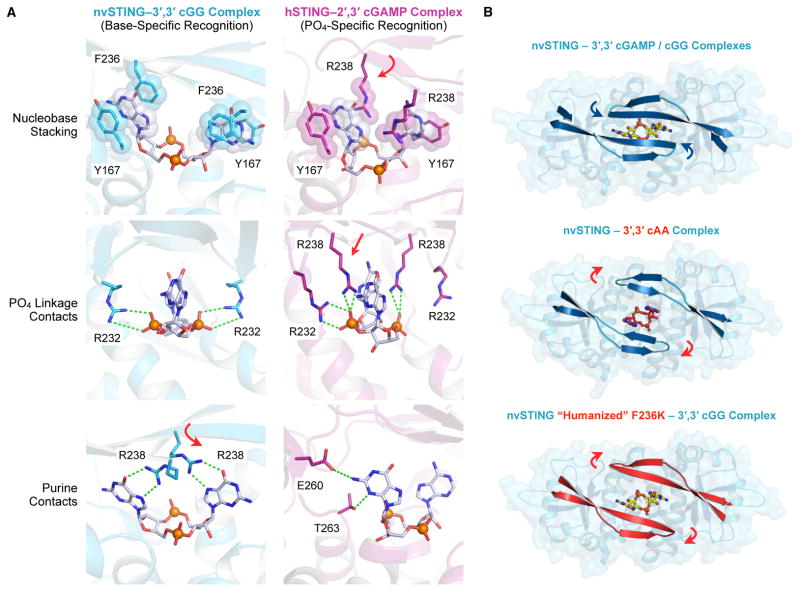
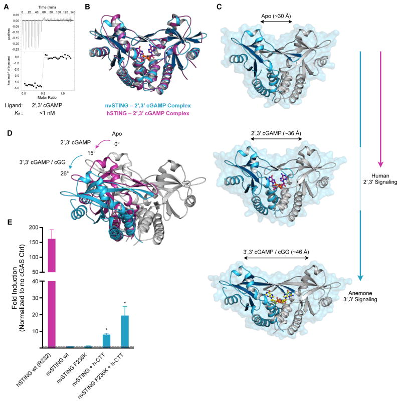
In humans, the cGAS-STING immunity pathway signals in response to cytosolic DNA via 2',3' cGAMP, a cyclic dinucleotide (CDN) second messenger containing mixed 2'-5' and 3'-5' phosphodiester bonds. Prokaryotes also produce CDNs, but these are exclusively 3' linked, and thus the evolutionary origins of human 2',3' cGAMP signaling are unknown. Here we illuminate the ancient origins of human cGAMP signaling by discovery of a functional cGAS-STING pathway in Nematostella vectensis, an anemone species >500 million years diverged from humans. Anemone cGAS appears to produce a 3',3' CDN that anemone STING recognizes through nucleobase-specific contacts not observed in human STING. Nevertheless, anemone STING binds mixed-linkage 2',3' cGAMP indistinguishably from human STING, trapping a unique structural conformation not induced by 3',3' CDNs. These results reveal that human mixed-linkage cGAMP achieves universal signaling by exploiting a deeply conserved STING conformational intermediate, providing critical insight for therapeutic targeting of the STING pathway.


Figures

Figure 1
Figure 1. STING Cyclic Dinucleotide Recognition Predates Modern Innate Immunity
(A) Gel-shift assay measuring complex formation between recombinant STING proteins and radiolabeled CDNs. Each panel shows reactions in order: (1) 3′,3′ cAA, (2) 3′,3′ cGAMP, (3) 2′,3′ cGAMP and (4) 3′,3′ cGG. (B) Phylogenetic alignment of animal STING proteins colored by amino-acid conservation. Key CDN interacting residues are indicated. (C) Cartoon schematic of human and anemone STING proteins. Predicted transmembrane domains are indicated in grey, and the crystallized CDN receptor domain is indicated in magenta or blue. Notably, the cterminal tail required for hSTING interferon signaling is absent in nvSTING. (D,E) Crystal structure of apo nvSTING CDN receptor domain in “rotated” and “unrotated” states. Overlaid structures compare the apo nvSTING structures (blue) and apo hSTING (PDB 4F9E) or apo mouse STING (PDB 4KC0) (magenta) revealing conformational dynamics and an ancestrally conserved STING fold. See also Figure S1.
Figure 2
Figure 2. Anemone cGAS Signals Via a 3′,3′-Linked Cyclic Dinucleotide
(A) Cell culture assay using wildtype hSTING (R232) and an interferon β luciferase reporter to measure CDN synthase activity in transfected human cells. Titrated synthases include human cGAS (h-cGAS), Vibrio cholerae DncV (Vc DncV), Pseudomonas aeruginosa WspR D70E (Pa WspR*), Bacillus subtilis DisA (Bs DisA) and the product CDNs are indicated. (B) Cartoon schematic of candidate N. vectensis cGAS-like enzymes, and screen for CDN synthase activity using interferon β assay as in A. Activity is detected with gene nv-cGAS (nv-A7SFB5.1), but not in a catalytic inactive nv-cGAS control (E/D mutant) or in the closely related candidate gene nv-A7S0T1.1. (C) Cell interferon β luciferase as in A, using either a wildtype hSTING (R232) allele responsive to all CDNs, or a mutant hSTING (R232H) allele highly selective for 2′,3′ cGAMP. Production of 2′,3′ cGAMP by human cGAS activates both STING alleles, while 3′,3′ CDN production by bacterial synthases and nv-cGAS only activates wildtype hSTING. (D) Cell interferon β luciferase as in A, with supplementation of a 3′,3′ cGAMP specific phosphodiesterase (PDE) (Vibrio cholerae VCA0681). Vc DncV and nv-cGAS 3′,3′ cGAMP production is ablated by 3′,3′ cGAMP PDE co-expression, while other synthases are not significantly affected. (E) Cartoon schematic of proposed anemone 3′,3′ cGAMP and human 2′,3′ cGAMP second messengers. Data in C and D are normalized to hSTING wt and synthase control. Error bars represent the SE of the mean of at least three independent experiments (asterisk denotes p <0.002). See also Figures S2 and S3.
Figure 3
Figure 3. nvSTING Specifically Recognizes Guanine Bases in 3′,3′ Second Messengers
(A) Crystal structure of nvSTING in complex with 3′,3′ cGAMP, 3′,3′ cGG or 3′,3′ cAA as indicated. Zoomed-in cutaway includes simulated-annealing Fo–Fc omit maps of ligand density contoured to 5.0 σ (3′,3′ cGAMP, cGG) or 4.0 σ (cAA). (B) Top-down view of nvSTING–3′,3′ cAA crystal structure. The beta-strand lid domains are highlighted and colored according to range of movement compared to the closed nvSTING–3′,3′ cGAMP / cGG crystal structures. Red arrows denote the direction of beta-strand lid closure upon 3′,3′ cGAMP / cGG binding. (C) Crystal structures of nvSTING–3′,3′ cGAMP / cGG (blue) and hSTING–3′,3′ cGG (PDBs 45FY [R232] and 4F9G [H232]) (magenta) complexes. The beta-strand lid domain is highlighted illustrating the fully closed nvSTING lid domain compared with the disordered and loosely organized hSTING lid domain. (D) ITC measurements of nvSTING affinity for specific 3′,3′ CDN ligands as indicated. ITC data is representative of at least three independent experiments. See also Figure S4 and Table S1.
Figure 4
Figure 4. Structural Basis of nvSTING Guanine-Specific Cyclic Dinucleotide Recognition
(A) Detailed STING–CDN interactions in nvSTING–3′,3′ cGG (blue) and hSTING–2′,3′ cGAMP (magenta) complexes as described in the Results. nvSTING exhibits guanine base-specific ligand recognition while hSTING exhibits 2′,3′ phosphodiester linkage-specific ligand recognition. Variance between nvSTING and hSTING at position F236/K236 induces repositioning of R238 to support either guanine-specific contacts (nvSTING, red arrow) or phosphodiester-linkage contacts (hSTING, red arrows). For clarity, nvSTING amino acids are numbered according to hSTING sequence. (B) Top-down view of nvSTING–3′,3′ CDN crystal complexes and relative positioning of the beta-strand lid domain. The wildtype nvSTING lid domain (blue) is tightly closed over 3′,3′ cGAMP / cGG ligands (blue arrows) and in contrast the lid domain remains open in the 3′,3′ cAA bound structure (red cAA ligand and indicating red arrows). A humanizing F236K mutation in nvSTING prevents guanine-recognition and the nvSTING(F236K) lid domain (red) remains open when bound to 3′,3′ cGG. See also Figure S4.
Figure 5
Figure 5. 2′,3′ cGAMP Traps a Unique and Conserved STING Conformation
(A) ITC measurements of nvSTING affinity for 2′,3′ cGAMP. (B) Structural overlay of nvSTING–2′,3′ cGAMP complex (blue) and hSTING–2′,3′ cGAMP complex (magenta) (PDB 4KSY) demonstrating the monomer wing rotation and core CDN-interacting portion of nvSTING and hSTING are unchanged. (C) Structural comparison of nvSTING structures in various ligandbound complexes. CDN ligands lock STING in alternative conformations as measured by the distance between the apical monomer wing domains (monomer 1 in blue, monomer 2 in grey). nvSTING–3′,3′ CDN interactions result in complete monomer rotation (blue vertical line) while primary hSTING–2′,3′ cGAMP signaling traps a partially rotated intermediate structure (magenta line). (D) Endogenous anemone 3′,3′ second messengers trigger an ~26° rotation in monomer wing domains from the apo state (apo grey, 3′,3′-bound in blue), while human 2′,3′ cGAMP traps an ~15° rotated structural intermediate in the nvST ING and hSTING structures (2′,3′-bound in magenta). (E) Cell interferon β luciferase as in Figure 2A, using indicated STING plasmids stimulated with human cGAS overexpression. Error bars represent the SE of the mean of at least three independent experiments (asterisk denotes p <0.002). ITC data is representative of at least three independent experiments. See also Figure S5 and Table S1.
Figure 6
Figure 6. Ratchet Model of STING Activation
(A,B) Ratchet model of CDN-induced conformational change leading to STING activation as described in Results. Specific lid domain interactions allow STING to discriminate endogenously produced second-messengers in a species-specific manner. Correct CDN engagement transduces reorganization of the STING lid to create large conformation changes in the apical wing domains. The unique ability of 2′,3′ cGAMP to capture a conserved structural intermediate allows universal signaling independent of STING allele diversity. See also Movie S1.

References

    1. Ablasser A, Goldeck M, Cavlar T, Deimling T, Witte G, Rohl I, Hopfner KP, Ludwig J, Hornung V. cGAS produces a 2′-5′-linked cyclic dinucleotide second messenger that activates STING. Nature. 2013;498:380–384. - PMC - PubMed
    1. Blaauboer SM, Gabrielle VD, Jin L. MPYS/STING-mediated TNF-alpha, not type I IFN, is essential for the mucosal adjuvant activity of (3′-5′)-cyclic-di-guanosine-monophosphate in vivo. Journal of immunology. 2014;192:492–502. - PMC - PubMed
    1. Burdette DL, Monroe KM, Sotelo-Troha K, Iwig JS, Eckert B, Hyodo M, Hayakawa Y, Vance RE. STING is a direct innate immune sensor of cyclic di-GMP. Nature. 2011;478:515–518. - PMC - PubMed
    1. Chen H, Sun H, You F, Sun W, Zhou X, Chen L, Yang J, Wang Y, Tang H, Guan Y, et al. Activation of STAT6 by STING is critical for antiviral innate immunity. Cell. 2011;147:436–446. - PubMed
    1. Chin KH, Tu ZL, Su YC, Yu YJ, Chen HC, Lo YC, Chen CP, Barber GN, Chuah ML, Liang ZX, et al. Novel c-di-GMP recognition modes of the mouse innate immune adaptor protein STING. Acta crystallographica Section D, Biological crystallography. 2013;69:352–366. - PubMed

Publication types