. 2015 Sep 3;59(5):732-43.
doi: 10.1016/j.molcel.2015.07.014. Epub 2015 Aug 20.

Global Promoter Targeting of a Conserved Lysine Deacetylase for Transcriptional Shutoff during Quiescence Entry

Affiliations

Global Promoter Targeting of a Conserved Lysine Deacetylase for Transcriptional Shutoff during Quiescence Entry

Jeffrey N McKnight et al. Mol Cell. .

Abstract

Quiescence is a conserved cell-cycle state characterized by cell-cycle arrest, increased stress resistance, enhanced longevity, and decreased transcriptional, translational, and metabolic output. Although quiescence plays essential roles in cell survival and normal differentiation, the molecular mechanisms leading to this state are not well understood. Here, we determined changes in the transcriptome and chromatin structure of S. cerevisiae upon quiescence entry. Our analyses revealed transcriptional shutoff that is far more robust than previously believed and an unprecedented global chromatin transition, which are tightly correlated. These changes require Rpd3 lysine deacetylase targeting to at least half of gene promoters via quiescence-specific transcription factors including Xbp1 and Stb3. Deletion of RPD3 prevents cells from establishing transcriptional quiescence, leading to defects in quiescence entry and shortening of chronological lifespan. Our results define a molecular mechanism for global reprogramming of transcriptome and chromatin structure for quiescence driven by a highly conserved chromatin regulator.


Figures

Figure 1
Figure 1. Global Transcriptional Shutoff Accompanies Quiescence Entry
(A) Boxplot of transcript abundance for all genes (left) or ribosomal protein genes (right) during logarithmic growth in rich media (log), 2 hours after glucose exhaustion (DS), or purified quiescent cells from a 7 day culture (Q). (B) Browser shot of strand-specific, normalized transcript abundance across chromosome III. Blue indicates Watson strand and red indicates Crick strand. (C–E). Scatter plots comparing normalized transcript abundance between indicated cell cycle stages. Yellow dots indicate ribosomal protein transcripts. Red diagonal line indicates y=x axis to represent where expected transcript distribution should occur in absence of global transcription changes. Normalized FPKM denotes fragments per kilobase of exon per million fragments mapped determined by Cufflinks and scaled to normalize for standard-corrected RNA per cell. See also Figure S1 and Table S1
Figure 2
Figure 2. Global Changes in Chromatin Reflect Transciptional Repression in Q Cells
(A) Nucleosome dyad signal aligned at 4550 transcription start sites (TSS) (Nagalakshmi et al., 2008) for matched MNase digests from Q and log cells. (B, C) Nucleosome dyad signal for two different digestion levels in log cells (B) or Q cells (C) at TSS. Corresponding MNase digestion ladders are shown in Figure S2A. (D) Individual nucleosome dyad signal aligned at TSS, and sorted by increase and decrease in dyad signal at nucleosome depleted regions (NDR) as cells transition from log growth (left) to quiescence (right). (E) Western Blot using antibodies against acetylated histone tails as indicated in log, DS, or Q cells. Total H3 was used as a loading control. See also Figure S2
Figure 3
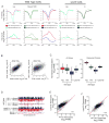
Figure 3. Tight Correlations Between Chromatin Transition and Transcriptional Shutoff
(A) Nucleosome dyad signal (left), histone H3 density (center), and hyperacetylated H4 signals (right) in log or Q cells at TSSs for top 500 genes most repressed in Q cells. Dyad signal is from 80% mononucleosome data set, H3 signal is first Z-score normalized, and H4 hyperacetylation signal is corrected for histone density. H3K23 acetylation profiles are show in Figure S3A and S3B. (B) Same as (A) but for the 500 least repressed (or induced) transcripts in Q cells. (C) The changes in signals between most and least repressed genes upon Q entry. (D) Correlation between changes in H3 occupancy (from ChIP-Seq) and changes in transcription between Q and log cells (left). Heat map of changed H3 signals (Q/log, normalized to input), sorted by changes in transcription between Q and log cells (right). (E) Same as (D) but for changes in H3-normalized, hyperacetylated H4ac levels. (F–H) Changes in MNase dyad signals (F, from 50% mononucleosome digest), H3 occupancy (G), or H4 hyperacetylation (H) at 166 transcription factor motifs from the JASPAR database (Mathelier et al., 2014) (see Supplemental Experimental Procedures) sorted by difference in signals. Green bars indicate motifs within indicated cluster corresponding to transcription factor with known interactions with Rpd3 (from BioGRID database: http://thebiogrid.org). Transcription factor identity and expanded Rpd3 interaction motifs are shown in Figure S3E–G. See also Figure S3
Figure 4
Figure 4. Rpd3 is Required for Quiescence Entry and Survival
(A) Representative image of WT (left) and Δrpd3 (right) stationary phase cultures after 7 days of growth prior to Q cell isolation, depicting distinct culture morphologies. (B) Growth analysis of WT and Δrpd3 cells through 48 hours in YPD. Arrows indicate time of glucose exhaustion. (C) Viability assay showing cell survival as cells enter the Q state in WT and Δrpd3 cells. (D) Representative image of Q cells purified from 7-day stationary phase cultures indicating similar gross morphology. (E) Western Blot of indicated acetylated histones in WT and Δrpd3 cells. H3 serves as a loading control. (F) Survival curve for purified Q cells, measured by colony forming units, after incubation in pure water for indicated time at 30 degrees C. Data are presented as mean +/− SEM. See also Figure S4
Figure 5
Figure 5. Rpd3 Plays the Key Role for Transcriptional Quiescence
(A) Histone H3 density (top) or hyperacetylated H4ac signal (bottom) for 500 most- and 500 least-repressed genes in Q state for indicated strains. Orange lines highlight the points of similarity between WT DS shift cells and Δrpd3 Q cells. (B) Scatterplot showing correlation of H4ac signal at TSSs between indicated strains. (C) Boxplot showing normalized transcript abundance globally (left) or for ribosomal protein transcripts (right). Orange lines highlight similarities between WT DS shift and Δrpd3 Q. (D) Browser shot comparing normalized transcription levels in WT DS, WT Q, and Δrpd3 Q cells. (E, F) Scatterplots showing correlation and global differences between the transcriptomes of the indicated strains. Red diagonal line indicates y=x axis, and yellow dots correspond to ribosomal protein genes. See also Figure S5.
Figure 6
Figure 6. Global Relocalization of Rpd3 Upon Quiescent Entry
(A) Browser shot of Rpd3 binding in log cells (light blue) and Q cells (dark blue) along the length of chromosome IX (top) or a zoomed in (translucent red rectangle) segment of chromosome IX (bottom). (B) Global distribution of Rpd3 localization at TSSs in aggregate (left) or individual genes ranked by Rpd3-Myc signal (right). (C) Browser shot of Rpd3-Myc ChIP-seq signals in WT (black), Δstb3 (blue), and Δxbp1 (orange) strains showing TF-dependence of Rpd3 binding. Gene names indicate promoters where Rpd3 binding is dictated by the associated TF. (D) Global comparison of Rpd3 promoter binding in Δstb3 and Δxbp1 strains. Rpd3 ChIP-seq signal ratio was calculated at 4550 TSSs. Blue indicates promoters where deletion of STB3 reduces Rpd3 binding while orange indicates promoters where XBP1 deletion reduces Rpd3 binding. (E) Scatterplot showing mutual exclusivity of Rpd3 binding and H4 hyperacetylation in Q cells at gene promoters. (F) Histone acetylation levels, relative transcriptional repression, and change in Pol II binding during the log-to-Q transition at annotated TSS ranked by Rpd3 binding in Q cells. Transcriptional repression is represented as log2 transcription ratio centered at median repression value. Acetylation difference is the log2 ratio normalized to H3, Pol II change is log2 ratio of Rpb3 (Pol II subunit) binding between log and Q cells. (G) Difference in acetylation and transcription between WT Q and Δrpd3 Q cells ranked by Rpd3 binding in Q cells. See also Figure S6
Figure 7
Figure 7. Model for Rpd3-Mediated Quiescence
(A) Cartoon representation of chromatin state in log and Q cells. See text for details. (B) Characteristic locus demonstrating Rpd3-mediated repression. The TKL1 gene is actively transcribed in log cells, where Rpd3 is absent (top). In Q cells, Xbp1 binds to a specific motif (green rectangle) and recruits Rpd3 upstream of the TKL1 promoter (red arrow), histones are deacetylated into the coding region (yellow shaded region), nucleosomes are stabilized (green shaded region), and transcription is extensively repressed.

Similar articles

Cited by

References

    1. Alejandro-Osorio AL, Huebert DJ, Porcaro DT, Sonntag ME, Nillasithanukroh S, Will JL, Gasch AP. The histone deacetylase Rpd3p is required for transient changes in genomic expression in response to stress. Genome biology. 2009;10:R57. - PMC - PubMed
    1. Allen C, Buttner S, Aragon AD, Thomas JA, Meirelles O, Jaetao JE, Benn D, Ruby SW, Veenhuis M, Madeo F, et al. Isolation of quiescent and nonquiescent cells from yeast stationary-phase cultures. The Journal of cell biology. 2006;174:89–100. - PMC - PubMed
    1. Aragon AD, Rodriguez AL, Meirelles O, Roy S, Davidson GS, Tapia PH, Allen C, Joe R, Benn D, Werner-Washburne M. Characterization of differentiated quiescent and nonquiescent cells in yeast stationary-phase cultures. Molecular biology of the cell. 2008;19:1271–1280. - PMC - PubMed
    1. Baker LA, Ueberheide BM, Dewell S, Chait BT, Zheng D, Allis CD. The yeast Snt2 protein coordinates the transcriptional response to hydrogen peroxide-mediated oxidative stress. Molecular and cellular biology. 2013;33:3735–3748. - PMC - PubMed
    1. Brauer MJ, Huttenhower C, Airoldi EM, Rosenstein R, Matese JC, Gresham D, Boer VM, Troyanskaya OG, Botstein D. Coordination of growth rate, cell cycle, stress response, and metabolic activity in yeast. Molecular biology of the cell. 2008;19:352–367. - PMC - PubMed

Publication types

MeSH terms

Associated data