. 2015 Sep 15;109(6):1282-94.
doi: 10.1016/j.bpj.2015.07.034. Epub 2015 Aug 20.

Cardiolipin Interactions with Proteins

Affiliations

Cardiolipin Interactions with Proteins

Joan Planas-Iglesias et al. Biophys J. .

Abstract

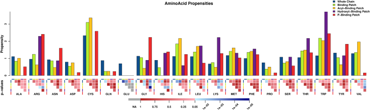
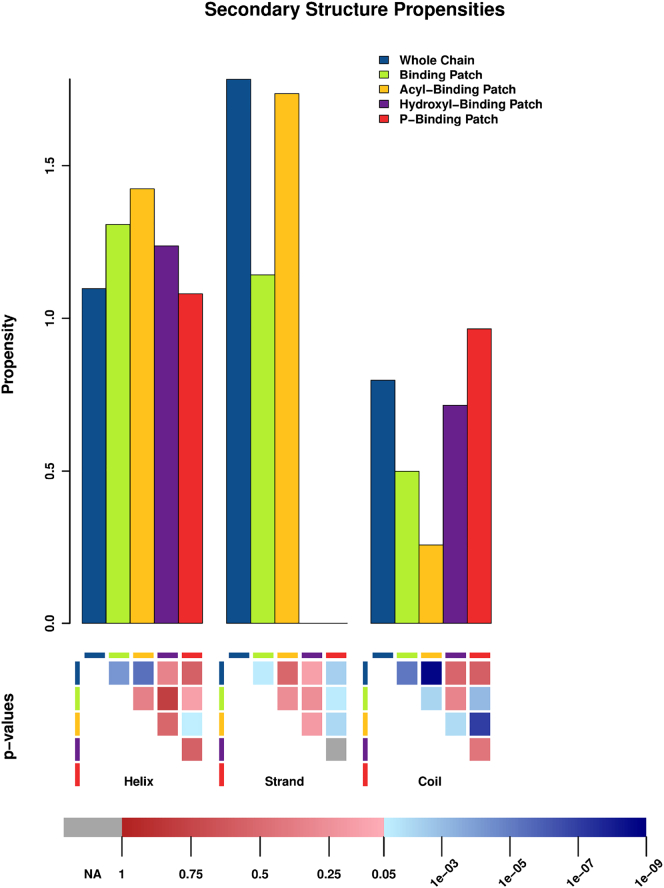
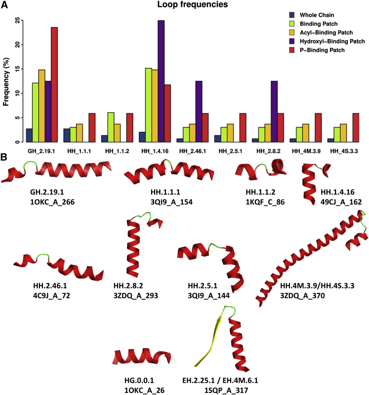
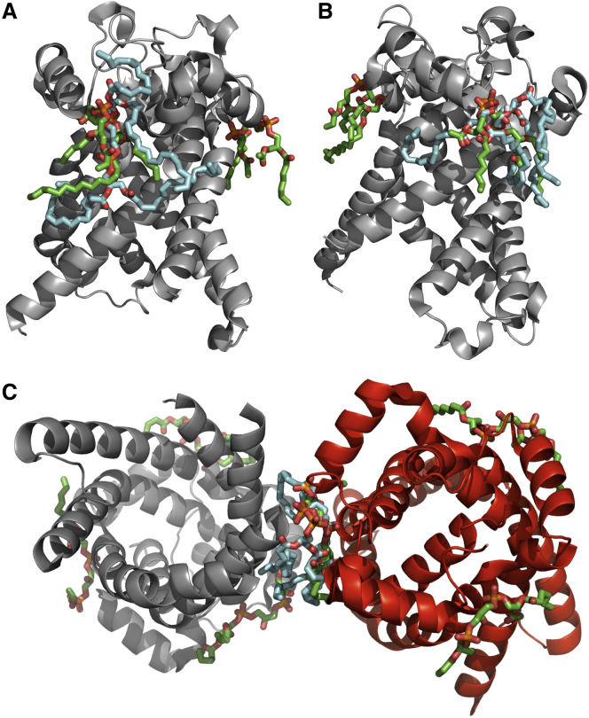
Cardiolipins (CL) represent unique phospholipids of bacteria and eukaryotic mitochondria with four acyl chains and two phosphate groups that have been implicated in numerous functions from energy metabolism to apoptosis. Many proteins are known to interact with CL, and several cocrystal structures of protein-CL complexes exist. In this work, we describe the collection of the first systematic and, to the best of our knowledge, the comprehensive gold standard data set of all known CL-binding proteins. There are 62 proteins in this data set, 21 of which have nonredundant crystal structures with bound CL molecules available. Using binding patch analysis of amino acid frequencies, secondary structures and loop supersecondary structures considering phosphate and acyl chain binding regions together and separately, we gained a detailed understanding of the general structural and dynamic features involved in CL binding to proteins. Exhaustive docking of CL to all known structures of proteins experimentally shown to interact with CL demonstrated the validity of the docking approach, and provides a rich source of information for experimentalists who may wish to validate predictions.


Figures

Figure 1
Figure 1
Propensities of observed amino acids in all protein chains analyzed (blue), BPs (green), ABPs (dark yellow), OBPs (purple), and PBPs (red). The p-values for the statistical significance of differences between propensities of each amino acid in different BPs are shown in the lower panel. NA (gray) indicates that two empty sets were compared and hence no statistics can be computed. In red and using a linear scale, nonsignificant p-values ranging from 1 (darker red, indicates that greater differences could always be observed in a random set) to 0.05 (lighter red) are shown. In blue and using a logarithmic scale, significant p-values are shown, from 0.05 (light blue, differences observed could be reproduced by random only in 5 over 100 cases) to p-values near 0 (dark blue, the probability to observe such differences by chance is the lowest).
Figure 2
Figure 2
Relative propensity of observed secondary structures in all protein chains (blue), BPs (green), ABPs (dark yellow), OBPs (purple), and PBPs (red). The p-values for the statistical significance of differences between propensities of each amino acid in different BPs are shown in the lower panel, using the same color legend described in Fig 1.
Figure 3
Figure 3
(A) Relative frequencies of observed supersecondary motifs (loops) in all protein chains (blue), BPs (green), ABPs (dark yellow), OBPs (purple), and PBPs (red). (B) Cartoon images of the loops present in PBPs. Different secondary structure elements are shown in color (helices in red, strands in yellow, and coiled-coil regions in green). Each motif is identified in the first text line below by the code of its corresponding loop type(s). The PDB file from where each loop structure shown was obtained, protein chain and starting residue of the supersecondary structure are provided in the second line below each cartoon.
Figure 4
Figure 4
Loops of ADP/ATP translocase 1 (PDB 1OKC chain A) interacting with two cardiolipin molecules. (A) One CL molecule (left) interacts with loop GH_2.19.1 (1OKC_A_66; residues 66–99; colored in yellow).The second CL molecule (right) interacts with a different instance of loop GH_2.19.1 (1OKC_A_266; residues 266–291; colored in cyan), showing that both binding sites adopt similar structural conformation. The second molecule also interacts with loop HH_1.1.2 (1OKC_A_4; residues 4–37; colored in light orange) and loop HG_0.1.1 (1OKC_A_26; residues 26–40; colored in pale yellow). Overlapping region between HH_1.1.2 and HG_0.1.1 (residues 26–37) is shown in brown). Positively charged amino acids Arg and Lys in loop GH_2.19.1 are shown in blue, and the conserved Gly in the loop motif is colored in purple. (B) Enlarged images of areas circled in (A) showing interaction between positively charged residues and the phosphate groups of CL in the two different binding sites, where CL binding is accomplished by different geometries. The geometry of binding is described by measured distances between two proximal oxygen atoms in the different CL phosphate groups to carbon beta and distal nitrogen in relevant Arg (4.6 Å and 7.7 Å, respectively, left) or Lys (4.9 Å and 13.4 Å, respectively, right).
Figure 5
Figure 5
Overlap of docked CL molecule with CL costructures of 1OKC and 4C9J. The docked CL is colored in cyan and the CL costructures are colored in green. (A) Overlap with CDL 800 molecule of 1OKC; (B) Overlap with CDL 801 molecule of 1OKC; (C) Overlap with CDL 802 molecule of 4C9J chain A (red).

References

    1. Daum G. Lipids of mitochondria. Biochim. Biophys. Acta. 1985;822:1–42. - PubMed
    1. Schlame M., Brody S., Hostetler K.Y. Mitochondrial cardiolipin in diverse eukaryotes. Comparison of biosynthetic reactions and molecular acyl species. Eur. J. Biochem. 1993;212:727–735. - PubMed
    1. Belikova N.A., Tyurina Y.Y., Kagan V.E. Heterolytic reduction of fatty acid hydroperoxides by cytochrome c/cardiolipin complexes: antioxidant function in mitochondria. J. Am. Chem. Soc. 2009;131:11288–11289. - PubMed
    1. Xu Y., Kelley R.I., Schlame M. Remodeling of cardiolipin by phospholipid transacylation. J. Biol. Chem. 2003;278:51380–51385. - PubMed
    1. Houtkooper R.H., Vaz F.M. Cardiolipin, the heart of mitochondrial metabolism. Cell. Mol. Life Sci. 2008;65:2493–2506. - PMC - PubMed

Publication types

LinkOut - more resources