

Occurrence and Management Strategies for Catheter Entrapment with Onyx Liquid Embolization
- PMID: 26301030
- PMCID: PMC4535605
Occurrence and Management Strategies for Catheter Entrapment with Onyx Liquid Embolization
Abstract
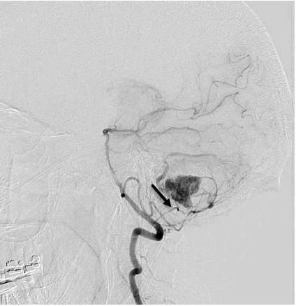
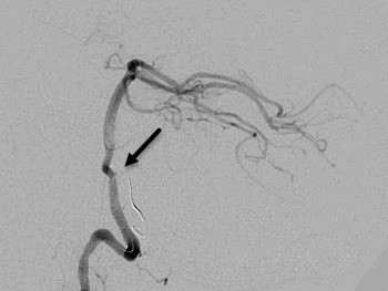
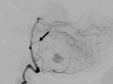
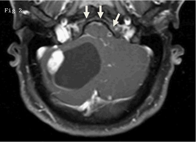
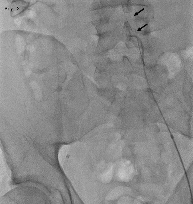
In June 2012, Food and Drug Administration (FDA) issued a warning about the risk of catheter entrapment associated with Onyx embolization. We used our experience, literature review, and FDA Manufacturer and User Facility Device Experience (MAUDE) data review to identify five strategies to address catheter entrapment: 1/. Surgical resection of vessel at point of entrapment of catheter and retraction from exterior portion at the femoral region; 2/. Advancing and closing the loop of snare over the entrapped catheter followed by retraction; 3/. Advancing the distal access catheter over the entrapped catheter and retraction with forward movement of the distal access catheters; 4/. Inflation of balloon catheter coaxial to the entrapped catheter with subsequent retraction; and 5/. Intravascular retention and internalization of microcatheter. In the MAUDE data, there were 77 reports of catheter entrapment with Onyx embolization; microcatheter was retracted by surgical excision in 15, endovascular snare or other retriever devices in 5, deliberately entrapped inside the vessel using stent in 1, and left without intervention within intravascular compartment in 27 patients.
Keywords: Catheter entrapment; Onyx; copolymer; intravascular; liquid embolic system; microcatheter.
Figures
Similar articles
-
Microcatheter entrapment retrieval from Onyx embolization in brain arteriovenous malformations: A technical note.Interv Neuroradiol. 2015 Oct;21(5):620-3. doi: 10.1177/1591019915583226. Epub 2015 Jul 31. Interv Neuroradiol. 2015. PMID: 26232252 Free PMC article.
-
Management of a fractured microcatheter during middle meningeal artery embolization.J Neurointerv Surg. 2024 Nov 22;16(12):1214. doi: 10.1136/jnis-2024-022279. J Neurointerv Surg. 2024. PMID: 39433325
-
Over-the-catheter retrieval of a retained microcatheter following Onyx embolization: a technical report.J Neurointerv Surg. 2012 Jul;4(4):e13. doi: 10.1136/neurintsurg-2011-010040. Epub 2011 Jun 16. J Neurointerv Surg. 2012. PMID: 21990504
-
Turnpike Catheter failure, causes and mechanisms: Insights from the MAUDE database.Ann Med Surg (Lond). 2022 Jun 5;78:103923. doi: 10.1016/j.amsu.2022.103923. eCollection 2022 Jun. Ann Med Surg (Lond). 2022. PMID: 35734685 Free PMC article. Review.
-
Major Complications and Failure Modes of the Angiosculpt Scoring Balloon Catheter: Analysis of the MAUDE Database.Curr Probl Cardiol. 2023 Apr;48(4):101557. doi: 10.1016/j.cpcardiol.2022.101557. Epub 2022 Dec 14. Curr Probl Cardiol. 2023. PMID: 36528205 Review.
Cited by
-
Emerging Embolic Agents in Endovascular Embolization: An Overview.Prog Biomed Eng (Bristol). 2020 Jan;2(1):012003. doi: 10.1088/2516-1091/ab6c7d. Epub 2020 Feb 12. Prog Biomed Eng (Bristol). 2020. PMID: 34553126 Free PMC article. No abstract available.
-
Transarterial embolization of peripheral high-flow arteriovenous malformation with ethylene vinyl alcohol copolymer (Onyx®): single-center 10-year experience.Radiol Med. 2019 Feb;124(2):154-162. doi: 10.1007/s11547-018-0948-6. Epub 2018 Oct 27. Radiol Med. 2019. PMID: 30368719
-
Onyx embolization with the Apollo detachable tip microcatheter: A single-center experience.Interv Neuroradiol. 2018 Jun;24(3):339-344. doi: 10.1177/1591019918758494. Epub 2018 Feb 26. Interv Neuroradiol. 2018. PMID: 29482458 Free PMC article.
-
Microsurgical Removal of Microcatheter in the Middle Cerebral Artery During Resection of an Arteriovenous Malformation Resection.Cureus. 2017 Apr 13;9(4):e1164. doi: 10.7759/cureus.1164. Cureus. 2017. PMID: 28507836 Free PMC article.
-
Photomodulated Extrusion as a Localized Endovascular Hydrogel Deposition Method.Adv Healthc Mater. 2023 May;12(12):e2202632. doi: 10.1002/adhm.202202632. Epub 2023 Feb 3. Adv Healthc Mater. 2023. PMID: 36681868 Free PMC article.
References
-
- Pierot L, Cognard C, Herbreteau D, Fransen WJ, Van Rooij E, Boccardi A, Beltramello N, Sourour K, Kupcs A, Biondi A, Bonafe W, Reith A, Casasco Endovascular treatment of brain arteriovenous malformations using a liquid embolic agent: results of a prospective, multicentre study (BRAVO) Eur Radiol. 2013;23(10):2838–2845. - PubMed
-
- Strauss I, Frolov V, Buchbut D, Gonen L, Malmon S. Critical appraisal of endovascular treatment of brain arteriovenous malformation using onyx in a series of 92 consecutive patients. Acta Neurochir (Wien) 2013;155(4):611–617. - PubMed
-
- Abud DG, Abud TG, Nakiri GS. Management of brain AVM procedural hemorrhagic complication by the “security” catheter technique. J Neuroradiol. 2013;40(1):45–49. - PubMed
LinkOut - more resources
Full Text Sources