Use of Intraoperative Computed Tomography for Revisional Procedures in Patients with Complex Maxillofacial Trauma
- PMID: 26301152
- PMCID: PMC4527637
- DOI: 10.1097/GOX.0000000000000455
Use of Intraoperative Computed Tomography for Revisional Procedures in Patients with Complex Maxillofacial Trauma
Abstract
Background: In patients with panfacial fractures and distorted anatomic landmarks of zygomatic and orbital complex, there is a risk of zygomaticomaxillary complex (ZMC) malpositioning even with the best efforts for surgical repair. This results in increased number of additional procedures to achieve accurate positioning.
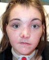
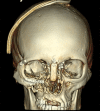
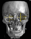
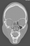
Methods: We describe the usage of intraoperative C-arm cone-beam computed tomographic (CT) scan for ZMC malpositioning in a representative patient with panfacial fractures.
Results: We have successfully used intraoperative CT scan for ZMC malpositioning in 3 patients. The representative patient had ZMC malposition after the initial attempt of surgical repair without any intraoperative imaging. On using intraoperative CT scan during the next attempt, we were able to reposition the ZMC accurately.
Conclusions: Intraoperative CT scan might improve the accuracy of ZMC positioning and decrease the chances of potential additional surgeries. In patients with distorted anatomical landmarks and panfacial fractures, it can be especially helpful toward correcting ZMC malposition.
Conflict of interest statement
Figures


References
-
- van den Bergh B, Karagozoglu KH, Heymans MW, et al. Aetiology and incidence of maxillofacial trauma in Amsterdam: a retrospective analysis of 579 patients. J Craniomaxillofac Surg. 2012;40:e165–e169. - PubMed
-
- van Hout WM, Van Cann EM, Abbink JH, et al. An epidemiological study of maxillofacial fractures requiring surgical treatment at a tertiary trauma centre between 2005 and 2010. Br J Oral Maxillofac Surg. 2013;51:416–420. - PubMed
-
- Forouzanfar T, Salentijn E, Peng G, et al. A 10-year analysis of the “Amsterdam” protocol in the treatment of zygomatic complex fractures. J Craniomaxillofac Surg. 2013;41:616–622. - PubMed
-
- Salentijn EG, Boverhoff J, Heymans MW, et al. The clinical and radiographical characteristics of zygomatic complex fractures: a comparison between the surgically and non-surgically treated patients. J Craniomaxillofac Surg. 2013;4:214–218. - PubMed
-
- Wilde F, Lorenz K, Ebner AK, et al. Intraoperative imaging with a 3D C-arm system after zygomatico-orbital complex fracture reduction. J Oral Maxillofac Surg. 2013;71:894–910. - PubMed
LinkOut - more resources
Full Text Sources
