. 2015:2015:642520.
doi: 10.1155/2015/642520. Epub 2015 Aug 2.

The Omega-3 Fatty Acid Docosahexaenoic Acid Modulates Inflammatory Mediator Release in Human Alveolar Cells Exposed to Bronchoalveolar Lavage Fluid of ARDS Patients

Affiliations

The Omega-3 Fatty Acid Docosahexaenoic Acid Modulates Inflammatory Mediator Release in Human Alveolar Cells Exposed to Bronchoalveolar Lavage Fluid of ARDS Patients

Paolo Cotogni et al. Biomed Res Int. 2015.

Abstract

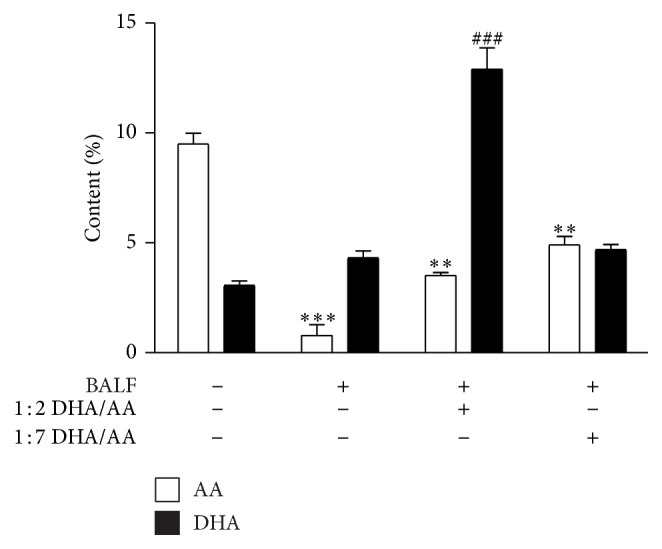
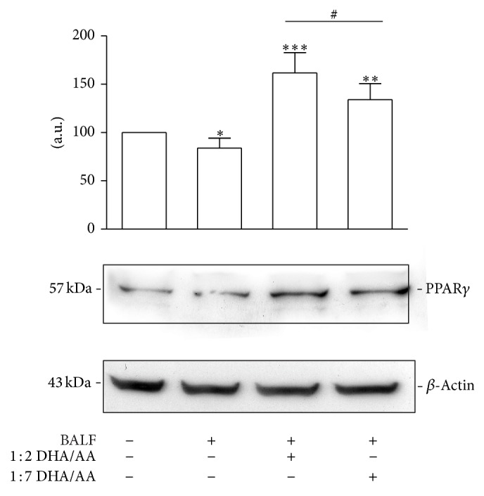
Background: This study investigated whether the 1 : 2 ω-3/ω-6 ratio may reduce proinflammatory response in human alveolar cells (A549) exposed to an ex vivo inflammatory stimulus (bronchoalveolar lavage fluid (BALF) of acute respiratory distress syndrome (ARDS) patients). Methods. We exposed A549 cells to the BALF collected from 12 ARDS patients. After 18 hours, fatty acids (FA) were added as docosahexaenoic acid (DHA, ω-3) and arachidonic acid (AA, ω-6) in two ratios (1 : 2 or 1 : 7). 24 hours later, in culture supernatants were evaluated cytokines (TNF-α, IL-6, IL-8, and IL-10) and prostaglandins (PGE2 and PGE3) release. The FA percentage content in A549 membrane phospholipids, content of COX-2, level of PPARγ, and NF-κB binding activity were determined.

Results: The 1 : 2 DHA/AA ratio reversed the baseline predominance of ω-6 over ω-3 in the cell membranes (P < 0.001). The proinflammatory cytokine release was reduced by the 1 : 2 ratio (P < 0.01 to <0.001) but was increased by the 1 : 7 ratio (P < 0.01). The 1 : 2 ratio reduced COX-2 and PGE2 (P < 0.001) as well as NF-κB translocation into the nucleus (P < 0.01), while it increased activation of PPARγ and IL-10 release (P < 0.001). Conclusion. This study demonstrated that shifting the FA supply from ω-6 to ω-3 decreased proinflammatory mediator release in human alveolar cells exposed to BALF of ARDS patients.


Figures

Figure 1
Figure 1
BALF cytokine pattern in ARDS. (a) IL-1β and TNF-α proinflammatory cytokine content in BALF collected from pulmonary (ARDSp, white bars) and extrapulmonary (ARDSexp, black bars) ARDS patients. (b) IL-6 and IL-8 cytokine content in BALF collected from pulmonary (ARDSp, white bars) and extrapulmonary (ARDSexp, black bars) ARDS patients. The results are expressed as picograms (pg) or nanograms (ng) of cytokines per mL as indicated. Data are presented as mean ± standard deviation of 12 independent determinations (n = 7 ARDSp and n = 5 ARDSexp). BALF, bronchoalveolar lavage fluid; ARDS, acute respiratory distress syndrome; IL, interleukin; TNF, tumor necrosis factor. * P < 0.05 ARDSp versus ARDSexp. ** P < 0.01 ARDSp versus ARDSexp. *** P < 0.001 ARDSp versus ARDSexp.
Figure 2
Figure 2
Effects of ω-3/ω-6 PUFA ratios on BALF induced cytokine release from A549 cells. (a) TNF-α proinflammatory cytokine release from A549 cells, stimulated with BALF and treated with 1 : 2 and 1 : 7 DHA/AA PUFA ratios. (b) IL-6 proinflammatory cytokine release from A549 cells stimulated with BALF and treated with 1 : 2 and 1 : 7 DHA/AA (ω-3/ω-6) PUFA ratios. (c) IL-8 proinflammatory cytokine release from A549 cells stimulated with BALF and treated with 1 : 2 and 1 : 7 DHA/AA PUFA ratios. (d) IL-10 anti-inflammatory cytokine release from A549 cells stimulated with BALF and treated with 1 : 2 and 1 : 7 DHA/AA PUFA ratios. In each panel, data are presented as picograms (pg) of the indicated cytokine per million cells. Data are presented as mean ± standard deviation of 12 independent determinations (n = 12). PUFA, polyunsaturated fatty acid; BALF, bronchoalveolar lavage fluid; TNF, tumor necrosis factor; DHA, docosahexaenoic acid; AA, arachidonic acid; IL, interleukin; LPS, lipopolysaccharide. *** P < 0.001 1 : 2 DHA/AA versus all. ** P < 0.01 1 : 2 DHA/AA versus all. ## P < 0.01 1 : 7 DHA/AA versus LPS and BALF.
Figure 3
Figure 3
Schematic representation of PUFA mechanism of action in BALF-stimulated A549 cells. PUFAs, polyunsaturated fatty acids; COX, cycloxygenase; PG, prostaglandin; PPAR, peroxisome proliferator-activated receptor; NF-κB, nuclear factor-kappa B; IκB, inhibitor of NF-κB.
Figure 4
Figure 4
Effects of ω-3/ω-6 PUFA ratios on the percentage content of AA and DHA in A549 membrane phospholipids. Relative percentage content of AA (white bars) and DHA (black bars) in phospholipids of A549 cell membranes stimulated with BALF and treated with 50 μM 1 : 2 or 1 : 7 DHA/AA PUFA ratios. Data are presented as percentage content of AA and DHA in the membrane phospholipids of unstimulated or stimulated A549 cells as indicated. Data are presented as mean ± standard deviation of 4 independent experiments (n = 4). PUFA, polyunsaturated fatty acid; AA, arachidonic acid; DHA, docosahexaenoic acid; BALF, bronchoalveolar lavage fluid. *** P < 0.001 BALF versus unstimulated. ** P < 0.01 1 : 2 and 1 : 7 DHA/AA versus all. ### P < 0.001 versus all.
Figure 5
Figure 5
Effects of ω-3/ω-6 PUFA ratios on PGE2 and PGE3 synthesis and release. (a) COX-2 relative protein content in A549 cells, stimulated with BALF and treated with 50 μM 1 : 2 and 1 : 7 DHA/AA PUFA ratios. Data are expressed as “a.u.” (arbitrary units) of the densitometric values, normalized on the corresponding β-actin. The value of unstimulated cells was arbitrarily set as 100. Data are presented as mean ± standard deviation of 6 independent determinations (n = 6). The image is representative of all the WB experiments. (b) PGE2 (white bars) and PGE3 (black bars) content in culture media of A549 cells, stimulated with BALFs and treated with 50 μM 1 : 2 or 1 : 7 DHA/AA ratio. Data are presented as picograms (pg) of the indicated PG per mL. Data are presented as mean ± standard deviation of 12 independent determinations (n = 12). PUFA, polyunsaturated fatty acid; PG, prostaglandin; COX, cycloxygenase; BALF, bronchoalveolar lavage fluid; DHA, docosahexaenoic acid; AA, arachidonic acid; WB, western blot. *** P < 0.001 BALF versus unstimulated cells and 1 : 2 DHA/AA. ** P < 0.01 1 : 7 DHA/AA versus all. * P < 0.05 1 : 2 DHA/AA versus unstimulated cells. ## P < 0.01 1 : 2 DHA/AA versus all.
Figure 6
Figure 6
Effects of ω-3/ω-6 PUFA ratios on NF-κB. (a) p65 NF-κB relative protein content in the cytoplasmic fraction of A549 cells, stimulated with BALF and treated with 50 μM 1 : 2 or 1 : 7 DHA/AA ratios. Data are expressed as “a.u.” (arbitrary units) of the densitometric values, normalized on the corresponding β-actin. The value of BALF was arbitrarily set as 100. Data are presented as mean ± standard deviation of 6 independent determinations (n = 6). The image is representative of all the WB experiments. (b) NF-κB relative protein content in the nuclear fraction of A549 cells, stimulated with BALF and treated with 50 μM 1 : 2 or 1 : 7 DHA/AA ratios. Data are expressed as “a.u.” (arbitrary units) of the densitometric values, normalized on the corresponding lamin A/C. The value of BALF was arbitrarily set as 100. Data are presented as mean ± standard deviation of 6 independent determinations (n = 6). (c) IκBα relative protein content in the cytoplasmic fraction of A549 cells, stimulated with BALF and treated with 50 μM 1 : 2 or 1 : 7 DHA/AA ratios. Data are expressed as “a.u.” (arbitrary units) of the densitometric values, normalized on the corresponding β-actin. The value of BALF was arbitrarily set as 100. Data are presented as mean ± standard deviation of 6 independent determinations (n = 6). The image is representative of all the WB experiments. PUFA, polyunsaturated fatty acid; NF-κB, nuclear factor-kappa B; BALF, bronchoalveolar lavage fluid; DHA, docosahexaenoic acid; AA, arachidonic acid; WB, western blot; IκB, inhibitor of NF-κB. * P < 0.05 1 : 2 DHA/AA versus all. ** P < 0.01 1 : 2 DHA/AA versus all. *** P < 0.001 1 : 2 DHA/AA versus all.
Figure 7
Figure 7
Effects of ω-3/ω-6 PUFA ratios on PPARγ expression. PPARγ relative protein content in A549 cells stimulated with BALF and treated with 50 μM 1 : 2 or 1 : 7 DHA/AA ratios. Data are expressed as “a.u.” (arbitrary units) of the densitometric values, normalized on the corresponding β-actin. The value of unstimulated cells was arbitrarily set as 100. Data are presented as mean ± standard deviation of 6 independent determinations (n = 6). The image is representative of all the WB experiments. PUFA, polyunsaturated fatty acid; PPAR, peroxisome proliferator-activated receptor; BALF, bronchoalveolar lavage fluid; DHA, docosahexaenoic acid; AA, arachidonic acid; WB, western blot. * P < 0.05 BALF versus unstimulated cells. ** P < 0.01 1 : 7 DHA/AA versus BALF and unstimulated cells. *** P < 0.001 1 : 2 DHA/AA versus BALF and unstimulated cells. # P < 0.05 1 : 2 DHA/AA versus 1 : 7 DHA/AA.

Similar articles

Cited by

References

    1. Ferguson N. D., Fan E., Camporota L., et al. The Berlin definition of ARDS: an expanded rationale, justification, and supplementary material. Intensive Care Medicine. 2012;38(10):1573–1582. doi: 10.1007/s00134-012-2682-1. - DOI - PubMed
    1. ARDS Definition Task Force, Ranieri V. M., Rubenfeld G. D., et al. Acute respiratory distress syndrome: the Berlin definition. The Journal of the American Medical Association. 2012;307(23):2526–2533. doi: 10.1001/jama.2012.5669. - DOI - PubMed
    1. Terragni P., Faggiano C., Ranieri Marco V. Extracorporeal membrane oxygenation in adult patients with acute respiratory distress syndrome. Current Opinion in Critical Care. 2014;20(1):86–91. doi: 10.1097/MCC.0000000000000053. - DOI - PubMed
    1. Pugin J., Verghese G., Widmer M.-C., Matthay M. A. The alveolar space is the site of intense inflammatory and profibrotic reactions in the early phase of acute respiratory distress syndrome. Critical Care Medicine. 1999;27(2):304–312. doi: 10.1097/00003246-199902000-00036. - DOI - PubMed
    1. Mayer K., Gokorsch S., Fegbeutel C., et al. Parenteral nutrition with fish oil modulates cytokine response in patients with sepsis. American Journal of Respiratory and Critical Care Medicine. 2003;167(10):1321–1328. doi: 10.1164/rccm.200207-674oc. - DOI - PubMed

Publication types

MeSH terms