. 2015 Oct;47(10):1206-11.
doi: 10.1038/ng.3383. Epub 2015 Aug 24.

Genetic association analyses highlight biological pathways underlying mitral valve prolapse

Affiliations

Genetic association analyses highlight biological pathways underlying mitral valve prolapse

Christian Dina et al. Nat Genet. 2015 Oct.

Abstract

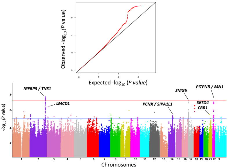
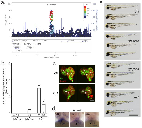
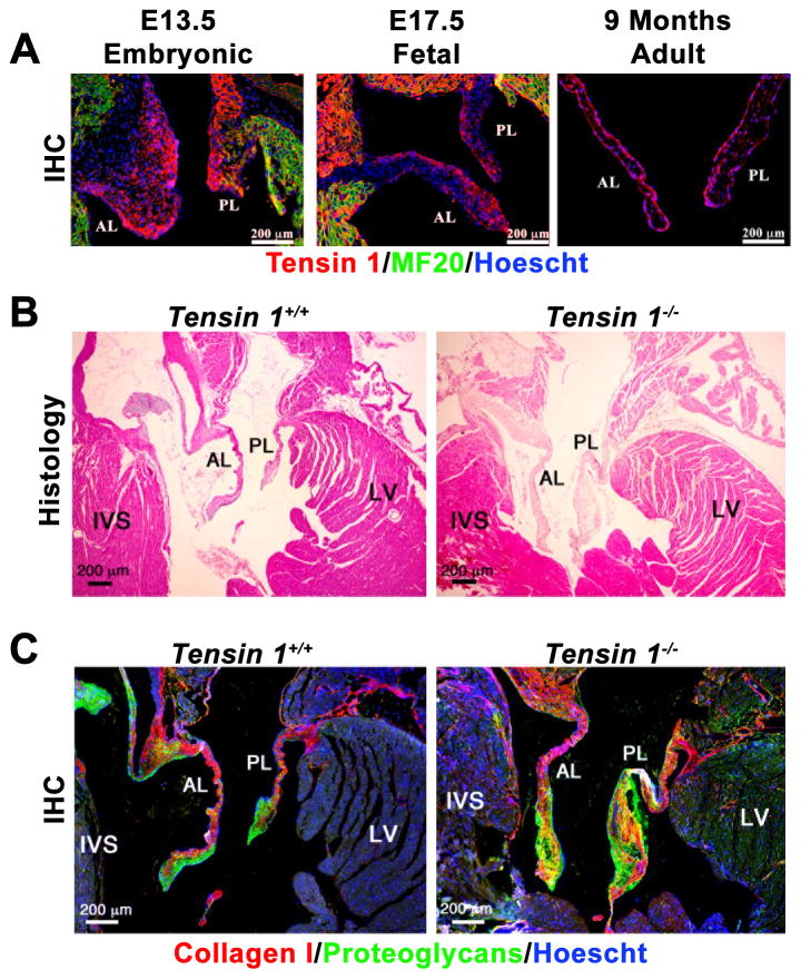
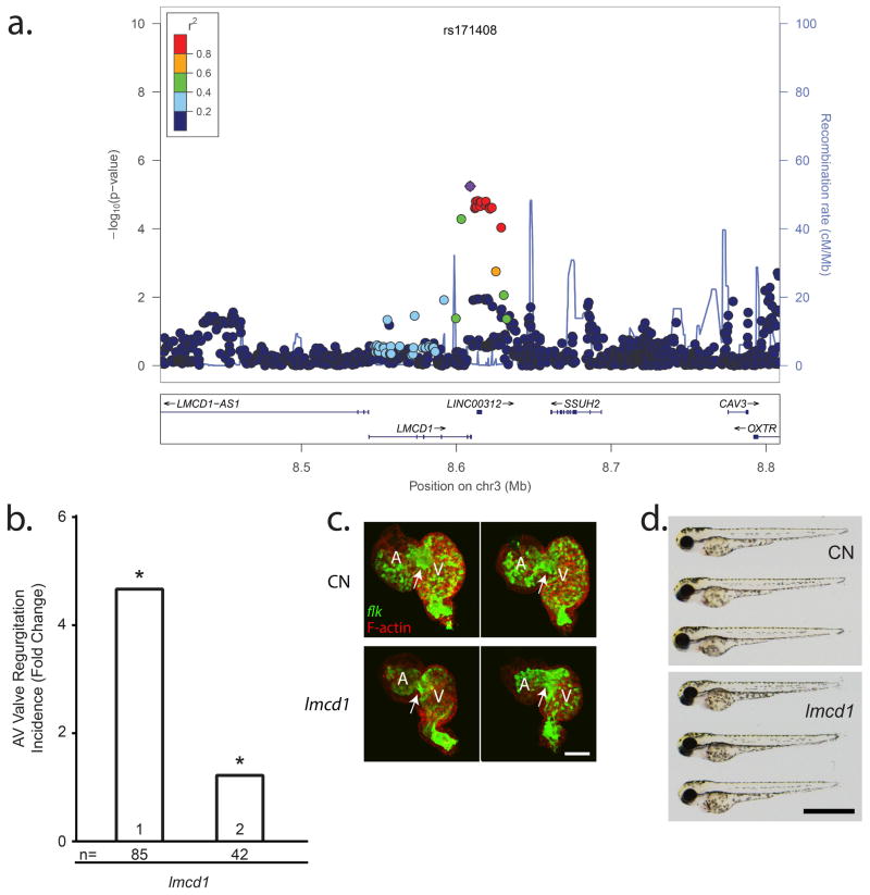
Nonsyndromic mitral valve prolapse (MVP) is a common degenerative cardiac valvulopathy of unknown etiology that predisposes to mitral regurgitation, heart failure and sudden death. Previous family and pathophysiological studies suggest a complex pattern of inheritance. We performed a meta-analysis of 2 genome-wide association studies in 1,412 MVP cases and 2,439 controls. We identified 6 loci, which we replicated in 1,422 cases and 6,779 controls, and provide functional evidence for candidate genes. We highlight LMCD1 (LIM and cysteine-rich domains 1), which encodes a transcription factor and for which morpholino knockdown of the ortholog in zebrafish resulted in atrioventricular valve regurgitation. A similar zebrafish phenotype was obtained with knockdown of the ortholog of TNS1, which encodes tensin 1, a focal adhesion protein involved in cytoskeleton organization. We also showed expression of tensin 1 during valve morphogenesis and describe enlarged posterior mitral leaflets in Tns1(-/-) mice. This study identifies the first risk loci for MVP and suggests new mechanisms involved in mitral valve regurgitation, the most common indication for mitral valve repair.


Conflict of interest statement

Competing financial interests.

The authors declare no competing financial interests.

Figures

Figure 1
Figure 1. Quantile-quantile (A) and manhattan (B) plots representing the association of 4.8 million SNPs at the GWAS meta-analysis
Red line indicates genome-wide significant threshold (P < 5 × 10⁻⁸) and blue line the p-value thershold used for follow-up (P < 10−5).
Figure 2
Figure 2. Cardiac regurgitation in zebrafish morpholino knockdown for candidate genes on Chr2q35
a) Genomic context of the association signal observed in the GWAS meta-analysis. The regional association plot was generated using locus zoom and displays surrounding genes, with TNS1, IGFBP2 and IGFBP5 identified as best potential candidates at this locus. b) Mitral regurgitation observed at 72 hours post fertilization (hpf) in zebrafish embryos after morpholino mediated knockdown. All results are presented as fold change compared to clutchmate controls. n=number of biological replicates per morpholino. (*) indicates p<0.05. c) 2-dimensional projections of z-series image stacks taken on excised control (CN) and tns1 knockdown zebrafish hearts. Green denotes EGFP expression, a marker of endothelium under the control of the flk promoter. Red staining indicates the distribution of F-actin, which is highly expressed in the functional myocardium. Scale bar represents 50μm. d) anti-bmp-4 probe labels the valve and surrounding myocardium in CN and tns1 knockdown embryos. Scale bar represents 50μm. e) Brightfield micrographs displaying gross morphology of 72hpf zebrafish morphants. Scale bar represents 1mm. Body axis length of morpholino-injected fish is slightly reduced compared to wild-types.
Figure 3
Figure 3. Murine Tensin 1 expression during developing valves and knockout phenotype at 9 months
A) Tensin1 expression in the mouse developing heart. IHC was performed for Tensin1 (red) at E13.5 (complete epithelial mesenchymal transformation), E17.5 (valve sculpting and elongation) and 9 months of age. MF20 (green) labels myocytes, Hoescht (Blue) labels nuclei. (B) Tensin1 knockout mice exhibit enlarged mitral leaflets. Hematoxylin and Eosin (H&E) histological staining was performed on Wild-type (Tensin+/+) and Tensin knockout (Tensin1−/−) mice. Scale bars are denoted. (C) Tensin1 knockout mice exhibit myxomatous mitral leaflets. Immunohistochemistry (IHC) for collagen (red), proteoglycans (green) show failure of normal matrix stratification and expansion of proteoglycan expression in the tensin1−/− mitral leaflets indicative of a myxomatous phenotype. AL= Anterior Leaflet, PL= Posterior Leaflet, LV=Left Ventricle, IVS=interventricular septum. Scale bars are denoted.
Figure 4
Figure 4. Cardiac regurgitation in zebrafish morpholino knockdown for Lmcd1 on Chr3p13
a) Genomic context of the association signal observed in the GWAS meta-analysis. The regional association plot was generated using locus zoom and displays surrounding genes, with LMCD1 identified as best potential candidate as the signal is intronic to LMCD1. b) Mitral regurgitation observed at 72 hours post fertilization (hpf) in zebrafish embryos after morpholino mediated knockdown. All results are presented as fold change compared to clutchmate controls. n=number of biological replicates per morpholino. (*) indicates p<0.05. c) 2-dimensional projections of z-series image stacks taken on excised 72hpf control (CN) and lmcd1 knockdown zebrafish hearts. Green denotes EGFP expression, a marker of endothelium under the control of the flk promoter. Red staining indicates the distribution of F-actin, which is highly expressed in the functional myocardium. Scale bar represents 50μm. d) Brightfield micrographs displaying gross morphology of 72hpf embryos following lmcd1 knockdown. Scale bar represents 1mm. CN=control morpholino injected embryos. No detectable morphological difference is observed between morpholino-injected fish and wild-types.

Similar articles

  • Genome-Wide Association Study-Driven Gene-Set Analyses, Genetic, and Functional Follow-Up Suggest GLIS1 as a Susceptibility Gene for Mitral Valve Prolapse.
    Yu M, Georges A, Tucker NR, Kyryachenko S, Toomer K, Schott JJ, Delling FN, Fernandez-Friera L, Solis J, Ellinor PT, Levine RA, Slaugenhaupt SA, Hagège AA, Dina C, Jeunemaitre X, Milan DJ, Norris RA, Bouatia-Naji N. Yu M, et al. Circ Genom Precis Med. 2019 May;12(5):e002497. doi: 10.1161/CIRCGEN.119.002497. Circ Genom Precis Med. 2019. PMID: 31112420 Free PMC article.
  • Genome-Wide Association Meta-Analysis Supports Genes Involved in Valve and Cardiac Development to Associate With Mitral Valve Prolapse.
    Yu M, Kyryachenko S, Debette S, Amouyel P, Schott JJ, Le Tourneau T, Dina C, Norris RA, Hagège AA, Jeunemaitre X, Bouatia-Naji N. Yu M, et al. Circ Genom Precis Med. 2021 Oct;14(5):e003148. doi: 10.1161/CIRCGEN.120.003148. Epub 2021 Aug 31. Circ Genom Precis Med. 2021. PMID: 34461747 Free PMC article.
  • Mutations in DCHS1 cause mitral valve prolapse.
    Durst R, Sauls K, Peal DS, deVlaming A, Toomer K, Leyne M, Salani M, Talkowski ME, Brand H, Perrocheau M, Simpson C, Jett C, Stone MR, Charles F, Chiang C, Lynch SN, Bouatia-Naji N, Delling FN, Freed LA, Tribouilloy C, Le Tourneau T, LeMarec H, Fernandez-Friera L, Solis J, Trujillano D, Ossowski S, Estivill X, Dina C, Bruneval P, Chester A, Schott JJ, Irvine KD, Mao Y, Wessels A, Motiwala T, Puceat M, Tsukasaki Y, Menick DR, Kasiganesan H, Nie X, Broome AM, Williams K, Johnson A, Markwald RR, Jeunemaitre X, Hagege A, Levine RA, Milan DJ, Norris RA, Slaugenhaupt SA. Durst R, et al. Nature. 2015 Sep 3;525(7567):109-13. doi: 10.1038/nature14670. Epub 2015 Aug 10. Nature. 2015. PMID: 26258302 Free PMC article.
  • Genetics of syndromic and non-syndromic mitral valve prolapse.
    Le Tourneau T, Mérot J, Rimbert A, Le Scouarnec S, Probst V, Le Marec H, Levine RA, Schott JJ. Le Tourneau T, et al. Heart. 2018 Jun;104(12):978-984. doi: 10.1136/heartjnl-2017-312420. Epub 2018 Jan 19. Heart. 2018. PMID: 29352010 Free PMC article. Review.
  • Genetic background of mitral valve prolapse.
    Guicciardi NA, De Bonis M, Di Resta C, Ascione G, Alfieri O, Maisano F, Vergara P. Guicciardi NA, et al. Rev Cardiovasc Med. 2022 Mar 12;23(3):96. doi: 10.31083/j.rcm2303096. Rev Cardiovasc Med. 2022. PMID: 35345263 Review.

Cited by

References

    1. Freed LA, et al. Mitral valve prolapse in the general population: the benign nature of echocardiographic features in the Framingham Heart Study. J Am Coll Cardiol. 2002;40:1298–304. - PubMed
    1. Disse S, et al. Mapping of a first locus for autosomal dominant myxomatous mitral-valve prolapse to chromosome 16p11.2-p12.1. Am J Hum Genet. 1999;65:1242–51. - PMC - PubMed
    1. Freed LA, et al. A locus for autosomal dominant mitral valve prolapse on chromosome 11p15.4. Am J Hum Genet. 2003;72:1551–9. - PMC - PubMed
    1. Kyndt F, et al. Mapping of X-linked myxomatous valvular dystrophy to chromosome Xq28. Am J Hum Genet. 1998;62:627–32. - PMC - PubMed
    1. Nesta F, et al. New locus for autosomal dominant mitral valve prolapse on chromosome 13: clinical insights from genetic studies. Circulation. 2005;112:2022–30. - PubMed

Publication types