. 2015 Sep;125(9):3704-13.
doi: 10.1172/JCI82462. Epub 2015 Aug 24.

Halting progressive neurodegeneration in advanced retinitis pigmentosa

Halting progressive neurodegeneration in advanced retinitis pigmentosa

Susanne F Koch et al. J Clin Invest. 2015 Sep.

Abstract

Hereditary retinal degenerative diseases, such as retinitis pigmentosa (RP), are characterized by the progressive loss of rod photoreceptors followed by loss of cones. While retinal gene therapy clinical trials demonstrated temporary improvement in visual function, this approach has yet to achieve sustained functional and anatomical rescue after disease onset in patients. The lack of sustained benefit could be due to insufficient transduction efficiency of viral vectors ("too little") and/or because the disease is too advanced ("too late") at the time therapy is initiated. Here, we tested the latter hypothesis and developed a mouse RP model that permits restoration of the mutant gene in all diseased photoreceptor cells, thereby ensuring sufficient transduction efficiency. We then treated mice at early, mid, or late disease stages. At all 3 time points, degeneration was halted and function was rescued for at least 1 year. Not only do our results demonstrate that gene therapy effectively preserves function after the onset of degeneration, our study also demonstrates that there is a broad therapeutic time window. Moreover, these results suggest that RP patients are treatable, despite most being diagnosed after substantial photoreceptor loss, and that gene therapy research must focus on improving transduction efficiency to maximize clinical impact.


Figures

Figure 8
Figure 8. Photoreceptor degeneration is halted at all treatment time points.
Quantification of ONL thickness from WT (blue), mutant (black), and treated mice. Treated mice were tamoxifen injected at 2 weeks (yellow, T2), 4 weeks (green, T4), and 8 weeks (red, T8) of age. Arrows indicate the treatment time points. Each symbol represents an individual mouse (n = 3 for each group at 52 weeks). Data for the untreated mutant at weeks 2, 4, and 8 are taken from Supplemental Figure 1B; data for week 20 are taken from Figure 4L.
Figure 7
Figure 7. Stable rescue of retinal structure following treatment at a late disease stage.
At 8 weeks of age, mutant mice were tamoxifen injected (T8) or not (Mut). Mice were then sacrificed at 10, 20, or 32 weeks of age, and retinae were sectioned and immunostained; nuclei were stained with Hoechst dye (blue). (AF) Images show anti-rhodopsin Ab (red) labeling of rod OSs. (GL) Images show anti-cone arrestin Ab (green) labeling of cones. Scale bars: 10 μm.
Figure 6
Figure 6. Stable rescue of visual function following treatment at a late disease stage.
At 8 weeks of age, mutant mice were tamoxifen injected or not; at 10 and 32 weeks of age, ERG responses were recorded. (A)Rod-specific b-wave, (B) photopic cone-specific b-wave, (C) mixed rod-cone-specific a-wave, and (D) mixed rod-cone–specific b-wave. Red dots represent individual treated mutant mice (n = 3 for each time point). Gray triangles represent individual untreated mutants (n = 10 and 4 at 10 and 32 weeks of age, respectively). Red solid lines and black dashed lines connect the group means of the treated and untreated mutant mice, respectively. For each mouse, the 2 eyes were averaged. A paired t test was used to compare weeks 10 and 32 for the treated group. A 2-sample t test was used to compare weeks 10 and 32 for the untreated mutant group. ***P < 0.001 for significant differences between weeks 10 and 32.
Figure 5
Figure 5. Absence of gliosis in treated mutant retina.
WT, mutant, and treated retinae were sectioned and immunostained at week 20. The treated mice were tamoxifen injected at 2 weeks (T2), 4 weeks (T4), and 8 weeks (T8) of age. (AE) Anti-GFAP Ab (red) was used to label reactive phenotype in Müller glia; nuclei were stained with Hoechst dye (blue). IHC was performed on 3 mice for every group. A representative immunostaining is shown. Scale bar: 30 μm.
Figure 4
Figure 4. Preservation of retinal structure after gene therapy administered at early, mid, and late disease stages.
WT, mutant, and treated retinae were sectioned and immunostained at week 20. Treated mice were tamoxifen injected at 2 weeks (T2), 4 weeks (T4), and 8 weeks (T8) of age. (AE) Images show anti-rhodopsin Ab (red) labeling of rod OSs. (FJ) Images show anti-cone arrestin Ab (green) labeling of cones. Nuclei were stained with Hoechst dye (blue). (KM) Quantification of rod OS length (K), ONL thickness (L), and cone OS length (M). Each gray dot represents an individual mouse (n = 3 for each group). Horizontal lines represent the group means. A linear regression model was fit to compare groups. *P < 0.05 and ***P < 0.001 for significant differences between treated and untreated mutant groups using linear regression model. Scale bars: 30 μm.
Figure 3
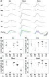
Figure 3. Preservation of retinal function after gene therapy administered at early, mid, and late disease stages.
WT, mutant, and treated mice were measured at week 18. (A) Representative ERG responses for rods, mixed rods and cones, and cones from WT (black, WT), mutant (red, Mut), and treated mice. The treated mice were tamoxifen injected at 2 weeks (dark blue, T2), 4 weeks (green, T4), and 8 weeks (light blue, T8) of age. (BE) Statistical analysis of ERG amplitudes for the scotopic rod-specific b-wave (B), the photopic cone-specific b-wave (C), the mixed rod-cone–specific a-wave (D), and the mixed rod-cone–specific b-wave (E). Gray dots represent individual mice (for each mouse, the 2 eyes were averaged; n values are shown below each group), and horizontal lines represent the group means. A linear regression model was fit to compare groups. *P < 0.05, **P < 0.01, and ***P < 0.001 for significant differences between treated and untreated mutant groups using linear regression model.
Figure 2
Figure 2. Restoration of PDE6b expression at early, mid, and late stages of RP.
Two-, four-, and eight-week-old mutant mice were treated with tamoxifen (T2, T4, and T8, respectively). When mice were 20 weeks old, retinae were lysed, and Western blotting was performed for PDE6b. Blots for WT and untreated mutant retinae are also shown. Immunoblotting was performed in triplicate. A representative immunoblot is shown. β-actin was used as a loading control.
Figure 1
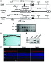
Figure 1. Inducible rod-specific Pde6gCreERT2 driver line.
(A) Targeting strategy for the generation of Pde6gCreERT2 mice. Exons 2–4 are represented by black boxes. Exons 2 and 3 were replaced by CreERT2. The targeting vector contains a neo/tk cassette (Neo). After homologous recombination, CreERT2 is placed under Pde6g locus control. (B) Southern blot analysis of EcoRI- and EcoRV-digested genomic DNA from 8 ES clones hybridized to the probe shown in A. The 8.5- and 6.6-kb bands represent WT and targeted Pde6g alleles, respectively. (C) X-gal–stained retinal section from a 4-week-old ROSA26-lacZ Pde6gCreERT2 mouse injected with tamoxifen at 2 weeks of age. Blue, lacZ reporter gene signal. Scale bar: 50 μm. RPE, retinal pigment epithelium. (D) PCR analysis of ONL DNA isolated from 4-week-old retinae identified a 284-bp fragment for WT, 284- and 415-bp fragments for an untreated mutant, and 284-, 415-, and 362-bp fragments for a mutant treated with tamoxifen at 2 weeks of age. (E) Retinal sections from 4-week-old mice immunostained for PDE6b; the treated mutant was tamoxifen injected at 2 weeks of age. Scale bar: 30 μm.

Comment in

References

    1. Hartong DT, Berson EL, Dryja TP. Retinitis pigmentosa. Lancet. 2006;368(9549):1795–1809. doi: 10.1016/S0140-6736(06)69740-7. - DOI - PubMed
    1. Berson EL. Retinitis pigmentosa. The Friedenwald Lecture. Invest Ophthalmol Vis Sci. 1993;34(5):1659–1676. - PubMed
    1. Davis RJ, et al. Therapeutic margins in a novel preclinical model of retinitis pigmentosa. J Neurosci. 2013;33(33):13475–13483. doi: 10.1523/JNEUROSCI.0419-13.2013. - DOI - PMC - PubMed
    1. Sahel JA, Roska B. Gene therapy for blindness. Annu Rev Neurosci. 2013;36:467–488. doi: 10.1146/annurev-neuro-062012-170304. - DOI - PubMed
    1. Petrs-Silva H, Linden R. Advances in gene therapy technologies to treat retinitis pigmentosa. Clin Ophthalmol. 2014;8:127–136. - PMC - PubMed

Publication types

MeSH terms