

Enucleation after Embolization of Liver Failure-Causing Giant Liver Hemangioma
- PMID: 26301888
- PMCID: PMC4554336
- DOI: 10.12659/AJCR.893298
Enucleation after Embolization of Liver Failure-Causing Giant Liver Hemangioma
Abstract
Background: Hepatic hemangioma is a congenital tumor of the mesenchymal tissues of the liver. While typically benign, these tumors can occasionally grow to sufficient size to cause a number of symptoms, including pain, severe hepatic dysfunction, or, rarely, consumptive coagulopathy. In such instances, surgical treatment may be warranted.
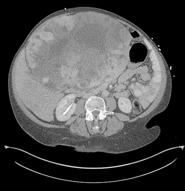
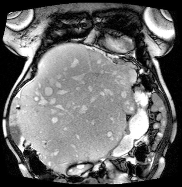
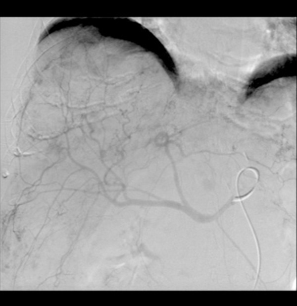
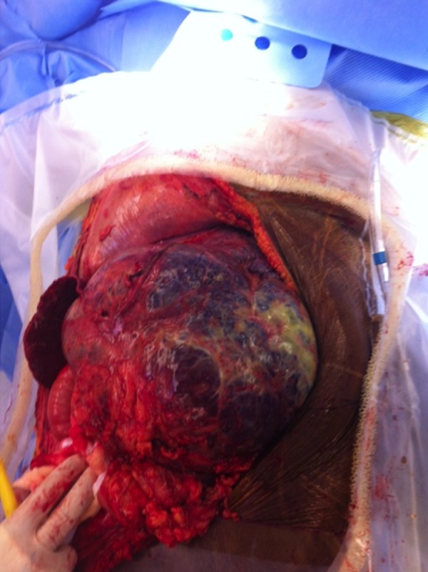
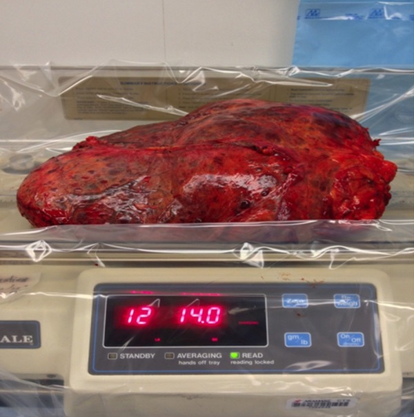
Case report: We present a case of a symptomatic giant hepatic hemangioma in an elderly patient who presented with impending liver failure. She was successfully treated with a combination of surgical enucleation and liver resection after preoperative arterial embolization. We also provide a brief discussion of current treatment options for giant hepatic hemangiomas.
Conclusions: Early referral to experienced surgical centers before the onset of dire complications such as severe hepatic dysfunction and liver failure is recommended.
Figures
References
-
- Giuliante F, Ardito F, Vellone M, et al. Reappraisal of surgical indications and approach for liver hemangioma: single-center experience on 74 patients. Am J Surg. 2011;201(6):741–48. - PubMed
-
- Hall GW. Kasabach-Merritt syndrome: pathogenesis and management. Br J Haematol. 2001;112(4):851–62. - PubMed
-
- Concejero AM, Chen CL, Chen TY, et al. Giant cavernous hemangioma of the liver with coagulopathy: adult Kasabach-Merritt syndrome. Surgery. 2009;145(2):245–47. - PubMed
-
- Yildiz S, Kantarci M, Kizrak Y. Cadaveric liver transplantation for a giant mass. Gastroenterology. 2014;146(1):e10–1. - PubMed
Publication types
MeSH terms
LinkOut - more resources
Full Text Sources
Medical
