. 2015 Oct;17(10):1317-26.
doi: 10.1038/ncb3233. Epub 2015 Aug 24.

Pyruvate carboxylation enables growth of SDH-deficient cells by supporting aspartate biosynthesis

Affiliations

Pyruvate carboxylation enables growth of SDH-deficient cells by supporting aspartate biosynthesis

Simone Cardaci et al. Nat Cell Biol. 2015 Oct.

Abstract

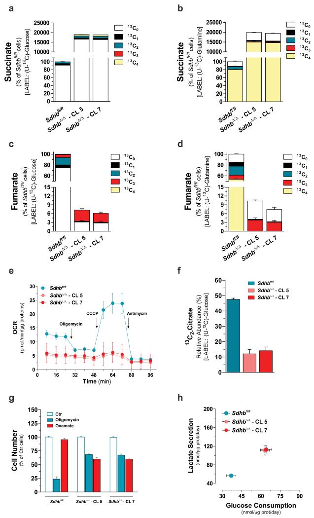
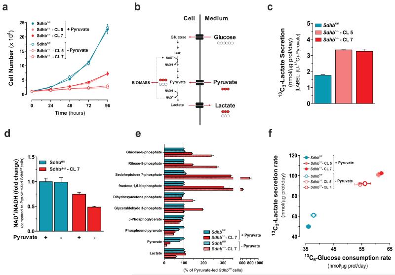
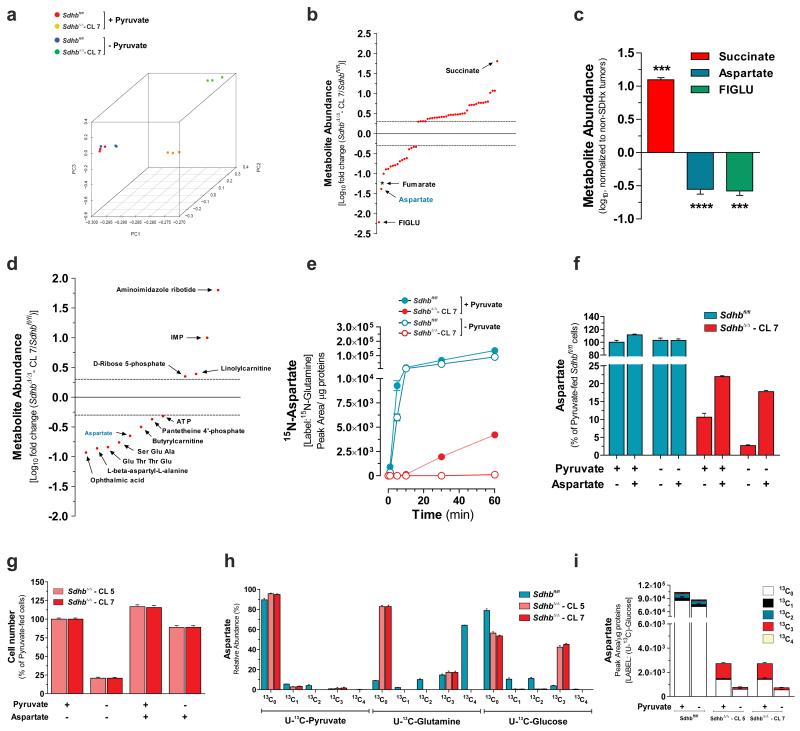
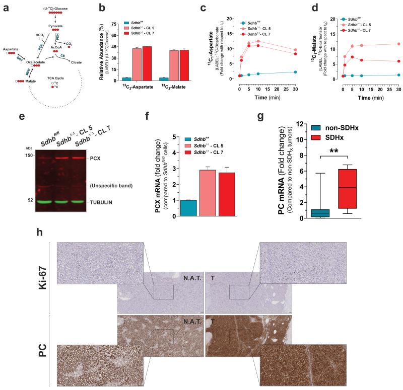
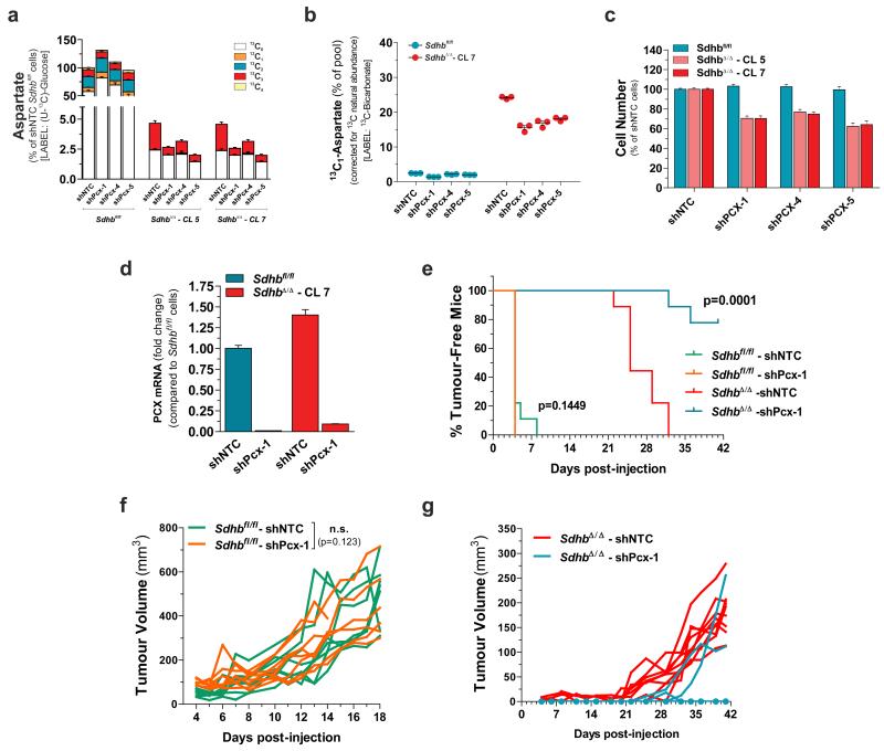
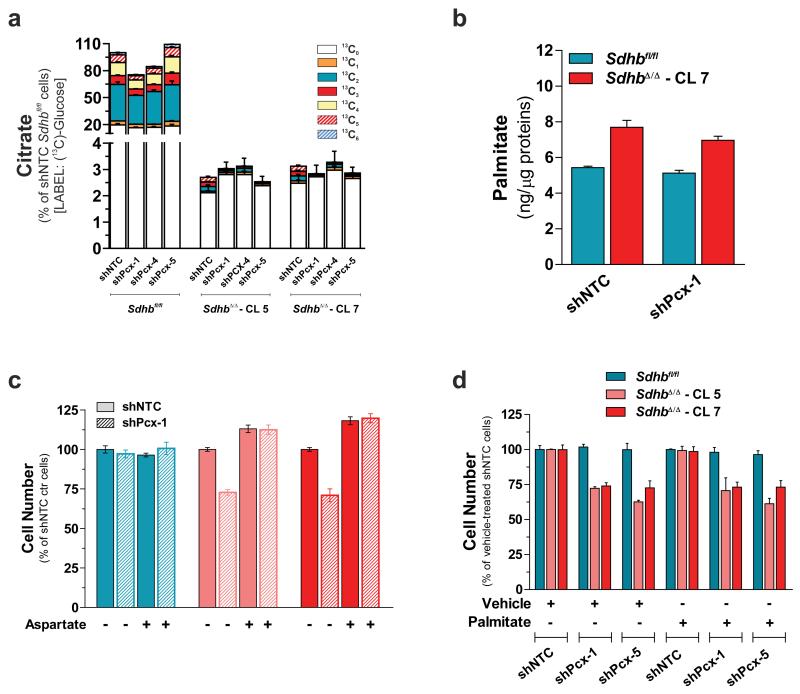
Succinate dehydrogenase (SDH) is a heterotetrameric nuclear-encoded complex responsible for the oxidation of succinate to fumarate in the tricarboxylic acid cycle. Loss-of-function mutations in any of the SDH genes are associated with cancer formation. However, the impact of SDH loss on cell metabolism and the mechanisms enabling growth of SDH-defective cells are largely unknown. Here, we generated Sdhb-ablated kidney mouse cells and used comparative metabolomics and stable-isotope-labelling approaches to identify nutritional requirements and metabolic adaptations to SDH loss. We found that lack of SDH activity commits cells to consume extracellular pyruvate, which sustains Warburg-like bioenergetic features. We further demonstrated that pyruvate carboxylation diverts glucose-derived carbons into aspartate biosynthesis, thus sustaining cell growth. By identifying pyruvate carboxylase as essential for the proliferation and tumorigenic capacity of SDH-deficient cells, this study revealed a metabolic vulnerability for potential future treatment of SDH-associated malignancies.


Figures

Figure 1
Figure 1. Loss of Sdhb is sufficient to truncate TCA cycle and disable mitochondrial respiration
Isotopologues distribution of intracellular succinate and fumarate after incubation for 24h with either U-13C-glucose (a, c) or U-13C-glutamine (b, d). All data are presented as mean±s.e.m of one representative experiment (n=3 wells) independently replicated twice (c, d) and three times (a, b). (e) Respiratory profile of the specified cells. Arrows indicate incubation of cells with the indicated compounds. Data are presented as mean±s.e.m of n=27 (Sdhbfl/fl), n=31 (SdhbΔ/Δ – CL 5) and n=29 (SdhbΔ/Δ – CL 7) wells pooled from four independent experiments. (f) Fraction of intracellular citrate pool containing two 13C atoms as result of pyruvate dehydrogenase activity. Data are presented as mean±s.e.m of one representative experiment (n=3 wells) independently replicated twice. (g) Cell number of the indicated cells measured after 96h of treatment with 0.5 μM oligomycin or 5 mM oxamate. Data are presented as mean±s.e.m of n=12 wells pooled from four independent experiments. (h) Rates of the indicated metabolites upon 48h of incubation of Sdhbfl/fl and SdhbΔ/Δ cells with U-13C-glucose. The sum of all isotopologues is reported for clarity. Data are presented as mean±s.e.m of n=18 wells pooled from three independent experiments. In this and all other figures, “wells” represent technical replicate samples set up and assessed under identical conditions and in parallel within a single experiment. Raw data of independently repeated experiments are provided in Supplementary Table 3.
Figure 2
Figure 2. SDH deficiency commits cells to consume extracellular pyruvate to maintain maximal glycolytic flux and cell growth
(a) Number of cells cultured in presence (+) or absence (−) of pyruvate measured at the indicated time points. Data are presented as mean±s.e.m of one representative experiment (n=4 wells), independently replicated three times. (b) Schematic representation of effects of pyruvate consumption on maintenance of NAD+/NADH redox state and glycolytic flux. Red and white circles indicate 13C and 12C carbon unit, respectively. (c) Secretion rate of exogenous pyruvate-derived lactate (13C3-lactate) of Sdhbfl/fl and SdhbΔ/Δ cells cultured for 48h with U-13C-Pyruvate. Data are presented as mean±s.e.m of n=6 wells pooled from two independent experiments. (d) Measurement of NAD+/NADH ratio expressed as fold change compared to pyruvate-fed Sdhbfl/fl cells (e) Pentose phosphate pathway and glycolytic intermediates in cells cultured for 24h in presence (+) or absence (−) of pyruvate. Data in d, e are presented as mean±s.e.m of one representative experiment (n=3 wells), independently replicated twice. (f) Exchange rates of the indicated labeled metabolites upon 48h of incubation of Sdhbfl/fl and SdhbΔ/Δ cells with U-13C-glucose in presence (+) or absence (−) of pyruvate. Data are presented as mean±s.e.m of n=18 wells pooled from three independent experiments. Raw data of independently repeated experiments are provided in Supplementary Table 3.
Figure 3
Figure 3. Glucose-dependent aspartate biosynthesis dictates pyruvate dependency of Sdhb-deficient cells
(a) PCA of the untargeted endo-metabolomic screening of Sdhbfl/fl and SdhbΔ/Δ cells cultured for 24h in presence (+) or absence (−) of pyruvate. (b) Untargeted metabolic profiling of Sdhb-null cells. Each dot represents a metabolite with a fold change >2 or <0.5 and a FDR-corrected p-value <0.0001. Only metabolites that meet the above-mentioned criteria in two out of three independent experiments (n=3 wells per experiment) are presented. * Since in this untargeted technique fumarate could not be measured, the fold change of fumarate are derived from results presented in Fig.1 c and d. FIGLU, Formiminoglutamic acid. (c) Targeted metabolites profiling of n=4 human SDHx-mutated paraganglioma/pheochromocytoma specimens. Data are expressed as log10 fold change with respect to n=18 non-SDHx-mutated tumours. ***P < 0.001, ****P < 0.0001, two-tailed Student’s t-test. (d) Untargeted metabolic profiling of pyruvate-deprived SdhbΔ/Δ cells. Each point represents a metabolite with a fold change > 2 or < 0.5 and a FDR-corrected p-value <0.0001. Only known endo-metabolites that meet the above-mentioned criteria in two out of three independent experiments (n=3 wells each) are presented. (e) Accumulation of 15N-aspartate in cells incubated with α-15N-glutamine for the indicated time in presence/absence of pyruvate. Data are presented as mean±s.e.m (n=3 wells) of one experiment, performed once. (f) Aspartate levels in cells cultured for 24h in presence or absence of pyruvate and/or 2.5 mM aspartate. Data are presented as mean±s.e.m (n=3 wells) of one representative experiment, independently replicated twice. (g) Cell number measured after 96h of culture in presence/absence of pyruvate and/or 2.5 mM aspartate. Data are presented as mean±s.e.m of n=16 wells pooled from four independent experiments. (h) Isotopologues labelling profile (mean±s.e.m.) of aspartate in cells cultured for 24h in presence of U-13C-pyruvate, U-13C-glutamine (n=6 wells for both conditions, pooled from two independent experiments) or U-13C-glucose (n=9 wells pooled from three independent experiments). (i) Aspartate levels in cells cultured for 24h with U-13C-glucose in presence/absence of pyruvate. Data are presented as mean±s.e.m (n=3 wells) of one representative experiment, independently replicated twice. Raw data of independently repeated experiments are provided in Supplementary Table 3.
Figure 4
Figure 4. Pyruvate carboxylase expression is induced in SDH-defective cells and SDHx-mutated human tumours
(a) Predicted labeling pattern of the indicated metabolites in cells cultured with U-13C-glucose. Red and white circles indicate 13C and 12C carbon unit, respectively. PDH, Pyruvate Dehydrogenase; PCX, Pyruvate Carboxylase; CS, Citrate Synthase; MDH, Malate Dehydrogenase; GOT, Glutamate-Oxalacetate Transaminase. (b) Abundance of 13C3-Aspartate and 13C3-malate in U-13C-glucose-cultured cells. Data in b are presented as mean±s.e.m of n=9 wells pooled from three independent experiments. Accumulation of 13C1-aspartate (c) and 13C1-Malate (d) in cells incubated with 13C-bicarbonate for the indicated time. Data in c, d are presented as mean±s.e.m of n=3 wells of one representative experiment, independently replicated twice. (e) Western blot analysis of PCX protein levels in the indicated cells. Image representative of two independent experiments. (f) qPCR analysis of PCX mRNA levels in the indicated cells. Data are presented as mean±s.e.m of n=9 wells pooled from three independent experiments. (g) qPCR analysis of PC mRNA levels in the n=4 human SDHx-mutated paraganglioma/pheochromocytoma specimens. Data are expressed as fold change with respect to n=18 non-SDHx mutated tumours. **P < 0.01. two-tailed Student’s t-test. (h) Immuhistochemical assessment of PC levels in a human SDHB-mutated RCC (Tumour ID 21542, details are reported in Supplementary Table 2). The cell proliferation marker Ki-67 identifies iperproliferative neoplastic areas of the section. N.A.T., normal adjacent tissue; T., tumour. Scale bars = 100 μM. Raw data of independently repeated experiments are provided in Supplementary Table 3.
Figure 5
Figure 5. Pyruvate carboxylation is a vulnerable adaptation to Sdhb loss
(a) Aspartate abundance in PCX-silenced cells cultured for 24h with U-13C-glucose. Data are presented as mean±s.e.m of n=9 wells pooled from three independent experiments. (b) Relative abundance of 13C1-aspartate in PCX-silenced cells incubated with 13C-bicarbonate for 10 min. Data are presented as mean±s.e.m of n=3 wells of one representative experiment, independently replicated twice. (c) Data are presented as mean±s.e.m of n=12 wells pooled from three independent experiments.. (d) PCX expression in H-RasV12-transformed cells infected with lentiviruses expressing either a shNTC or shPcx-1 sequence prior to injection into nude mice. Data are presented as mean±s.e.m (n=3 wells) of one experiment, performed once. (e-g) In vivo growth of cells described in (d) xenografted in athymic nude mice. The % of tumour-free mice over time (e) and the tumour volumes of each xenografted mouse (f, g) are presented (n=8 Sdhbfl/fl-shNTC, n=8 Sdhbfl/fl-shPcx1, n=9 SdhbΔ/Δ-shNTC, n=9 SdhbΔ/Δ-shPcx1). The Log-rank (Mantel-Cox) test was used to calculate the statistical significance between curves in (e). A statistical permutation test was used to compare the statistical significance between curves of the selected genotypes in (f), as described in Methods. Data derive from one experiment, performed once. Raw data of independently repeated experiments are provided in Supplementary Table 3.
Figure 6
Figure 6. Pyruvate carboxylase supports growth of Sdhb-null cells by sustaining aspartate biosynthesis
(a) Citrate abundance in PCX-silenced cells cultured for 24h with U-13C-glucose. Data are presented as mean±s.e.m of n=9 wells pooled from three independent experiments. (b) Effect of PCX silencing on the total intracellular palmitate levels. Data are presented as mean±s.e.m (n=3 wells) of one experiment, performed once. (c) Number of PCX-silenced cells in the presence (+) or absence (−) of 2.5 mM aspartate measured after 96h of culture. Data are presented as mean±s.e.m (n=4 wells) of one experiment, performed once. (d) Number of control and PCX-silenced cells supplemented with 50 μM palmitate measured after 96h of culture. Data are presented as mean±s.e.m (n=4 wells) of one experiment, performed once. Raw data of independently repeated experiments are provided in Supplementary Table 3.

References

    1. Gaude E, Frezza C. Defects in mitochondrial metabolism and cancer. Cancer Metab. 2014;2:10. - PMC - PubMed
    1. Baysal BE, et al. Mutations in SDHD, a mitochondrial complex II gene, in hereditary paraganglioma. Science. 2000;287:848–851. - PubMed
    1. Niemann S, Müller U. Mutations in SDHC cause autosomal dominant paraganglioma, type 3. Nature Genet. 2000;26:268–270. - PubMed
    1. Astuti D, et al. Gene mutations in the succinate dehydrogenase subunit SDHB cause susceptibility to familial pheochromocytoma and to familial paraganglioma. Am. J. Hum. Genet. 2001;69:49–54. - PMC - PubMed
    1. Baysal BE, et al. Prevalence of SDHB, SDHC, and SDHD germline mutations in clinic patients with head and neck paragangliomas. J. Med. Genet. 2002;39:178–183. - PMC - PubMed

MeSH terms