Clinical Trial
. 2015 Oct;72(10):1139-47.
doi: 10.1001/jamaneurol.2015.1807.

Nerve Growth Factor Gene Therapy: Activation of Neuronal Responses in Alzheimer Disease

Affiliations
Clinical Trial

Nerve Growth Factor Gene Therapy: Activation of Neuronal Responses in Alzheimer Disease

Mark H Tuszynski et al. JAMA Neurol. 2015 Oct.

Abstract

Importance: Alzheimer disease (AD) is the most common neurodegenerative disorder and lacks effective disease-modifying therapies. In 2001, we initiated a clinical trial of nerve growth factor (NGF) gene therapy in AD, the first effort at gene delivery in an adult neurodegenerative disorder. This program aimed to determine whether a nervous system growth factor prevents or reduces cholinergic neuronal degeneration in patients with AD. We present postmortem findings in 10 patients with survival times ranging from 1 to 10 years after treatment.

Objective: To determine whether degenerating neurons in AD retain an ability to respond to a nervous system growth factor delivered after disease onset.

Design, setting, and participants: Patients in this anatomicopathological study were enrolled in clinical trials from March 2001 to October 2012 at the University of California, San Diego, Medical Center in La Jolla. Ten patients with early AD underwent NGF gene therapy using ex vivo or in vivo gene transfer. The brains of all 8 patients in the first phase 1 ex vivo trial and of 2 patients in a subsequent phase 1 in vivo trial were examined.

Main outcomes and measures: Brains were immunolabeled to evaluate in vivo gene expression, cholinergic neuronal responses to NGF, and activation of NGF-related cell signaling. In 2 patients, NGF protein levels were measured by enzyme-linked immunosorbent assay.

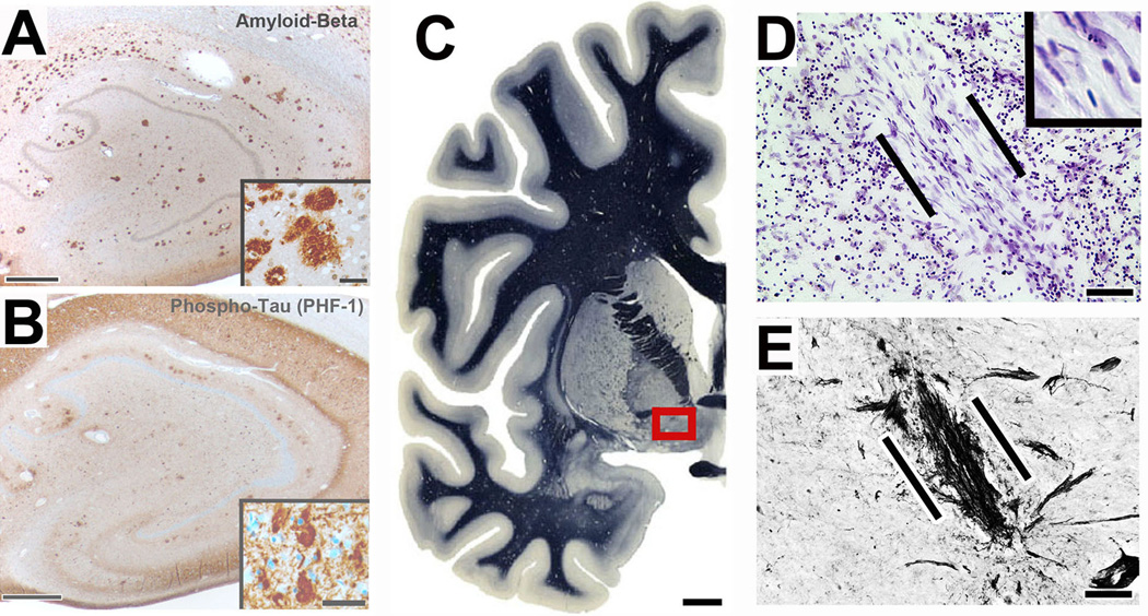
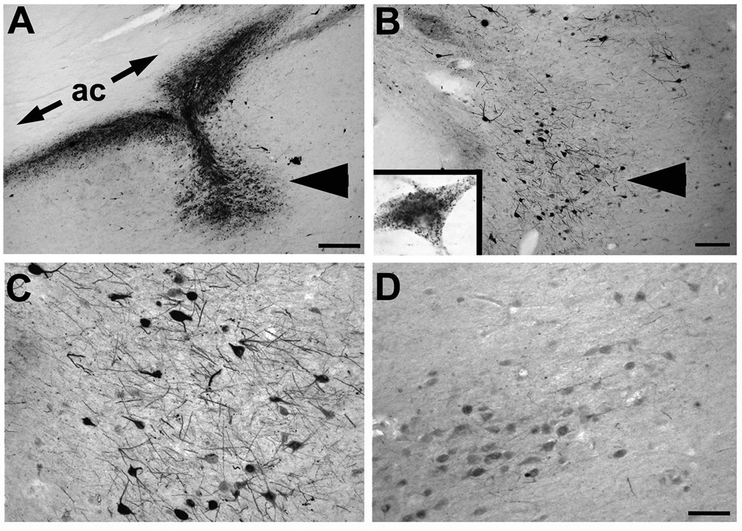
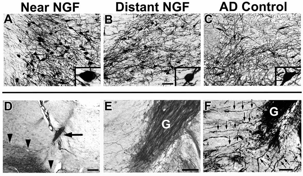
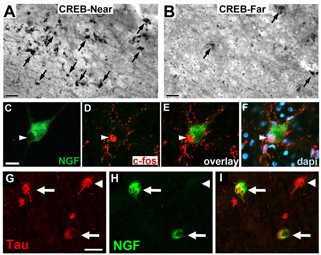
Results: Among 10 patients, degenerating neurons in the AD brain responded to NGF. All patients exhibited a trophic response to NGF in the form of axonal sprouting toward the NGF source. Comparing treated and nontreated sides of the brain in 3 patients who underwent unilateral gene transfer, cholinergic neuronal hypertrophy occurred on the NGF-treated side (P < .05). Activation of cellular signaling and functional markers was present in 2 patients who underwent adeno-associated viral vectors (serotype 2)-mediated NGF gene transfer. Neurons exhibiting tau pathology and neurons free of tau expressed NGF, indicating that degenerating cells can be infected with therapeutic genes, with resultant activation of cell signaling. No adverse pathological effects related to NGF were observed.

Conclusions and relevance: These findings indicate that neurons of the degenerating brain retain the ability to respond to growth factors with axonal sprouting, cell hypertrophy, and activation of functional markers. Sprouting induced by NGF persists for 10 years after gene transfer. Growth factor therapy appears safe over extended periods and merits continued testing as a means of treating neurodegenerative disorders.


Figures

Figure 1
Figure 1. AD pathology and site of gene delivery
All subjects in this study exhibited Alzheimer’s pathology; a case seven years post-gene transfer is shown. (A) Hippocampus shows numerous plaques. Inset shows diffuse and dense-core plaques. (B) Tangles and dense neurofibrillary tau pathology are present, evident also in inset. (C) Overview of left half of normal human brain; boxed region indicates region containing neurons of nucleus basalis of Meynert that are targeted by NGF gene delivery. Myelin stain reproduced by permission from Mai et al., Atlas of the Human Brain, 2003, Elsevier . (D) Nissl stain of graft of autologous fibroblasts, genetically modified to secrete human NGF and injected into the nucleus basalis. Cells were injected seven years previously; graft is indicated between lines. Cells survive and exhibit typical fibroblast morphology (inset). (E) In the adjacent section of Panel D, p75 neurotrophin receptor immunolabeling shows basal forebrain cholinergic axons penetrating into the graft in a linear fashion and this concentration of p75 fibers are only present at the sites of grafts (confirmed by Nissl staining). Scale bar A, 300µm (Inset 50µm); B, 300µm (Inset 50µm); C, 6mm; D, 30µm; E, 25µm.
Figure 2
Figure 2. AAV2-NGF gene expression
(A) NGF labeling shows site of NGF gene delivery in nucleus basalis of Meynert (arrowhead), under anterior commissure (ac). Patient injected three years previously. (B) Another site from same brain showing NGF uptake in neurons in region of nucleus basalis (arrowhead). Inset shows single neuron with granular intraneuronal labeling. (C) Higher magnification of NGF-expressing neurons, compared to (D) less intense labeling in nucleus basalis neurons located 3mm from injection site. Scale bar A 325µm; B 250µm; C–D, 100µm.
Figure 3
Figure 3. Cholinergic neuronal hypertrophy and sprouting
Labeling for p75, a neurotrophin receptor expressed on cholinergic neurons of the nucleus basalis of Meynert. A-C, images three years after AAV2-NGF delivery; D-F, images seven years after ex vivo gene transfer. (A) Cholinergic neurons labeled for p75 within the zone of NGF transduction, (B) 3mm from the zone of transduction, and (C) in control AD brain of the same Braak stage. Cells near to NGF transduction region appear larger. Inset, higher magnification views of typical neuron from each region. (D) Graft of fibroblasts transduced to secrete NGF (arrow), adjacent to nucleus basalis of Meynert (indicated by arrowheads). (E) Graft (g) at higher magnification is densely penetrated by p75-labeled axons arising from nucleus basalis of Meynert. These axons sprouting toward graft, a classic trophic response. (F) F) p75-labeled axons from nucleus basalis of Meynert sprouting toward graft. Individual axons coursing toward graft are evident (arrows). Scale bars A–C 125µm; D 500µm; E–F 100µm.
Figure 4
Figure 4. Cell activation after NGF gene delivery
(A, B) In regions of NGF expression (CREB-Near), there is induction of phospho-CREB expression, a canonical mediator of downstream neurotrophin signaling and cell activation , compared to regions of the NBM 3mm distant from site of NGF injection (CREB-Far). Patient received AAV2-NGF three years earlier. Similar response is evident in patient who received AAV2-NGF and survived one year (not shown). (C–F) An individual neuron expressing NGF (green) also expresses c-fos, another canonical marker of neurotrophin-mediated activation of cell signaling . c-fos labeling is nuclear (arrowheads), as expected. This image is also from the brain of the patient that underwent AAV2-NGF gene transfer three years earlier. (G–I) NGF expression in degenerating neurons expressing Tau Pathology. (G) Neurons in nucleus basalis of Meynert exhibit characteristic Tau pathology, illustrated by tau phosphorylation (AT180 antibody); (H) One of the neurons from panel A is also expressing NGF, shown in the merge panel; (I) These findings indicate that actively degenerating neurons can be infected by AAV2 vectors to express a potentially therapeutic gene product. Scale bars A–B 50µm, C–F 35µm, G–I 25 µm.

Comment in

References

    1. Nagahara AH, Merrill DA, Coppola G, et al. Neuroprotective effects of brain-derived neurotrophic factor in rodent and primate models of Alzheimer's disease. Nat Med. 2009;15:331–337. - PMC - PubMed
    1. Kordower JH, Winn SR, Liu YT, et al. The aged monkey basal forebrain: rescue and sprouting of axotomized basal forebrain neurons after grafts of encapsulated cells secreting human nerve growth factor. Proc Nat Acad Sci. 1994;91:10898–10902. - PMC - PubMed
    1. Tuszynski MH, U HS, Amaral DG, Gage FH. Nerve growth factor infusion in the primate brain reduces lesion-induced cholinergic neuronal degeneration. J Neurosci. 1990;10:3604–3614. - PMC - PubMed
    1. Conner JM, Darracq MA, Roberts J, Tuszynski MH. Nontropic actions of neurotrophins: subcortical nerve growth factor gene delivery reverses age-related degeneration of primate cortical cholinergic innervation. Proc Nat Acad Sci. 2001;98:1941–1946. - PMC - PubMed
    1. Nagahara AH, Mateling M, Kovacs I, et al. Early BDNF Treatment Ameliorates Cell Loss in the Entorhinal Cortex of APP Transgenic Mice. J Neurosci. 2013;33:15596–15602. - PMC - PubMed

Publication types