. 2015 Nov:485:492-501.
doi: 10.1016/j.virol.2015.07.018. Epub 2015 Aug 21.

Viral precursor protein P3 and its processed products perform discrete and essential functions in the poliovirus RNA replication complex

Affiliations

Viral precursor protein P3 and its processed products perform discrete and essential functions in the poliovirus RNA replication complex

Allyn Spear et al. Virology. 2015 Nov.

Abstract

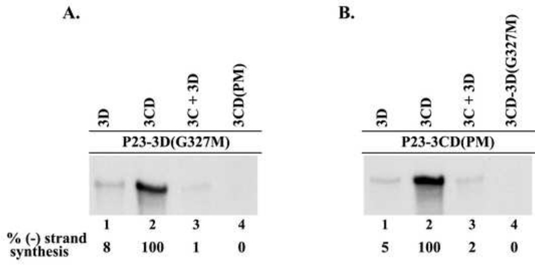
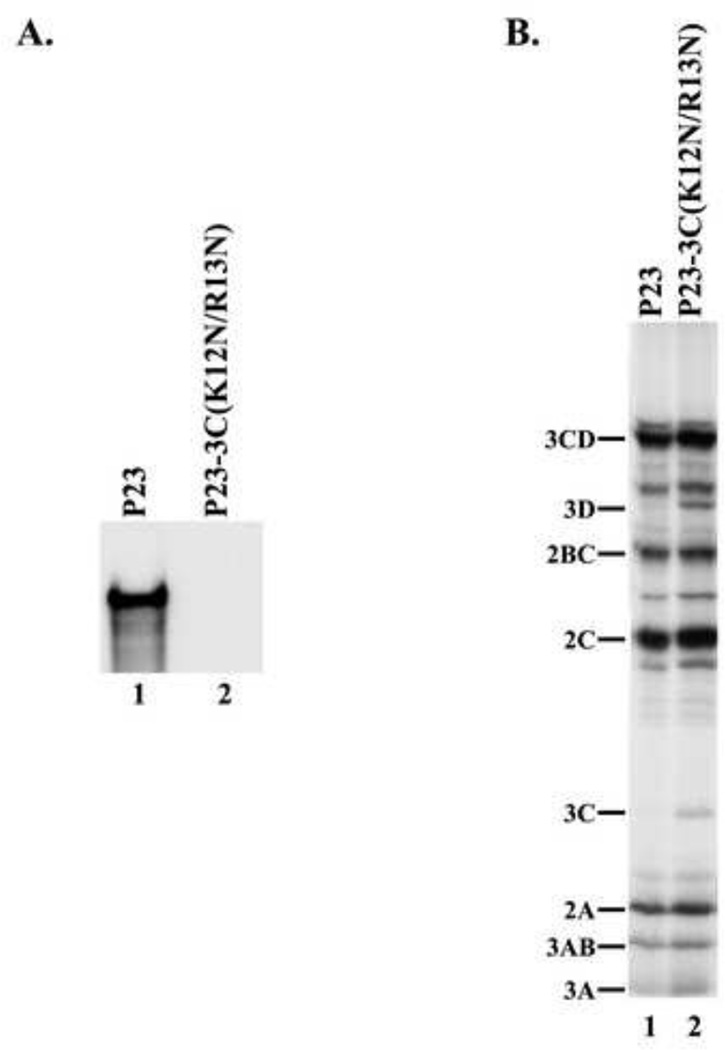
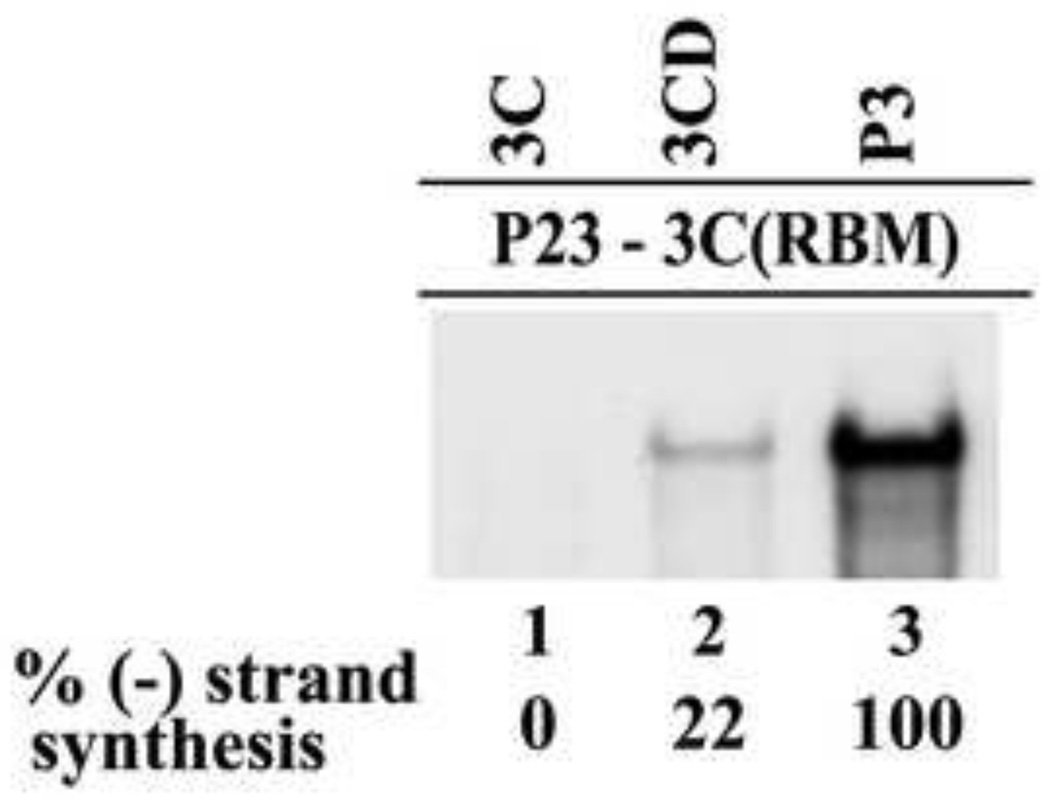
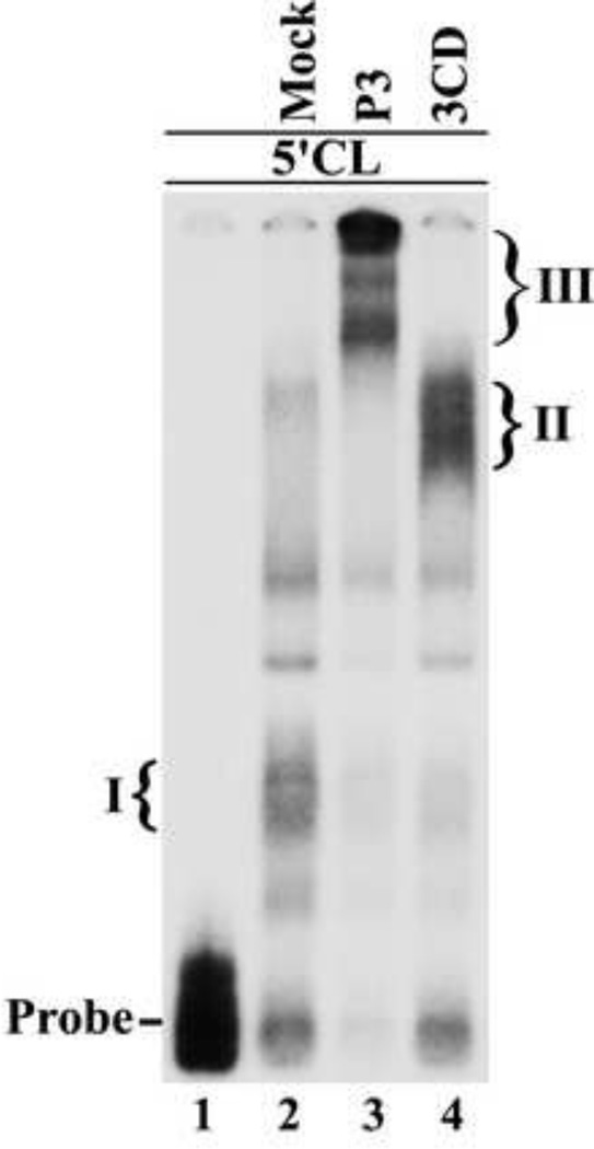
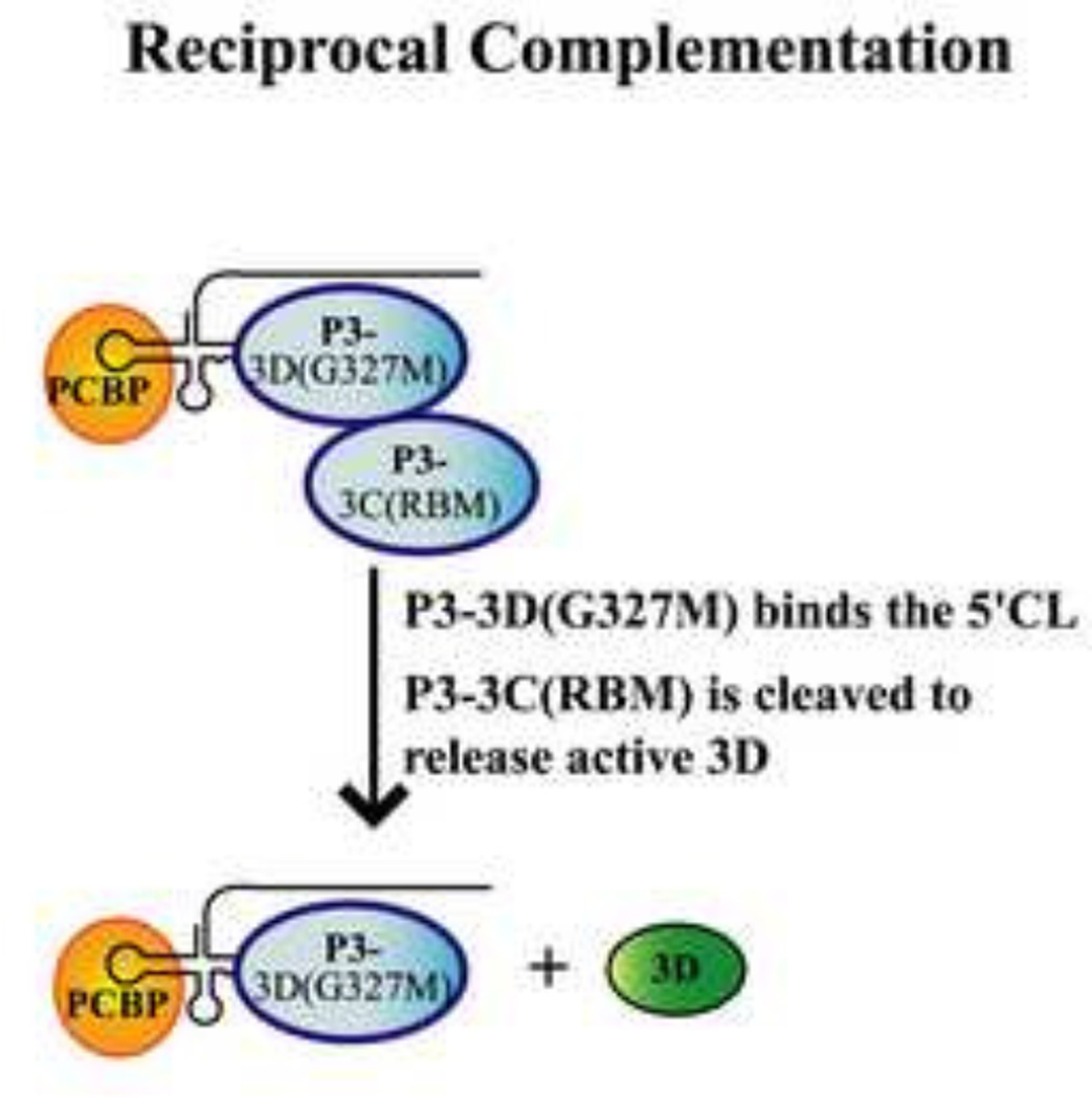
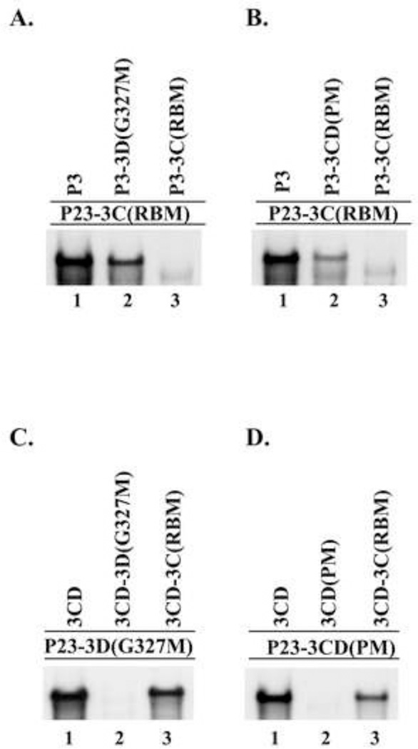
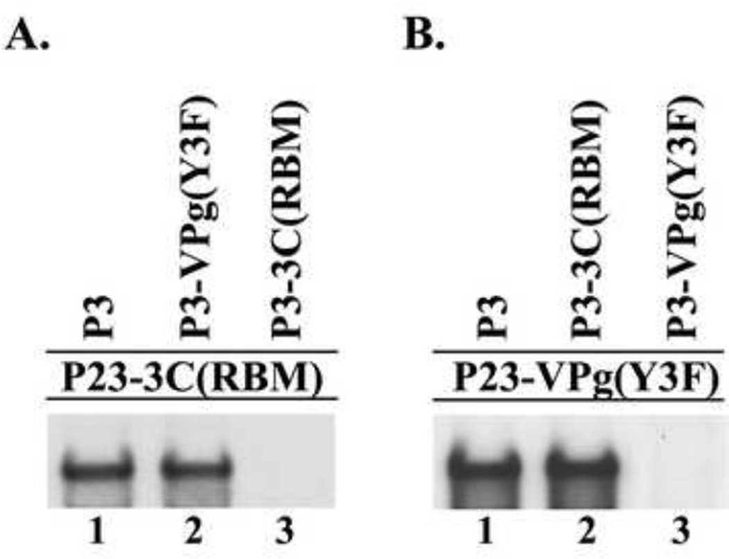
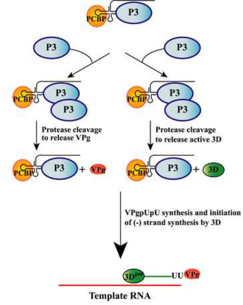
The differential use of protein precursors and their products is a key strategy used during poliovirus replication. To characterize the role of protein precursors during replication, we examined the complementation profiles of mutants that inhibited 3D polymerase or 3C-RNA binding activity. We showed that 3D entered the replication complex in the form of its precursor, P3 (or 3CD), and was cleaved to release active 3D polymerase. Furthermore, our results showed that P3 is the preferred precursor that binds to the 5'CL. Using reciprocal complementation assays, we showed that one molecule of P3 binds the 5'CL and that a second molecule of P3 provides 3D. In addition, we showed that a second molecule of P3 served as the VPg provider. These results support a model in which P3 binds to the 5'CL and recruits additional molecules of P3, which are cleaved to release either 3D or VPg to initiate RNA replication.

Keywords: (–) Strand RNA; 3C-RNA binding activity; 3D polymerase; 5′Cloverleaf (5′CL); Poliovirus (PV); Poliovirus 3CD; Poliovirus P3; RNA replication complex; Reciprocal complementation; VPg.


Figures

Fig.1
Fig.1
Diagram of the poliovirus P23 polyprotein processing cascade showing the precursor and processed viral proteins. The P23 polyprotein is cleaved by the viral protease 3C/3CD at the cleavage sites shown as filled diamonds (formula image).
Fig.2
Fig.2
Schematic of the PV1 RNA transcripts used in this study. (A) PV1(A)80 transcript RNA contains the entire poliovirus genomic RNA sequence. This RNA encodes all of the viral proteins and serves as a template for (−) strand synthesis. (B) P23 transcript RNA contains a deletion of the P1 capsid coding region, but encodes the viral replication proteins and serves as a replicon RNA. Mutations constructed in 3C/3D/3CD are indicated by arrows and include the 3C-RNA binding mutant (RBM; K12N/R13N), the 3CD processing mutant (PM; T181K, Q182D, S183G, Q184N), and the 3D polymerase activity mutant (G327M). (C) The viral protein expression RNA contains the specific protein coding sequence flanked by the authentic PV1 5’ NTR, IRES and 3’ NTR and poly(A) tail.
Fig.3
Fig.3
Characterization of mutations inhibiting 3D polymerase activity. (A) 32P-labeled (−) strand RNA synthesis was assayed using PIRCs isolated from HeLa S10 reactions as described in Materials and methods. Reactions contained subgenomic wildtype P23 RNA or P23 RNA with a mutation either in 3D (3D(G327M)) or in 3CD coding region (3CD (PM)). Full length labeled product RNA was analyzed by denaturing CH3HgOH gel electrophoresis and autoradiography. (B) Portions of the HeLa S10 reactions described in (A) were labeled with [35S] methionine to assay for protein synthesis. These reactions were analyzed by SDS-PAGE and autoradiography. Viral proteins are indicated at left.
Fig.4
Fig.4
Viral proteins 3CD or P3 were required to rescue replication of 3D(G327M) or 3CD(PM) mutant RNAs. (−) Strand RNA synthesis was assayed using PIRCs isolated from HeLa S10 reactions as described in Materials and methods. (A) Reactions contained P23-3D(G327M) RNA as a template and a second complementing RNA expressing the indicated protein. (B) Reactions contained P23-3CD(PM) RNA as template and a second RNA expressing the indicated protein. All complementing RNAs contain the ΔGUA3 mutation which inhibits (−) strand synthesis. Labeled product RNA was analyzed as described in Fig.3. The amount of the labeled product RNAs synthesized in each reaction was quantitated using ImageJ as described in Materials and methods.
Fig.5
Fig.5
The presence of additional 3C did not enhance the ability of 3D to restore (−) strand synthesis. (−) Strand synthesis was assayed using PIRCs isolated from HeLa S10 reactions as described in Materials and methods. Reactions contained P23-3D(G327M) RNA (A) or P23-3CD(PM) RNA (B) and complementing RNAs expressing the indicated protein(s). Labeled product RNA was analyzed as described in Fig.3 and quantitated as described in Fig.4.
Fig.6
Fig.6
Characterization of a 3C-RNA binding mutant. (A) (−) Strand synthesis was assayed using PIRCs isolated from HeLa S10 reactions as described in Materials and methods. Reactions contained either wildtype P23 RNA or a 3C-RNA binding mutant (P23-3C(K12N/R13N)). Labeled product RNA was analyzed as described in Fig.3. (B) Translation of the transcript RNA was measured as described in Fig.3B.
Fig.7
Fig.7
Viral protein P3 was required to rescue replication of P23-3C(RBM). (−) Strand synthesis was assayed using PIRCs isolated from HeLa S10 reactions as described in Materials and methods. Reactions contained P23 RNA containing a 3C-RNA binding mutation (3C(K12N/R13N or 3C(RBM)) and a complementing RNA expressing the indicated protein. Labeled product RNA was analyzed as described in Fig.3 and quantitated as described in Fig.4.
Fig.8
Fig.8
Viral protein P3 protein binds the 5’CL RNA. Electrophoretic mobility shift assay (EMSA) using labeled 5’CL RNA probe as described in Materials and methods. The position of the RNA probe is shown in lane 1. The probe was incubated with an aliquot of a mock reaction (lane 2) or with P3 (lane 3) or 3CD (lane 4). The P3 and 3CD used in these reaction contained mutations in the active site of 3C protease to inhibit processing of P3 and 3CD. The previously described RNP complex formed with the 5’CL RNA and PCBP is labeled as complex I (lane 2). The RNP complex formed with the 5’CL RNA and 3CD is labeled as complex II (lane 4).The complex formed with the 5’CL RNA and P3 is labeled as complex III (lane 3).
Fig.9
Fig.9
Model showing reciprocal complementation between a 3C-RNA binding mutant and a 3D polymerase mutant in the same reaction. In this model, the following multi-step process is predicted to occur during reciprocal complementation: (1) the P3-3D(G327M) provides the RNA binding activity and binds the 5’CL along with PCBP. (2) The P3-3C(RBM) is recruited to this complex and provides 3D polymerase activity after undergoing proteolytic processing.
Fig.10
Fig.10
Reciprocal complementation between a 3C-RNA binding mutant and a 3D polymerase mutant. (−) Strand synthesis was assayed using PIRCs isolated from HeLa S10 reactions as described in Materials and methods. (A) and (B) Reactions contain P23-3C(RBM) template RNA and a second complementing RNA expressing the indicated protein. (C) Reactions contained P23-3D(G327M)) template RNA and a second RNA expressing the indicated protein. (D) Reactions contained P23-3CD(PM) template RNA and a second RNA expressing the indicated protein. Labeled product RNA was analyzed as described in Fig.3.
Fig.11
Fig.11
Reciprocal complementation between a 3C-RNA binding mutant and a VPg-linkage site mutant. (−) Strand synthesis was assayed using PIRCs isolated from HeLa S10 reactions as described in Materials and methods. (A) Reactions contain P23-3C(RBM) template RNA and a second complementing RNA expressing the indicated protein. (B) Reactions contain P23-VPg(Y3F) template RNA and a second RNA expressing the indicated protein. Labeled product RNA was analyzed as described in Fig.3.
Fig.12
Fig.12
Model showing the role of P3 precursor protein in the multi-step process of replication complex assembly. The cellular protein, PCBP, and the viral precursor protein, P3, bind to the 5’CL and form the 5’CL-RNP complex. Pathway on the right: A second molecule of P3 is recruited to the 5’CL-RNP complex via protein-protein interactions. This P3 serves as the 3D provider after being cleaved to release active 3D polymerase in the replication complex. Pathway on the left: A second molecule of P3, which serves as the VPg provider, is recruited to the 5’CL-RNP complex. This P3 is cleaved to release VPg. The 3D polymerase is then used to uridylylate VPg and to initiate and elongate VPgpUpU-primed (−) strand synthesis.

Similar articles

Cited by

References

    1. Andino R, Rieckhof GE, Achacoso PL, Baltimore D. Poliovirus RNA synthesis utilizes an RNP complex formed around the 5'-end of viral RNA. EMBO J. 1993;12:3587–3598. - PMC - PubMed
    1. Andino R, Rieckhof GE, Baltimore D. A functional ribonucleoprotein complex forms around the 5' end of poliovirus RNA. Cell. 1990a;63:369–380. - PubMed
    1. Andino R, Rieckhof GE, Trono D, Baltimore D. Substitutions in the protease (3Cpro) gene of poliovirus can suppress a mutation in the 5' noncoding region. J. Virol. 1990;64:607–612. - PMC - PubMed
    1. Barton DJ, Black EP, Flanegan JB. Complete replication of poliovirus in vitro: preinitiation RNA replication complexes require soluble cellular factors for the synthesis of VPg-linked RNA. J. Virol. 1995;69:5516–5527. - PMC - PubMed
    1. Barton DJ, Morasco BJ, Flanegan JB. Assays for poliovirus polymerase, 3Dpol, and authentic RNA replication in HeLa S10 extracts. Methods Enzymol. 1996;275:35–57. - PubMed

Publication types

LinkOut - more resources