. 2015 Aug 25;13(1):255.
doi: 10.1186/s12957-015-0669-4.

Reduced USP39 expression inhibits malignant proliferation of medullary thyroid carcinoma in vitro

Affiliations

Reduced USP39 expression inhibits malignant proliferation of medullary thyroid carcinoma in vitro

Yong An et al. World J Surg Oncol. .

Abstract

Background: Medullary thyroid carcinoma (MTC) constitutes approximately 5 % of all thyroid cancers and carries a worse prognosis than other differentiated thyroid cancers. Targeted therapies are being investigated for systemic treatment of MTC. Ubiquitin-specific peptidase 39 (USP39) functions in pre-mRNA splicing as a component of the U4/U6-U5 tri-snRNP and also participates in spindle checkpoint and cytokinesis. In this study, we aimed to evaluate the potential role in MTC.

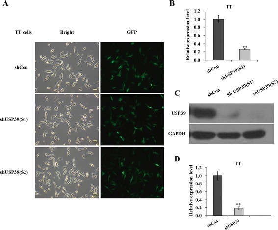
Methods: We used lentivirus-delivered short hairpin RNA (shRNA) to silence USP39 expression in one MTC cell line TT. USP39 expression was detected by qPCR and Western blot. For functional analysis, MTT assay was performed to evaluate the proliferation activity, and FACS was used to assess the cell distribution in the cell cycle. Moreover, the expressions of cell cycle-related proteins were examined by Western blot.

Results: Both two shRNA sequences against USP39 could efficiently reduce its expression in TT cells. Knockdown of USP39 significantly decreased cell proliferation and caused cell cycle arrest at G2/M phase. Moreover, G2/M phase-associated proteins, Cyclin B1 and CDK1, were obviously down-regulated in TT cells after USP39 silencing.

Conclusions: Therefore, knockdown of USP39 is likely to provide a novel alternative to targeted therapy of MTC and deserves further investigation.


Figures

Fig. 1
Fig. 1
Lentivirus-delivered shRNA targeting USP39 depleted its endogenous expression in TT cells. a Evaluation of the lentivirus transduction rate, which was more than 80 % as calculated by cellular enumeration using fluorescence and light microscopy. b Quantitative analysis of USP39 knockdown efficiency (S1) in TT cells was assessed by qRT-PCR. β-actin gene was used as an internal control. c Representative immunoblot showing USP39 knockdown efficiency determined in TT cells. GAPDH protein was used as an internal control. d Quantitative analysis of USP39 knockdown efficiency (S2) in TT cells was assessed by qRT-PCR. β-actin gene was used as an internal control. Each point represents the mean ± SD of three independent repeats. The significance was determined by t test. **p < 0.01; scale bar, 10 μm
Fig. 2
Fig. 2
Knockdown of USP39 inhibited proliferation of TT cells. a MTT showing growth curves determined in TT cells. The number of viable cells was much fewer in shUSP39(S1)/(S2) groups than in shCon group. b Comparison of the cell population in G0/G1, S, and G2/M phase between shCon and shUSP39(S1) groups was assessed by flow cytometry. c The percentage of cells in G2/M phase was significantly higher in the shUSP39(S1) group than in the shCon group, while the percentages of cells in S phase was simultaneously reduced. d Quantitative analysis of Aurora B expression alteration in TT cells was assessed by qRT-PCR. β-actin gene was used as an internal control. Each point represents the mean ± SD of three independent repeats. The significance was determined by t test. **p < 0.01
Fig. 3
Fig. 3
Knockdown of USP39 down-regulates G2/M-related cell cycle regulators. a Quantitative analysis of Cyclin B1 expression alteration in TT cells was assessed by qRT-PCR. β-actin gene was used as an internal control. b Representative immunoblot showing CDK1 and p-CDK1(Tyr15) protein levels determined in TT cells. GAPDH protein was used as an internal control. Each point represents the mean ± SD of three independent repeats. The significance was determined by t test. *p < 0.05

References

    1. Sippel RS, Kunnimalaiyaan M, Chen H. Current management of medullary thyroid cancer. Oncologist. 2008;13:539–47. doi: 10.1634/theoncologist.2007-0239. - DOI - PubMed
    1. Pitt SC, Moley JF. Medullary, anaplastic, and metastatic cancers of the thyroid. Semin Oncol. 2010;37:567–79. doi: 10.1053/j.seminoncol.2010.10.010. - DOI - PubMed
    1. Burke JF, Schlosser L, Harrison AD, Kunnimalaiyaan M, Chen H. MK-2206 causes growth suppression and reduces neuroendocrine tumor marker production in medullary thyroid cancer through Akt inhibition. Ann Surg Oncol. 2013;20:3862–8. doi: 10.1245/s10434-013-3168-2. - DOI - PMC - PubMed
    1. Mulligan LM, Eng C, Healey CS, Clayton D, Kwok JB, Gardner E, et al. Specific mutations of the RET proto-oncogene are related to disease phenotype in MEN 2A and FMTC. Nat Genet. 1994;6:70–4. doi: 10.1038/ng0194-70. - DOI - PubMed
    1. Moline J, Eng C. Multiple endocrine neoplasia type 2: an overview. Genet Med. 2011;13:755–64. doi: 10.1097/GIM.0b013e318216cc6d. - DOI - PubMed

LinkOut - more resources