. 2015 Nov;83(11):4335-48.
doi: 10.1128/IAI.00277-15. Epub 2015 Aug 24.

Phobalysin, a Small β-Pore-Forming Toxin of Photobacterium damselae subsp. damselae

Affiliations

Phobalysin, a Small β-Pore-Forming Toxin of Photobacterium damselae subsp. damselae

Amable J Rivas et al. Infect Immun. 2015 Nov.

Abstract

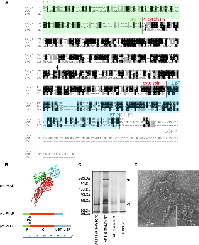
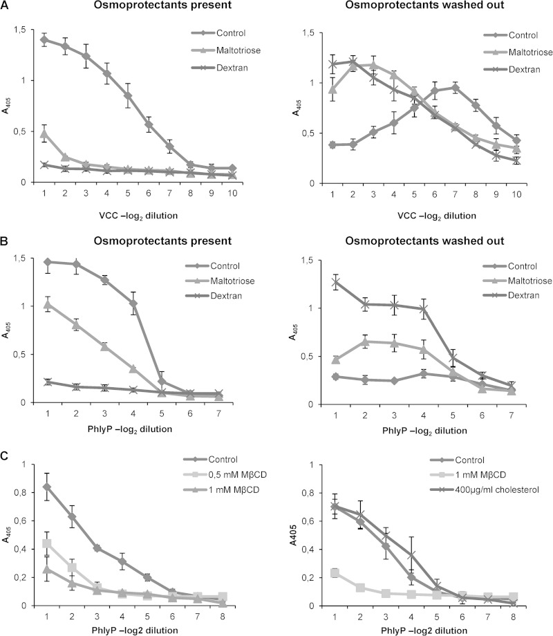
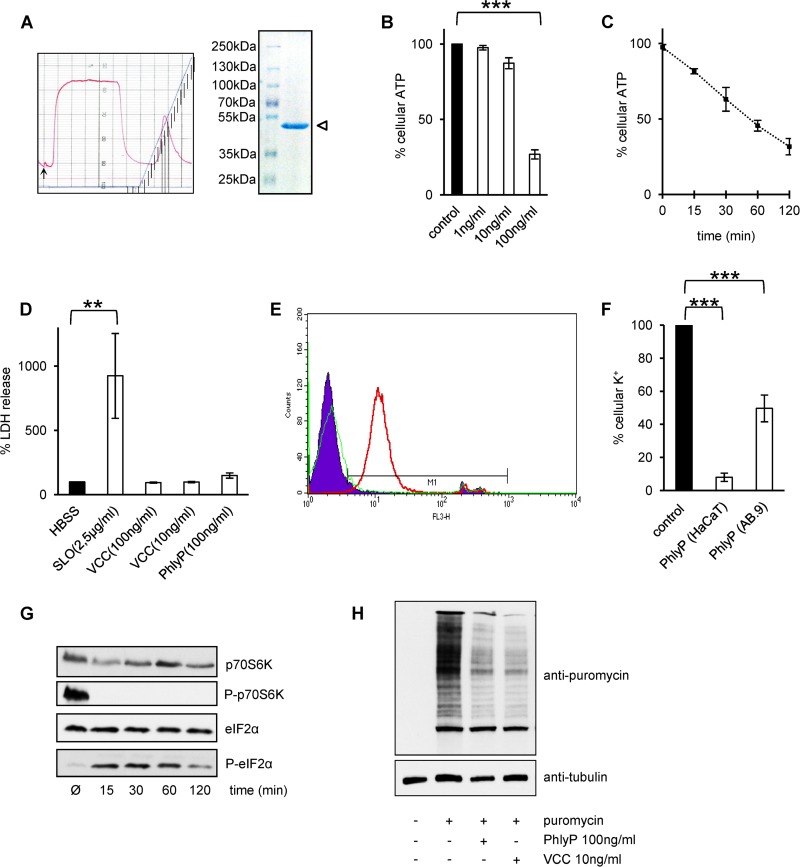
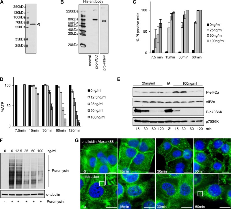
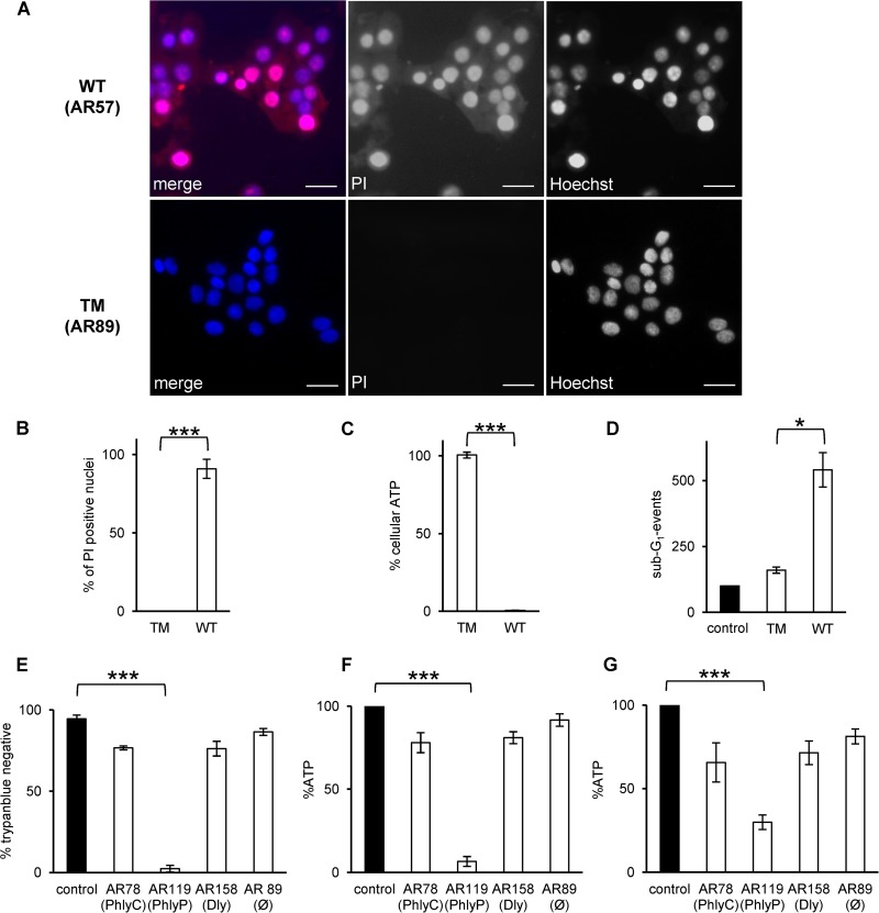
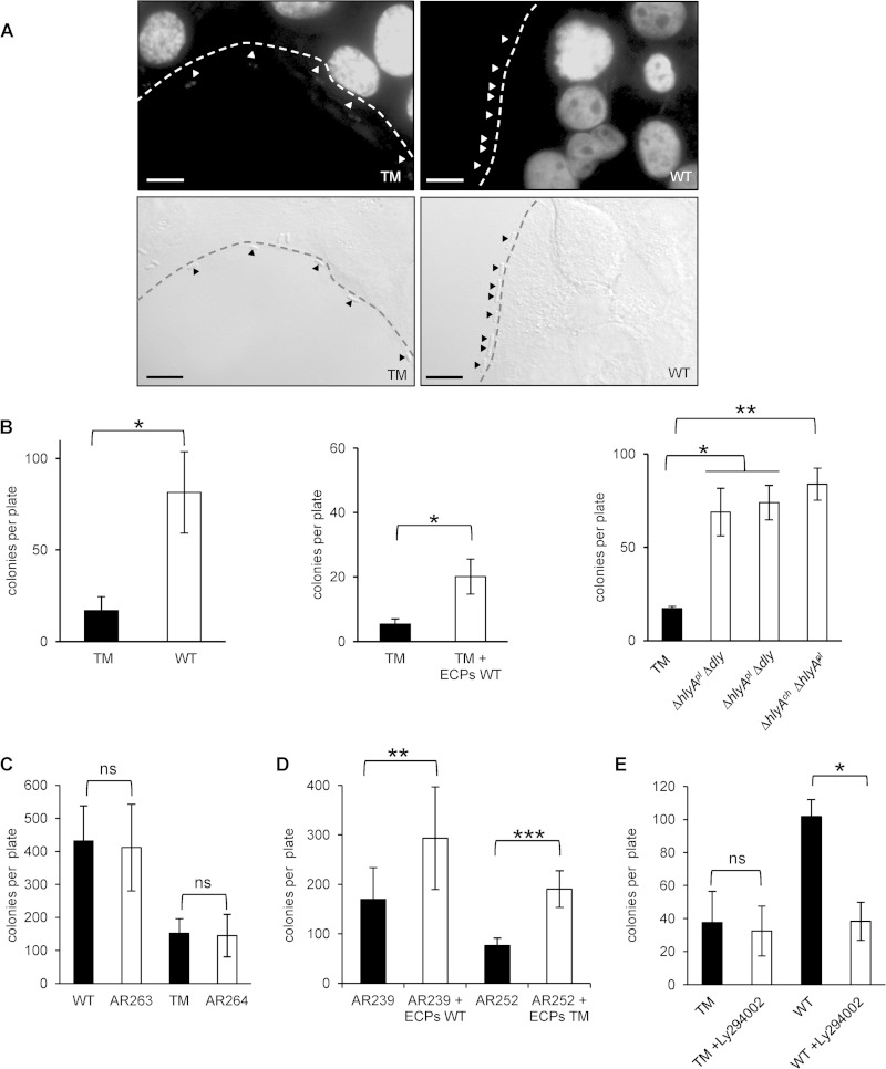
Photobacterium damselae subsp. damselae, an important pathogen of marine animals, may also cause septicemia or hyperaggressive necrotizing fasciitis in humans. We previously showed that hemolysin genes are critical for virulence of this organism in mice and fish. In the present study, we characterized the hlyA gene product, a putative small β-pore-forming toxin, and termed it phobalysin P (PhlyP), for "photobacterial lysin encoded on a plasmid." PhlyP formed stable oligomers and small membrane pores, causing efflux of K(+), with no significant leakage of lactate dehydrogenase but entry of vital dyes. The latter feature distinguished PhlyP from the related Vibrio cholerae cytolysin. Attack by PhlyP provoked a loss of cellular ATP, attenuated translation, and caused profound morphological changes in epithelial cells. In coculture experiments with epithelial cells, Photobacterium damselae subsp. damselae led to rapid hemolysin-dependent membrane permeabilization. Unexpectedly, hemolysins also promoted the association of P. damselae subsp. damselae with epithelial cells. The collective observations of this study suggest that membrane-damaging toxins commonly enhance bacterial adherence.


Figures

FIG 1
FIG 1
PhlyP, a new β-pore-forming toxin. (A) Alignment of amino acid sequences of pro-PhlyP, pro-VCC, and VVC (the prodomain of VVC is not contiguously expressed with the hemolysin domain and the lectin domain). Sequences were aligned by use of ClustalW 2.0 (68) and displayed with version 3.21 of BOXSHADE, written by K. Hofmann and M. Baron (http://www.ch.embnet.org/software/BOX_form.html). The prodomain of PhlyP is shaded in green, and the L-βT domain is shown in blue. The cytolysin domain is labeled in red, and the L-βP domain is labeled in gray. The black arrow points at W318 of pVCC. (B) (Top) Similarity-based structure prediction for pPhlyP, performed by using the I-TASSER server (34). The C score for alignment of mature PhlyP and VCC was 0.99; the score for the protoxins was negative due to the lack of the L-βP domain in pPhlyP. The model as shown here was created with the aid of UCSF Chimera (http://www.cgl.ucsf.edu/chimera). (Bottom) Comparison of domain structures of pPhlyP and pVCC. The scale bar shows numbers of amino acid residues. PAS, proteolytic activation site. (C) Erythrocyte ghosts were incubated with the indicated ECPs, treated with trypsin, and separated by SDS-PAGE, and proteins were silver stained. The filled triangle on the right indicates the presumed PhlyP oligomer, and the open triangle indicates the monomer. (D) Rabbit erythrocyte ghosts were loaded with PhlyP (ECPs of strain AR119) and analyzed by TEM.
FIG 2
FIG 2
PhlyP forms small membrane pores, and its hemolytic activity requires cholesterol in cell membranes. (A and B) Osmoprotection experiments were performed with pVCC (A) and PhlyP (ECPs of AR119) (B). The left graphs show hemolysis in the presence of osmoprotectants, and the right graphs show hemolysis after washout of osmoprotectants. Data show mean values ± SE (n = 3). (C) RRCs were depleted of cholesterol by using methyl-β-cyclodextrin (MβCD) and were challenged with ECPs of strain AR119 (left). Replenishment of cholesterol was conducted by adding soluble cholesterol to cholesterol-depleted RRCs (right). Data are mean values ± SE (n = 3).
FIG 3
FIG 3
P. damselae subsp. damselae extracellular products contain mature PhlyP. (A) The left panel shows the elution profile from the Mono S column; note the single peak in the right half of the profile, which contained the hemolytic activity. The right panel shows an SDS-PAGE gel with purified PhlyP (two peak fractions from the elution profile), indicated by an open triangle. (B) ATP was measured after incubation of HaCaT cells with various concentrations of purified PhlyP for 2 h. Data are mean values ± SE (n = 3). ***, P ≤ 0.001 in two-sided, unpaired Student's t test. (C) Results of ATP assays after incubation of HaCaT cells with purified PhlyP (100 ng/ml) for various times. Data shown are percentages of the level in untreated controls and are mean values ± SE (n = 3). (D) HaCaT cells were treated for 10 min with the indicated toxins, and lactate dehydrogenase (LDH) was measured in supernatants. Control, saline without toxin (HBSS). Data are mean values ± SE (n = 3). **, P ≤ 0.01 in two-sided, unpaired Student's t test. (E) HaCaT cells in suspension were incubated with PhlyP (100 ng/ml) for 4 min, PI (50 μg/ml) was added, and samples were analyzed after 1 min at 37°C by flow cytometry. FL3H, fluorescence intensity (log) for PI; counts, number of events per channel. Red line, PhlyP; green line, VCC; filled area, untreated cells. (F) Cellular K+ levels were determined by flame photometry in HaCaT or AB.9 cell lysates after exposure of cells to PhlyP. Cells were incubated with purified PhlyP (100 ng/ml) (HaCaT cells) or ECPs from strain AR119 (produces only PhlyP) (AB.9 cells). Intracellular levels of potassium were determined after 10 min. Data are percentages of the levels in untreated controls and are mean values ± SE (n ≥ 3). ***, P ≤ 0.001 in two-sided, unpaired Student's t tests. (G) HaCaT cells were treated with purified PhlyP (100 ng/ml) for the indicated times at 37°C and were analyzed by Western blotting for phosphorylation of p70S6K or eIF2α. (H) HaCaT cells were incubated with purified PhlyP (100 ng/ml) or VCC (10 ng/ml) for 10 min at 37°C. Subsequently, cells were incubated with 10 μg/ml puromycin (1 h at 37°C), which is incorporated into nascent proteins (69). Finally, cells were analyzed by Western blotting using antibodies directed against puromycin.
FIG 4
FIG 4
Recombinant pro-PhlyP (pPhlyP) reproduces cytotoxic effects observed with native PhlyP. (A) SDS-PAGE analysis of affinity-purified N-terminally His-tagged pPhlyP (indicated by an open triangle). (B) Purified N-terminally His-tagged pVCC and pPhlyP were analyzed by Western blotting with anti-His antibodies. (C) HaCaT cells were incubated as indicated with different doses of pPhlyP (recombinant) for the indicated times and then stained with PI. The graph shows percentages of PI-positive cells out of the total cell number (mean values ± SE [n = 3]). (D) HaCaT cells were incubated with the indicated doses of His-tagged pPhlyP. Cellular ATP levels were determined at the indicated times. Data are percentages of the values in untreated controls and are mean values ± SE (n ≥ 5 for 15, 30, 60, and 120 min; n = 4 for 7.5 min). (E) HaCaT cells were incubated or not with 25 ng/ml or 100 ng/ml His-tagged pPhlyP for the indicated times. Lysates were analyzed by Western blotting for P-eIF2α, eIF2α, P-p70S6K, and p70S6K. (F) HaCaT cells were incubated or not with different doses of His-tagged pPhlyP for 1 h. Subsequently, cells were incubated or not for 1 h at 37°C with 10 μg/ml puromycin, which is incorporated into nascent proteins (69). Cells were analyzed by Western blotting for puromycin and α-tubulin. (G) HaCaT cells were incubated for the indicated times with 100 ng/ml His-tagged pPhlyP and then either washed, fixed, and treated with 0.1% Triton X-100 prior to staining with phalloidin-Alexa 488 (upper panels) or washed and treated with MitoTracker Green FM (200 nM) for 30 min at 37°C (lower panels). Bars = 20 μm.
FIG 5
FIG 5
P. damselae subsp. damselae toxins cause fulminant membrane damage. (A) HaCaT cells were infected with washed wild-type (WT) or triple mutant (TM) P. damselae subsp. damselae at a multiplicity of infection (MOI) of 1:30, incubated for 30 min at 37°C, stained for 1 min with PI (50 μg/ml), and subsequently fixed and stained with Hoechst 33342. Bars = 50 μm. Representative images are shown. (B) HaCaT cells were incubated for 8 min with ECPs (6 μg/ml total protein) from the WT or TM strain and stained as described for panel A, and PI-positive cells in digital microscopic images were enumerated. The graph shows mean values ± SE for 4 independent experiments. ***, P ≤ 0.001 in two-sided, unpaired Student's t test. (C) HaCaT cells were incubated with ECPs of the wild-type or triple mutant strain. After 1 h, the cellular ATP level was determined. Data are mean values ± SE (n = 4). ***, P ≤ 0.001 in two-sided, unpaired Student's t test. (D) Cells were incubated with normal medium or ECPs for 48 h and then processed for flow cytometric analysis of DNA content. Columns show numbers of sub-G1 events. Data shown are mean values ± SE (n = 3). *, P ≤ 0.05 in two-sided, unpaired Student's t test. (E) HaCaT cells were incubated for 1 h at 37°C with ECPs containing single toxins, as indicated. Control, medium only. Cells were stained with trypan blue (TB), and TB-positive and -negative cells were counted in a Neubauer chamber. Data are mean values ± SE (n = 3). ***, P ≤ 0.001 in two-sided, unpaired Student's t test. HaCaT (F) and AB.9 (G) cells were treated as described for panel E prior to measurement of ATP. Graphs show mean values ± SE (n = 3). ***, P ≤ 0.001 in two-sided, unpaired Student's t test.
FIG 6
FIG 6
Hemolysins promote bacterial adherence to target cells. (A) HaCaT cells were infected with the P. damselae subsp. damselae WT (AR57) strain or TM (AR89) strain. The distribution of bacteria was analyzed by differential interference contrast microscopy. Bars = 10 μm. (B) Results of adherence assays based on CFU counts. (Left) HaCaT cells infected with P. damselae subsp. damselae TM or WT. (Middle) HaCaT cells infected with P. damselae subsp. damselae TM and treated (white columns) or not (black column) with ECPs obtained from the WT strain (6 μg/ml total protein). (Right) HaCaT cells infected with the TM (black column) or double mutant strains, each expressing one of the known hemolysins only (white columns). Data shown are mean values ± SE (n = 3). * (left and middle panels), P ≤ 0.05 as determined by two-sided, unpaired Student's t test; * and ** (right panel), P ≤ 0.05 and P ≤ 0.01, respectively, as determined by ANOVA. (C) Results of adherence assays based on CFU counts. The bars show mean numbers of CFU (±SE [n = 3]) determined for samples of HaCaT cells infected with P. damselae subsp. damselae WT or TM (black) and their respective cheA mutants. ns, nonsignificant, i.e., P ≥ 0.05, in two-sided, unpaired Student's t tests. (D) HaCaT cells were infected with P. damselae subsp. damselae AR239 (ΔpilD) or AR252 (ΔpilD ΔtadV) in the presence of ECPs from the WT strain. For determination of the numbers of adherent AR239 and AR252 bacteria, a 10-fold larger volume of the harvested coculture than that of all other strains analyzed here was plated for CFU counts. Data shown are mean values ± SE (n = 3). ** and ***, P ≤ 0.01 and P ≤ 0.001, respectively, in two-sided, unpaired Student's t tests. (E) HaCaT cells were infected with the TM and WT strains in the presence of Ly294002 (1 μM). Data are mean values ± SE (n = 3). ns, nonsignificant; *, P ≤ 0.05 in two-sided, unpaired Student's t test.

References

    1. Álvarez JR, Lamba S, Dyer KY, Apuzzio JJ. 2006. An unusual case of urinary tract infection in a pregnant woman with Photobacterium damsela. Infect Dis Obstet Gynecol 2006:80682. - PMC - PubMed
    1. Clarridge JE, Zighelboim-Daum S. 1985. Isolation and characterization of two hemolytic phenotypes of Vibrio damsela associated with a fatal wound infection. J Clin Microbiol 21:302–306. - PMC - PubMed
    1. Goodell KH, Jordan MR, Graham R, Cassidy C, Nasraway SA. 2004. Rapidly advancing necrotizing fasciitis caused by Photobacterium (Vibrio) damsela: a hyperaggressive variant. Crit Care Med 32:278–281. doi:10.1097/01.CCM.0000104920.01254.82. - DOI - PubMed
    1. Kim HR, Kim JW, Lee MK, Kim JG. 2009. Septicemia progressing to fatal hepatic dysfunction in an cirrhotic patient after oral ingestion of Photobacterium damsela: a case report. Infection 37:555–556. doi:10.1007/s15010-009-9049-8. - DOI - PubMed
    1. Yamane K, Asato J, Kawade N, Takahashi H, Kimura B, Arakawa Y. 2004. Two cases of fatal necrotizing fasciitis caused by Photobacterium damsela in Japan. J Clin Microbiol 42:1370–1372. doi:10.1128/JCM.42.3.1370-1372.2004. - DOI - PMC - PubMed

Publication types

MeSH terms

LinkOut - more resources