

Marine amoebae with cytoplasmic and perinuclear symbionts deeply branching in the Gammaproteobacteria
- PMID: 26303516
- PMCID: PMC4642509
- DOI: 10.1038/srep13381
Marine amoebae with cytoplasmic and perinuclear symbionts deeply branching in the Gammaproteobacteria
Abstract
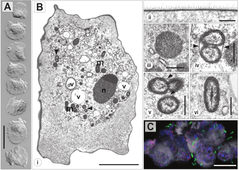
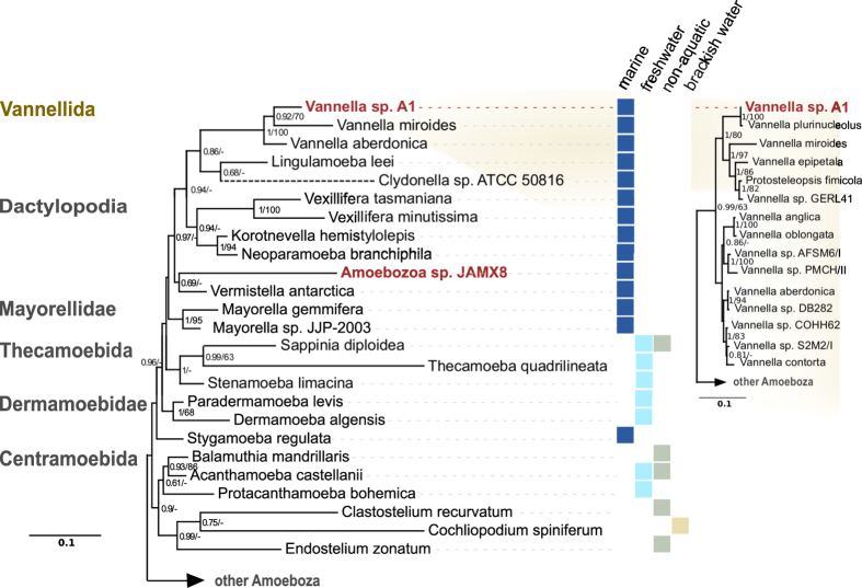
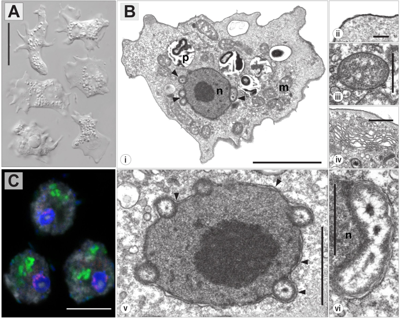
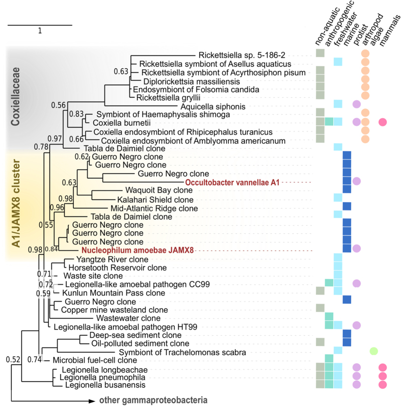
Amoebae play an important ecological role as predators in microbial communities. They also serve as niche for bacterial replication, harbor endosymbiotic bacteria and have contributed to the evolution of major human pathogens. Despite their high diversity, marine amoebae and their association with bacteria are poorly understood. Here we describe the isolation and characterization of two novel marine amoebae together with their bacterial endosymbionts, tentatively named 'Candidatus Occultobacter vannellae' and 'Candidatus Nucleophilum amoebae'. While one amoeba strain is related to Vannella, a genus common in marine habitats, the other represents a novel lineage in the Amoebozoa. The endosymbionts showed only low similarity to known bacteria (85-88% 16S rRNA sequence similarity) but together with other uncultured marine bacteria form a sister clade to the Coxiellaceae. Using fluorescence in situ hybridization and transmission electron microscopy, identity and intracellular location of both symbionts were confirmed; one was replicating in host-derived vacuoles, whereas the other was located in the perinuclear space of its amoeba host. This study sheds for the first time light on a so far neglected group of protists and their bacterial symbionts. The newly isolated strains represent easily maintainable model systems and pave the way for further studies on marine associations between amoebae and bacterial symbionts.
Figures
Similar articles
-
Infection and nuclear interaction in mammalian cells by 'Candidatus Berkiella cookevillensis', a novel bacterium isolated from amoebae.BMC Microbiol. 2019 May 9;19(1):91. doi: 10.1186/s12866-019-1457-z. BMC Microbiol. 2019. PMID: 31072343 Free PMC article.
-
Ménage-à-trois: the amoeba Nuclearia sp. from Lake Zurich with its ecto- and endosymbiotic bacteria.Protist. 2014 Sep;165(5):745-58. doi: 10.1016/j.protis.2014.08.004. Epub 2014 Aug 28. Protist. 2014. PMID: 25248027
-
'Candidatus Cochliophilus cryoturris' (Coxiellaceae), a symbiont of the testate amoeba Cochliopodium minus.Sci Rep. 2017 Jun 13;7(1):3394. doi: 10.1038/s41598-017-03642-8. Sci Rep. 2017. PMID: 28611430 Free PMC article.
-
Bacterial endosymbionts of free-living amoebae.J Eukaryot Microbiol. 2004 Sep-Oct;51(5):509-14. doi: 10.1111/j.1550-7408.2004.tb00278.x. J Eukaryot Microbiol. 2004. PMID: 15537084 Review.
-
Genetic and physiological interactions in the amoeba-bacteria symbiosis.J Eukaryot Microbiol. 2004 Sep-Oct;51(5):502-8. doi: 10.1111/j.1550-7408.2004.tb00277.x. J Eukaryot Microbiol. 2004. PMID: 15537083 Review.
Cited by
-
Entities inside one another - a matryoshka doll in biology?Environ Microbiol Rep. 2019 Feb;11(1):26-28. doi: 10.1111/1758-2229.12716. Epub 2018 Dec 26. Environ Microbiol Rep. 2019. PMID: 30588764 Free PMC article. No abstract available.
-
Free-Living Amoebae in Soil Samples from Santiago Island, Cape Verde.Microorganisms. 2021 Jul 7;9(7):1460. doi: 10.3390/microorganisms9071460. Microorganisms. 2021. PMID: 34361894 Free PMC article.
-
Identification and characterization of a large family of superbinding bacterial SH2 domains.Nat Commun. 2018 Oct 31;9(1):4549. doi: 10.1038/s41467-018-06943-2. Nat Commun. 2018. PMID: 30382091 Free PMC article.
-
Cell sorting reveals few novel prokaryote and photosynthetic picoeukaryote associations in the oligotrophic ocean.Environ Microbiol. 2021 Mar;23(3):1469-1480. doi: 10.1111/1462-2920.15351. Epub 2020 Dec 19. Environ Microbiol. 2021. PMID: 33295132 Free PMC article.
-
Cobaviruses - a new globally distributed phage group infecting Rhodobacteraceae in marine ecosystems.ISME J. 2019 Jun;13(6):1404-1421. doi: 10.1038/s41396-019-0362-7. Epub 2019 Feb 4. ISME J. 2019. PMID: 30718806 Free PMC article.
References
-
- Rodríguez-Zaragoza S. Ecology of free-living amoebae. Crit. Rev. Microbiol. 20, 225–241 (1994). - PubMed
-
- Khan N. A. Acanthamoeba: Biology and Pathogenesis. (Horizon Scientific Press, 2009).
-
- Cavalier-Smith T. et al.. Multigene phylogeny resolves deep branching of Amoebozoa. Mol. Phylogenet. Evol. 83, 293–304 (2015). - PubMed
-
- Smirnov A. V. in Encyclopedia of Microbiology. 558–577 (Elsevier, 2008).
Publication types
MeSH terms
Grants and funding
LinkOut - more resources
Full Text Sources
Other Literature Sources
Molecular Biology Databases
