. 2015 Nov;197(21):3463-71.
doi: 10.1128/JB.00352-15. Epub 2015 Aug 24.

In Vivo Formation of the Protein Disulfide Bond That Enhances the Thermostability of Diphosphomevalonate Decarboxylase, an Intracellular Enzyme from the Hyperthermophilic Archaeon Sulfolobus solfataricus

Affiliations

In Vivo Formation of the Protein Disulfide Bond That Enhances the Thermostability of Diphosphomevalonate Decarboxylase, an Intracellular Enzyme from the Hyperthermophilic Archaeon Sulfolobus solfataricus

Ai Hattori et al. J Bacteriol. 2015 Nov.

Abstract

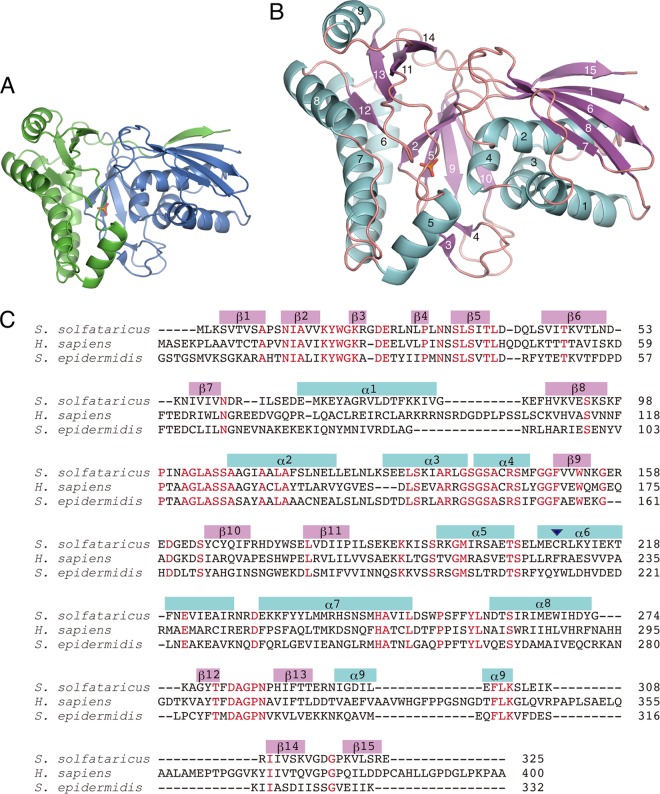
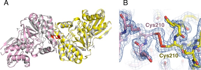
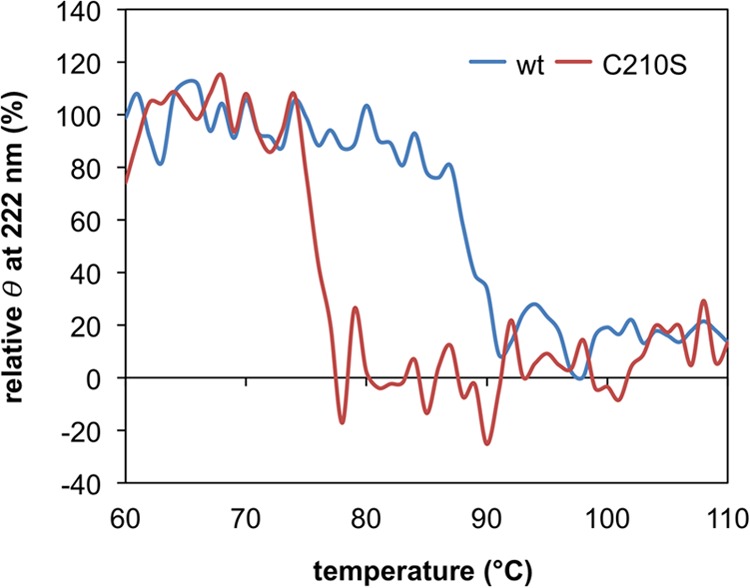
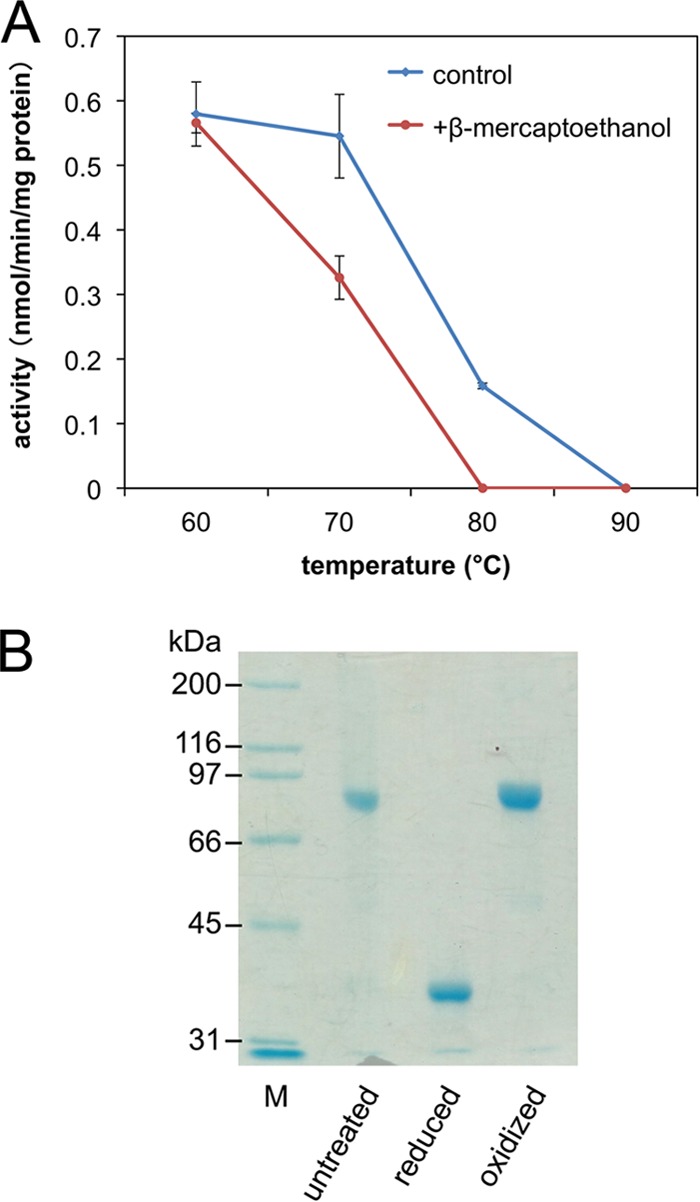
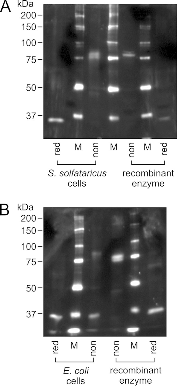
In the present study, the crystal structure of recombinant diphosphomevalonate decarboxylase from the hyperthermophilic archaeon Sulfolobus solfataricus was solved as the first example of an archaeal and thermophile-derived diphosphomevalonate decarboxylase. The enzyme forms a homodimer, as expected for most eukaryotic and bacterial orthologs. Interestingly, the subunits of the homodimer are connected via an intersubunit disulfide bond, which presumably formed during the purification process of the recombinant enzyme expressed in Escherichia coli. When mutagenesis replaced the disulfide-forming cysteine residue with serine, however, the thermostability of the enzyme was significantly lowered. In the presence of β-mercaptoethanol at a concentration where the disulfide bond was completely reduced, the wild-type enzyme was less stable to heat. Moreover, Western blot analysis combined with nonreducing SDS-PAGE of the whole cells of S. solfataricus proved that the disulfide bond was predominantly formed in the cells. These results suggest that the disulfide bond is required for the cytosolic enzyme to acquire further thermostability and to exert activity at the growth temperature of S. solfataricus.

Importance: This study is the first report to describe the crystal structures of archaeal diphosphomevalonate decarboxylase, an enzyme involved in the classical mevalonate pathway. A stability-conferring intersubunit disulfide bond is a remarkable feature that is not found in eukaryotic and bacterial orthologs. The evidence that the disulfide bond also is formed in S. solfataricus cells suggests its physiological importance.


Figures

FIG 1
FIG 1
Crystal structure of recombinant DMD from S. solfataricus. (A) Monomer subunit structure of DMD-P in ribbon representation. N- and C-terminal domains are colored blue and green, respectively. A phosphate molecule bound in a cleft in DMD-P is shown as a stick model. (B) Ribbon diagram of S. solfataricus DMD-P subunit. α-Helices and β-strands, labeled in the order of appearance, are colored light blue and pink, respectively. The phosphate molecule bound to DMD-P is shown as a stick model. (C) Amino acid sequences of DMD from S. solfataricus, H. sapiens, and S. epidermidis are aligned. Conserved residues are colored red. α-Helices and β-strands in S. solfataricus are indicated as boxes in blue and pink, respectively. An arrowhead in the region of α6 represents the Cys210 residue of S. solfataricus DMD, which is involved in intersubunit disulfide bond formation.
FIG 2
FIG 2
Quaternary structure of recombinant S. solfataricus DMD. (A) Dimer structures of S. solfataricus DMD-P and DMD-AS in ribbon representation. The subunits of DMD-P are shown in yellow and pink, and those of DMD-AS are shown in gray. Phosphate and glycerol molecules bound to DMD-P and sulfate molecules to DMD-AS are shown as stick models. Disulfide bonds between protomers are shown as red stick models. (B) Intersubunit disulfide bond in the DMD-P structure is shown as stick models with the 2FoFc electron density map at the 1.5σ level (blue). Stick models of the protomers are shown in the same colors used for panel A.
FIG 3
FIG 3
Heat-induced change in the secondary structure of the recombinant wild-type (wt) S. solfataricus DMD and the C210S mutant through far-UV CD spectroscopy analysis. The vertical axis represents the relative ellipticity (θ) at 222 nm, where the average of ellipticity values from 60 to 69°C for each measurement is set to 100%.
FIG 4
FIG 4
Thermostability of recombinant S. solfataricus DMD in the presence and absence of β-mercaptoethanol. (A) DMD activity remaining after 1 h of heat treatment at the indicated temperatures. (B) Nonreducing SDS-PAGE analysis of untreated, reduced, and oxidized S. solfataricus DMD. M, standard molecular mass markers.
FIG 5
FIG 5
Nonreducing SDS-PAGE and Western blot analysis of the cells of S. solfataricus (A) and E. coli expressing recombinant S. solfataricus DMD (B). Approximately 300 μg and 0.8 μg wet cells of S. solfataricus and E. coli, respectively, were applied on each lane. The cells were treated with nonreducing (non) or reducing (red) SDS-PAGE sample buffer. Purified recombinant S. solfataricus DMD, untreated (non) and reduced (red), was used as the controls. M, standard molecular mass markers. To avoid the effect of the reducing agent on the redox state of samples on adjacent lanes, the sample lanes were separated by those for markers.

References

    1. Kadokura H, Katzen F, Beckwith J. 2003. Protein disulfide bond formation in prokaryotes. Annu Rev Biochem 72:111–135. doi:10.1146/annurev.biochem.72.121801.161459. - DOI - PubMed
    1. Dombkowski AA, Sultana KZ, Craig DB. 2014. Protein disulfide engineering. FEBS Lett 588:206–212. doi:10.1016/j.febslet.2013.11.024. - DOI - PubMed
    1. Toth EA, Worby C, Dixon JE, Goedken ER, Marqusee S, Yeates TO. 2000. The crystal structure of adenylosuccinate lyase from Pyrobaculum aerophilum reveals an intracellular protein with three disulfide bonds. J Mol Biol 301:433–450. doi:10.1006/jmbi.2000.3970. - DOI - PubMed
    1. King NP, Lee TM, Sawaya MR, Cascio D, Yeates TO. 2008. Structures and functional implications of an AMP-binding cystathionine β-synthase domain protein from a hyperthermophilic archaeon. J Mol Biol 380:181–192. doi:10.1016/j.jmb.2008.04.073. - DOI - PMC - PubMed
    1. Boutz DR, Cascio D, Whitelegge J, Perry LJ, Yeates TO. 2007. Discovery of a thermophilic protein complex stabilized by topologically interlinked chains. J Mol Biol 368:1332–1344. doi:10.1016/j.jmb.2007.02.078. - DOI - PMC - PubMed

Publication types

LinkOut - more resources