. 2015 Oct 2;290(40):24547-60.
doi: 10.1074/jbc.M115.673558. Epub 2015 Aug 24.

Crystal structure of PnpCD, a two-subunit hydroquinone 1,2-dioxygenase, reveals a novel structural class of Fe2+-dependent dioxygenases

Affiliations

Crystal structure of PnpCD, a two-subunit hydroquinone 1,2-dioxygenase, reveals a novel structural class of Fe2+-dependent dioxygenases

Shiheng Liu et al. J Biol Chem. .

Abstract

Aerobic microorganisms have evolved a variety of pathways to degrade aromatic and heterocyclic compounds. However, only several classes of oxygenolytic fission reaction have been identified for the critical ring cleavage dioxygenases. Among them, the most well studied dioxygenases proceed via catecholic intermediates, followed by noncatecholic hydroxy-substituted aromatic carboxylic acids. Therefore, the recently reported hydroquinone 1,2-dioxygenases add to the diversity of ring cleavage reactions. Two-subunit hydroquinone 1,2-dioxygenase PnpCD, the key enzyme in the hydroquinone pathway of para-nitrophenol degradation, catalyzes the ring cleavage of hydroquinone to γ-hydroxymuconic semialdehyde. Here, we report three PnpCD structures, named apo-PnpCD, PnpCD-Fe(3+), and PnpCD-Cd(2+)-HBN (substrate analog hydroxyenzonitrile), respectively. Structural analysis showed that both the PnpC and the C-terminal domains of PnpD comprise a conserved cupin fold, whereas PnpC cannot form a competent metal binding pocket as can PnpD cupin. Four residues of PnpD (His-256, Asn-258, Glu-262, and His-303) were observed to coordinate the iron ion. The Asn-258 coordination is particularly interesting because this coordinating residue has never been observed in the homologous cupin structures of PnpCD. Asn-258 is proposed to play a pivotal role in binding the iron prior to the enzymatic reaction, but it might lose coordination to the iron when the reaction begins. PnpD also consists of an intriguing N-terminal domain that might have functions other than nucleic acid binding in its structural homologs. In summary, PnpCD has no apparent evolutionary relationship with other iron-dependent dioxygenases and therefore defines a new structural class. The study of PnpCD might add to the understanding of the ring cleavage of dioxygenases.

Keywords: biodegradation; cupin; dioxygenase; enzyme mechanism; metal coordination; metalloenzyme; ring cleavage; structure-function.


Figures

FIGURE 1.
FIGURE 1.
Metal ion specificity of PnpCD. Pro represents the purified PnpCD proteins after incubation with EDTA. Activity is defined as the number of micromoles of semialdehyde produced per min per ng of protein. The activity of PnpCD in the presence of Fe2+ is 19.7 μmol min−1 ng−1, which is equivalent to 100% enzyme activity. Error bars indicate the standard deviation.
FIGURE 2.
FIGURE 2.
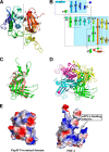
Structure of PnpD (α-subunit). A, schematic representation of the PnpD structure. The structure is colored in rainbow colors. B, protein topology of PnpD. C, structural comparison of PnpD N-terminal domain and plant transcription factor PBF-2. The N-terminal domain of PnpD is shown in salmon, and PBF-2 is shown in green. D, whirly structure of PBF-2. The nucleic acid-binding region is circled by the black dashed line. E, electric potential surface of PnpD N-terminal domain and PBF-2.
FIGURE 3.
FIGURE 3.
Cupin characterization of PnpD C-terminal domain and PnpC. A, DALI alignment of the PnpD cupin and its metal ion-binding cupin homologs. The two conserved cupin motifs are shown in the black boxes. The residues responsible for metal ion binding are assigned with the pentagrams. The red arrows indicate the cupin proteins with four-residue coordination environment. B, corresponding secondary structure of the cupin proteins shown in A. L represents loop, and E and H represent β-strand and α-helix, respectively. C, DALI alignment of PnpC and its metal ion-binding cupin homologs. D, corresponding secondary structure of the cupin proteins shown in C.
FIGURE 4.
FIGURE 4.
Structure of PnpC (β-subunit). A, schematic representation of the PnpC structure. The structure is colored in rainbow colors. B, protein topology of PnpC. C, structural comparison of PnpC and the C-terminal domain of PnpD. PnpC is shown as a green schematic. PnpD is shown as both schematic and surface mode and is colored in salmon.
FIGURE 5.
FIGURE 5.
Tightly associated heterotetramer of PnpCD. A, overall structure of the heterotetramer (αβ)2. α-Subunits are shown in salmon and slate, and β-subunits are shown in green and cyan. The noncrystallographic 2-fold axis is shown as a red dot. B, gel filtration analysis of PnpCD on “Superdex 200 Increase 10/300 GL.” Con, conalbumin; Ald, aldolase. C, residues that contribute to interactions between the two PnpCD molecules. D, surface representation of the PnpC-PnpD protein complex.
FIGURE 6.
FIGURE 6.
Multiple sequence alignment of HQDOs harboring two subunits. The alignment is carried out using T-Coffee, and the results are generated using ESPript. The residues conserved in all five sequences are highlighted in red. The four coordinating residues of PnpD are assigned with red triangles. WBC-3, Pseudomonas sp. strain WBC-3; DLL-E4, P. putida DLL-E4; TTNP3, Sphingomonas sp. strain TTNP3; ACB, Pseudomonas fluorescens ACB; SJ98, Burkholderia sp. strain SJ98.
FIGURE 7.
FIGURE 7.
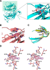
Active site of PnpCD. A, coordination sites of PnpCD-Fe3+. The coordinating atoms are assigned with black triangles. The FoFc maps contoured at 10σ show the electron density for metal ions. B, coordination sites of PnpCD-Cd2+-HBN. C, structural comparison of the iron-bound PnpCD and the iron-free PnpCDs. The iron-bound and iron-free states in PnpCD-Fe3+ are shown in cyan and yellow, respectively. The iron-free state in apo-PnpCD is shown in gray. D, stereo view of the substrate-binding site of PnpCD. The FoFc electron density map for hydroxybenzonitrile (HBN) is contoured at 3σ.
FIGURE 8.
FIGURE 8.
Activity, coordination, and substrate-binding assay on PnpCD. A, effect of active site residue mutants on enzyme activity of PnpCD. Activity is defined as the number of micromoles of semialdehyde produced per min/ng of protein. The activity of wild type PnpCD is 22.9 μmol min−1 ng−1, which is equivalent to 100% enzyme activity. Error bars indicate the standard deviation. B–E, effect of the Asn-258 mutants (N258D and N258A) on metal ion binding. F and G, effect of the mutant E248Q on substrate binding. The constant concentrations of dye NT647-labeled proteins were ∼0.8 μm for wild type PnpCD and ∼0.4 μm for the mutant E248Q, respectively. The initial concentration of hydroquinone was 5000 μm, which was serially diluted 15 times in a volume ratio of 1:1 with water each time. For sample preparation, each labeled protein (10 μl) was incubated with 16 concentrations of hydroquinone (10 μl), respectively.
FIGURE 9.
FIGURE 9.
Putative O2 binding pocket and proposed tunnels for substrate entry and product exit. A, putative O2 binding pocket in the PnpCD structures. B, surface representation of chain W (salmon) and chain Y (yellow) in the PnpCD-Fe3+ structure. The two tunnels that might be responsible for the substrate entry and product exit are circled by the white dashed line. C, residues that contribute to the formation of the two tunnels.
FIGURE 10.
FIGURE 10.
General ring cleavage mechanism proposed for PnpCD.

References

    1. Grube M., Muter O., Strikauska S., Gavare M., Limane B. (2008) Application of FT-IR spectroscopy for control of the medium composition during the biodegradation of nitro aromatic compounds. J. Ind. Microbiol. Biotechnol. 35, 1545–1549 - PubMed
    1. Figge K., Klahn J., Koch J. (1983) Testing of chemicals by evaluation of their distribution and degradation patterns in an environmental standard system. Regul. Toxicol. Pharmacol. 3, 199–215 - PubMed
    1. Zieris F. J., Feind D., Huber W. (1988) Long-term effects of 4-nitrophenol in an outdoor synthetic aquatic ecosystem. Arch. Environ. Contam. Toxicol. 17, 165–175
    1. Agency for Toxic Substances and Disease Registry (1992) Toxicological profile for nitrophenols: 2-nitrophenol and 4-nitrophenol. United States Department of Health and Human Services, Public Health Service, Atlanta
    1. Ju K. S., Parales R. E. (2010) Nitroaromatic compounds, from synthesis to biodegradation. Microbiol. Mol. Biol. Rev. 74, 250–272 - PMC - PubMed

Publication types

MeSH terms

LinkOut - more resources