. 2015 Sep 30;43(17):8416-34.
doi: 10.1093/nar/gkv838. Epub 2015 Aug 24.

Discovery of an essential nucleotidylating activity associated with a newly delineated conserved domain in the RNA polymerase-containing protein of all nidoviruses

Affiliations

Discovery of an essential nucleotidylating activity associated with a newly delineated conserved domain in the RNA polymerase-containing protein of all nidoviruses

Kathleen C Lehmann et al. Nucleic Acids Res. .

Abstract

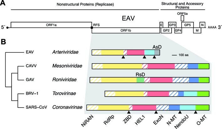
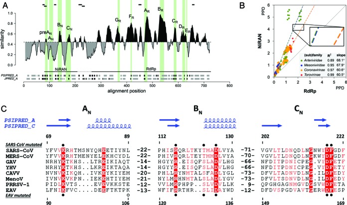
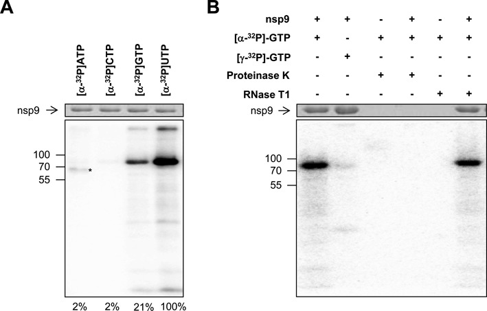
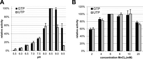
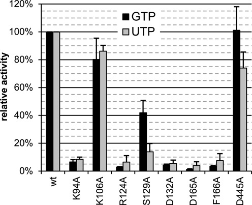
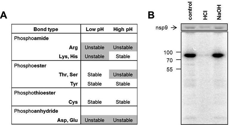
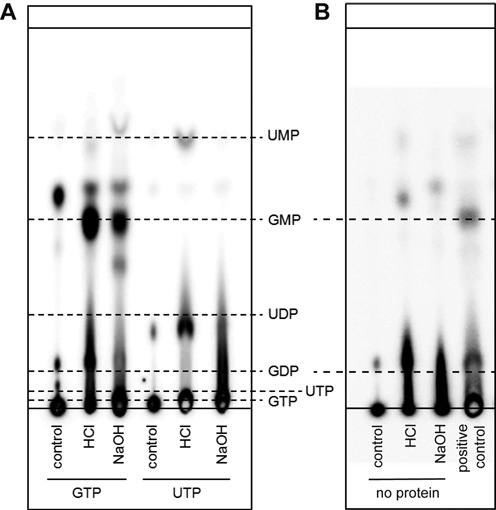
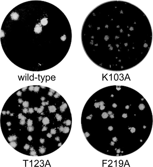
RNA viruses encode an RNA-dependent RNA polymerase (RdRp) that catalyzes the synthesis of their RNA(s). In the case of positive-stranded RNA viruses belonging to the order Nidovirales, the RdRp resides in a replicase subunit that is unusually large. Bioinformatics analysis of this non-structural protein has now revealed a nidoviral signature domain (genetic marker) that is N-terminally adjacent to the RdRp and has no apparent homologs elsewhere. Based on its conservation profile, this domain is proposed to have nucleotidylation activity. We used recombinant non-structural protein 9 of the arterivirus equine arteritis virus (EAV) and different biochemical assays, including irreversible labeling with a GTP analog followed by a proteomics analysis, to demonstrate the manganese-dependent covalent binding of guanosine and uridine phosphates to a lysine/histidine residue. Most likely this was the invariant lysine of the newly identified domain, named nidovirus RdRp-associated nucleotidyltransferase (NiRAN), whose substitution with alanine severely diminished the described binding. Furthermore, this mutation crippled EAV and prevented the replication of severe acute respiratory syndrome coronavirus (SARS-CoV) in cell culture, indicating that NiRAN is essential for nidoviruses. Potential functions supported by NiRAN may include nucleic acid ligation, mRNA capping and protein-primed RNA synthesis, possibilities that remain to be explored in future studies.


Figures

Figure 1.
Figure 1.
Genome organization and ORF1b-encoded enzymes and domains of nidoviruses. (A) The genome organization of Equine arteritis virus (EAV), including replicase open reading frames (ORFs) 1a and 1b, and 3′ ORFs encoding structural proteins, is shown. Genomes of other nidoviruses employ similar organizations while they may vary in respect to size of different regions and number of 3′ ORFs. RFS, ribosomal frameshift site. (B) ORF1b size and domain comparison between the five nidovirus (sub)families is shown for EAV (Arteriviridae), Cavally virus (CAVV, Mesoniviridae), Gill-associated virus (GAV, Roniviridae), Breda virus (BRV-1, Torovirinae) and Severe acute respiratory syndrome coronavirus (SARS-CoV, Coronavirinae); see Supplementary Table 1 for details regarding these viruses. NiRAN, nidovirus RdRp-associated nucleotidyltransferase; RdRp, RNA-dependent RNA polymerase; ZBD, Zn-binding domain; HEL1, helicase superfamily 1 core domain; ExoN, exoribonuclease; N-MT, N7-methyltransferase; NendoU, nidovirus uridylate-specific endoribonuclease; O-MT, 2′-O-methyltransferase; AsD, arterivirus-specific domain; RsD, ronivirus-specific domain. Depicted is a simplified domain organization since most enzymes are part of multidomain proteins. Note that viruses of the Torovirinae subfamily encode a truncated version of N-MT. Triangles, established cleavage sites by 3CLpro in two virus (sub)families; ORF1b-encoded proteins of other viruses may be proteolytically processed in a similar way. The order of emergence of different nidovirus (sub)families is presented by a simplified tree on the left.
Figure 2.
Figure 2.
Delineation and divergence of the NiRAN domain in the RdRp-containing proteins of nidoviruses. (A) Sequence variation, domain organization and secondary structure of the RdRp-containing protein of arteriviruses, and location of peptides identified by mass spectrometry after FSBG-labeling of arterivirus nsp9. Shown is the similarity density plot obtained for the multiple sequence alignment (MSA) of proteins including NiRAN and RdRp domains of arteriviruses. To highlight the regional deviation of conservation from that of the MSA average, areas above and below the mean similarity are shaded in black and grey, respectively. Uncertainty in respect to the domain boundary between NiRAN and RdRp is indicated by a dashed horizontal line. Positions of conserved sequence motifs of NiRAN and RdRp are indicated by vertical shading areas; motifs are labeled. Below the similarity density plot, secondary structure elements, predicted based on the arterivirus MSA using PSIPRED (PSIPRED_A) and Jpred 3 (JPRED_A), are presented in grey for α-helices, black for β-strands. (B) Relative scale of divergence of NiRAN versus RdRp in four different nidovirus (sub)families. Shown is scatter plot of PPDs of the NiRAN (y-axis) versus PPDs of RdRp (x-axis), which were calculated from the respective four PhyML trees. Dashed lines depict linear regressions fit in four differently highlighted PPD distributions, with its detail being magnified in the zoom-in; R2 and slope values of the regressions are listed in the inset panel. The solid diagonal line corresponds to the matching rate of PPDs for the two domains and is provided for comparison. (C) MSA of the three conserved NiRAN motifs of eight representative nidoviruses and their predicted secondary structures. Absolutely conserved residues are in white font, while partially conserved residues are highlighted. Secondary structure predictions were made with PSIPRED (64) based on arterivirus (PSIPRED_A) or coronavirus (PSIPRED_C) MSAs. Residues mutated in recombinant SARS-CoV (Coronaviridae) non-structural protein (nsp) 12 and recombinant EAV (Arteriviridae) nsp9 are indicated by filled (conserved) and empty (control) circles, above and below the alignment respectively. Mutated residues D445A in EAV and K103A, D618A in SARS-CoV are not shown. Amino acid numbers above and below the alignment refer to SARS-CoV nsp12 and EAV nsp9, respectively. MERS-CoV, Middle East respiratory syndrome coronavirus (Coronaviridae); GAV (Roniviridae); YHV, yellow head virus (Roniviridae); CAVV (Mesoniviridae); MenoV, Meno virus (Mesoniviridae); PRRSV-1, porcine reproductive and respiratory syndrome virus, European genotype (Arteriviridae). For other abbreviations, see Figure 1.
Figure 3.
Figure 3.
Establishing sequence conservation between NiRAN domains of different (sub)families. Shown are four pairwise dot-plots that compare HMM profiles of NiRAN domains of different origins using HHalign. For the entire set of dot-plots generated, please see Supplementary Figure S3. The bottom three plots correspond to steps used to produce the nidovirus-wide NiRAN MSA (Supplementary Figure S1), while the top plot is shown for comparison. Coordinates of query and target HMMs are presented on y-axis and x-axis, respectively. All local similarities between two profiles are depicted as black dots. Transparent fat dark and light gray lines on the dot-plot show paths of HHalign alignments, obtained in local and global modes, respectively. The E-value of the top local alignment is specified below each dot-plot. In the profile–profile alignment produced in global mode, conserved amino acids of NiRAN motifs may have been properly aligned or not. If conserved residues of a motif were aligned, the corresponding region of the alignment path is labeled with the respective motif name without an asterisk. If the misalignment of conserved residues was limited to a shift of one or two residues (HMM–HMM alignment columns), the corresponding region of the alignment path is labeled with the respective motif name plus an asterisk.
Figure 4.
Figure 4.
Comparison of nidovirus-wide conserved domains with sequence databases. Shown are histograms depicting E-values of the best non-nidovirus hits obtained during HMMER-mediated profile-sequence (A) and HHsearch-mediated profile–profile (B) searches of the GenBank and PFAM A databases, respectively, using MSA profiles of five nidovirus-wide conserved domains encoded by four nidovirus families. The identity of the non-nidoviral top-hit in the respective databases is specified. Stars indicate hits whose homologous relationship with the respective query is also supported by the functional and/or structural annotation of the respective targets.
Figure 5.
Figure 5.
EAV nsp9 has nucleotidylation activity. Purified recombinant EAV nsp9 (78 kDa) was incubated with the indicated 32P-labeled NTP in the presence of MnCl2. After denaturing SDS-PAGE, reaction products were visualized by Coomassie brilliant blue staining (top panels) and phosphor imaging (bottom panels). Positions of molecular weight markers are depicted on the left in kDa. (A) Uridylylation and guanylylation activity as revealed by covalent binding of the respective radioactive nucleotide to nsp9. Note that the protein indicated with an asterisk likely is an Escherichia coli­-derived impurity reacting with ATP. Relative band intensities are shown at the bottom. (B) Guanylylation was distinguished from RNA polymerization by incubating the products generated during the nucleotidylation assay with proteinase K (1 mg/ml) or with RNase T1 (0.5 U), which cleaves single-stranded RNA after G residues, for 30 min at 37°C.
Figure 6.
Figure 6.
EAV nsp9 guanylylation has a slightly broader or shifted pH optimum compared to uridylylation while the metal ion requirement is identical. (A) The pH optimum in the range from 5.5 to 9.5 was determined using the buffers listed in ‘Materials and Methods’ section. (B) Assessment of the optimal MnCl2 concentration for nucleotidylation. Error bars represent the standard deviation of the mean based on three independent experiments.
Figure 7.
Figure 7.
Conserved NiRAN residues are essential for the nucleotidylation activity. Alanine substitution of conserved NiRAN residues dramatically decreased the nucleotidylation activity of nsp9. In contrast, mutation of the non-conserved K106 in the NiRAN domain or the conserved D445 in the RdRp domain had only a mild effect on activity. Error bars represent the standard deviation of the mean based on three independent experiments.
Figure 8.
Figure 8.
A phosphoamide bond is formed between nsp9 and the guanosine phosphate. (A) Chemical stability of different phosphoamino acid bonds. Adapted from (91). (B) The protein was labeled with [α-32P]GTP and subsequently incubated at pH 8.5 (control) or under acidic or alkaline conditions. Reaction products were visualized after denaturing SDS-PAGE by Coomassie brilliant blue staining (top panel) and phosphor imaging (bottom panel). Size markers are depicted on the left in kDa.
Figure 9.
Figure 9.
GMP is released from labeled EAV nsp9 under acidic conditions. (A) nsp9 was labeled with [α-32P]GTP or [α-32P]UTP and was incubated at pH 8.5 (control) or under acidic or alkaline conditions after removal of non-incorporated nucleotides. Resulting products were separated with PEI-cellulose TLC. Solid lines represent the position where samples have been spotted (bottom) and the running front (top). Dashed lines represent the respective mobilities of the indicated nucleotides. (B) [α-32P]GTP was incubated under the same conditions as in (A) but omitting nsp9. An nsp9-containing sample treated with HCl served as positive control.
Figure 10.
Figure 10.
Plaque phenotypes of viable SARS-CoV NiRAN mutants. Progeny virus harvested at 3 days post transfection was used for plaque assays (see ‘Materials and Methods’ section) on Vero-E6 cell monolayers, which were fixed and stained after 3 days to visualize virus-induced plaques.

References

    1. de Groot R.J., Cowley J.A., Enjuanes L., Faaberg K.S., Perlman S., Rottier P.J., Snijder E.J., Ziebuhr J., Gorbalenya A.E. Order Nidovirales. In: King AMQ, Adams MJ, Carstens EB, Lefkowitz EJ, editors. Virus Taxonomy. Ninth Report of the International Committee on Taxonomy of Viruses. Amsterdam: Elsevier Academic Press; 2012. pp. 785–795.
    1. Lauber C., Ziebuhr J., Junglen S., Drosten C., Zirkel F., Nga P.T., Morita K., Snijder E.J., Gorbalenya A.E. Mesoniviridae: a proposed new family in the order Nidovirales formed by a single species of mosquito-borne viruses. Arch. Virol. 2012;157:1623–1628. - PMC - PubMed
    1. Neumann E.J., Kliebenstein J.B., Johnson C.D., Mabry J.W., Bush E.J., Seitzinger A.H., Green A.L., Zimmerman J.J. Assessment of the economic impact of porcine reproductive and respiratory syndrome on swine production in the United States. J. Am. Vet. Med. Assoc. 2005;227:385–392. - PubMed
    1. Coleman C.M., Frieman M.B. Coronaviruses: important emerging human pathogens. J. Virol. 2014;88:5209–5212. - PMC - PubMed
    1. Hilgenfeld R., Peiris M. From SARS to MERS: 10 years of research on highly pathogenic human coronaviruses. Antiviral Res. 2013;100:286–295. - PMC - PubMed

Publication types

MeSH terms