

Case Reports
doi: 10.1007/s12105-015-0650-0.
Epub 2015 Aug 25.
Cavernous Hemangioma of the External Canal, Tympanic Membrane, and Middle Ear Cleft: A Case Report
Affiliations
- PMID: 26304856
- PMCID: PMC4838959
- DOI: 10.1007/s12105-015-0650-0
Item in Clipboard
Case Reports
Cavernous Hemangioma of the External Canal, Tympanic Membrane, and Middle Ear Cleft: A Case Report
Head Neck Pathol.
2016 Jun.
Abstract
Cavernous hemangioma involving the external canal, tympanic membrane, and middle ear cavity is extremely rare. We present a case of a 45-year-old woman who had progressive right sided decreased hearing, pulsatile tinnitus, and aural fullness of 7 months duration. Microscopic examination, imaging studies, surgical treatment, and histological evaluation are reported. To the best of our knowledge, this is the first case of cavernous hemangioma with simultaneous involvement of the external ear, tympanic membrane, middle ear, and attic reported in English literature.
Keywords: Cavernous hemangioma; External ear; Middle ear; Vascular lesion.
Figures

a Endoscopic view of right ear shows a red-colored, mass occupying the medial portion of the external canal and pushing posterior part of the tympanic membrane. b Endoscopic view of right ear after elevation of tympano-meatal flap shows the vascular mass resting over posterior wall of the external canal and filling the middle ear cavity
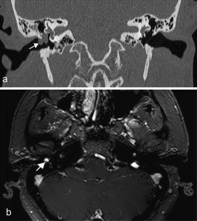
a Computed tomography scan of temporal bone, coronal view shows right side soft tissue mass involving medial part of the external canal, tympanic membrane, middle ear, and reaching the attic (white arrow). b Magnetic resonance imaging T1 with gadolinium contrast (axial view) shows hyperintense lesion in the right ear (white arrow)

Histopathologic examination of the specimen showed a cavernous hemangioma composed of dilated, thin walled vascular spaces of variable diameter filled with red blood cells, and lined by flat endothelial cells (H&E, ×200)
Similar articles
-
Cavernous hemangioma of the tympanic membrane and external ear canal.Am J Otolaryngol. 2007 May-Jun;28(3):180-3. doi: 10.1016/j.amjoto.2006.03.012. Am J Otolaryngol. 2007. PMID: 17499135 Review.
-
Cavernous hemangioma of the external ear canal.J Otolaryngol. 1987 Feb;16(1):40-2. J Otolaryngol. 1987. PMID: 3560305
-
Cavernous hemangioma of the external ear canal.Laryngoscope. 2002 Oct;112(10):1750-2. doi: 10.1097/00005537-200210000-00007. Laryngoscope. 2002. PMID: 12368608
-
[A rare case of endotympanic hemangioma].Acta Otorhinolaryngol Ital. 1994 Jul-Aug;14(4):457-62. Acta Otorhinolaryngol Ital. 1994. PMID: 7817750 Italian.
-
[Cavernous hemangioma of the tympanic membrane. Otoscopic image].Rev Invest Clin. 1999 Jan-Feb;51(1):39-42. Rev Invest Clin. 1999. PMID: 10344166 Review. Spanish.
Cited by
-
Resection of a Large Cavernous Hemangioma Following Preoperative Embolization in a Child's Temporal Bone.J Int Adv Otol. 2021 May;17(3):269-274. doi: 10.5152/iao.2021.8755. J Int Adv Otol. 2021. PMID: 34100755 Free PMC article.
-
Tissue engineering of human ear pinna.Cell Tissue Bank. 2022 Sep;23(3):441-457. doi: 10.1007/s10561-022-09991-7. Epub 2022 Feb 1. Cell Tissue Bank. 2022. PMID: 35103863 Review.
References
-
- Rahbar R, McGill TJ, Mulliken JB, et al. Vascular tumors and malformations of the head and neck. In: Cummings C, Flint PW, Haughey BH, et al., editors. Otolaryngology: head & neck surgery. 4. Philadelphia: Elsevier; 2005. pp. 4013–4020.
Publication types
MeSH terms
LinkOut - more resources
Full Text Sources
Other Literature Sources
