. 2015 Nov;1853(11 Pt A):2897-906.
doi: 10.1016/j.bbamcr.2015.08.012. Epub 2015 Aug 21.

Mitochondrial DNA has a pro-inflammatory role in AMD

Affiliations

Mitochondrial DNA has a pro-inflammatory role in AMD

Bernard Dib et al. Biochim Biophys Acta. 2015 Nov.

Erratum in

Abstract

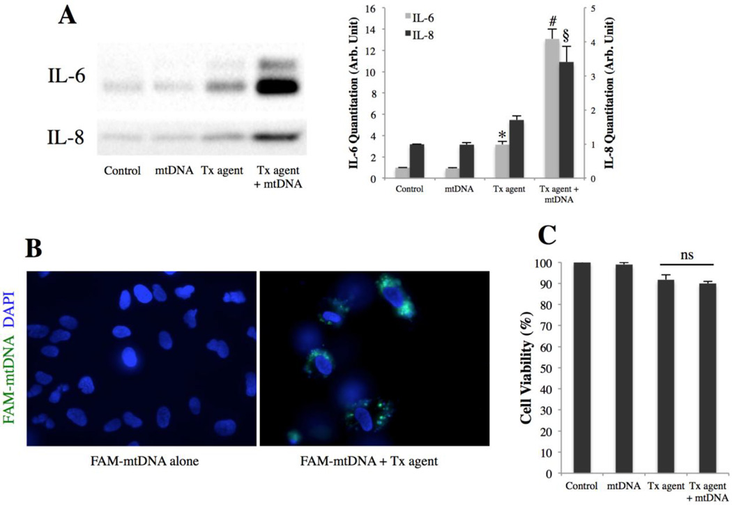
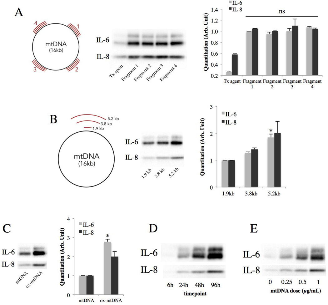
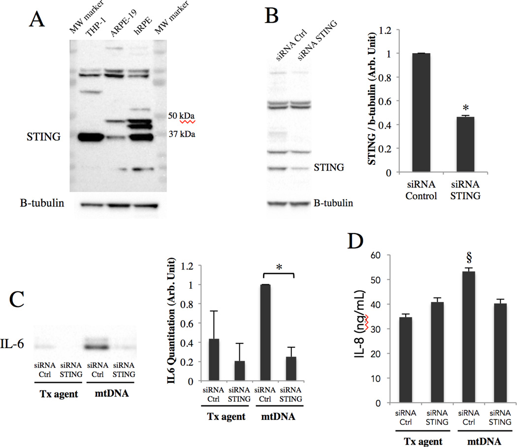
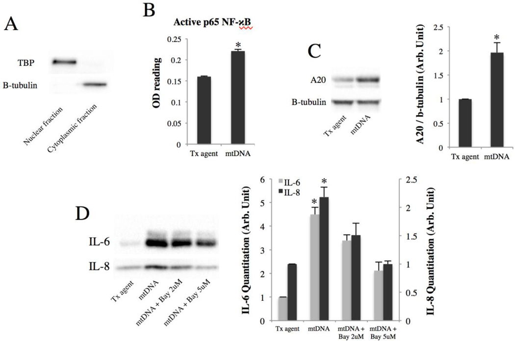
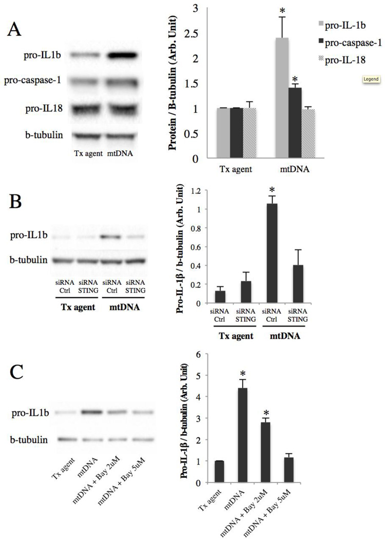
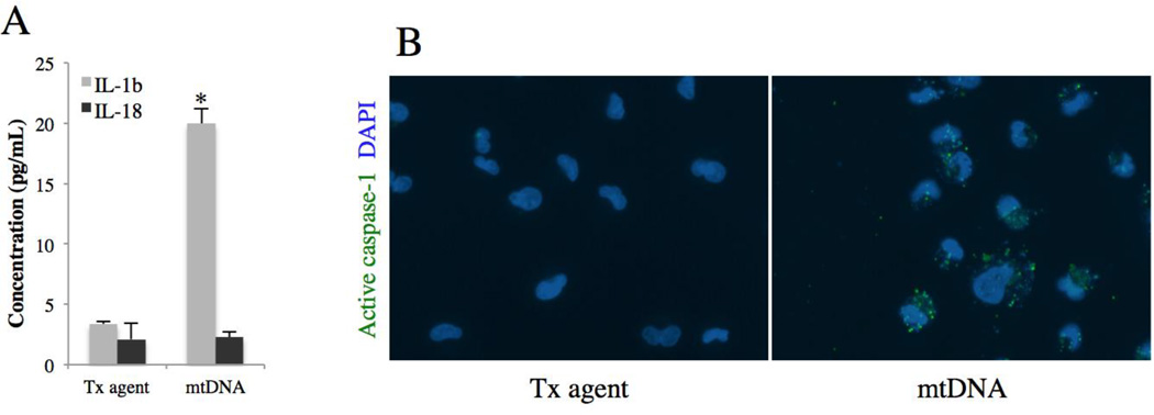
Age-related macular degeneration (AMD) is the leading cause of irreversible blindness in the elderly of industrialized nations, and there is increasing evidence to support a role for chronic inflammation in its pathogenesis. Mitochondrial DNA (mtDNA) has been recently reported to be pro-inflammatory in various diseases such as Alzheimer's and heart failure. Here, we report that intracellular mtDNA induces ARPE-19 cells to secrete inflammatory cytokines IL-6 and IL-8, which have been consistently associated with AMD onset and progression. The induction was dependent on the size of mtDNA, but not on specific sequence. Oxidative stress plays a major role in the development of AMD, and our findings indicate that mtDNA induces IL-6 and IL-8 more potently when oxidized. Cytokine induction was mediated by STING (Stimulator of Interferon Genes) and NF-κB as evidenced by abrogation of the cytokine response with the use of specific inhibitors (siRNA and BAY 11-7082, respectively). Finally, mtDNA primed the NLRP3 inflammasome. This study contributes to our understanding of the potential pro-inflammatory role of mtDNA in the pathogenesis of AMD.

Keywords: Age-related macular degeneration; Inflammation; Mitochondrial DNA; NLRP3 inflammasome; Retinal pigment epithelium.


Figures

Figure 1
Figure 1. mtDNA induces IL-6 and IL-8 secretion from ARPE-19 cells
(A) IL-6 and IL-8 secretion from ARPE-19 as determined by western blot of culture media 48h post-transfection with 1µg/mL mtDNA. Right, quantitation by densitometry. *p < 0.05 versus control and mtDNA groups; #p < 0.001 versus all other groups; §p < 0.01 versus all other groups. (B) ARPE-19 cells 24h after treatment with FAM-tagged mtDNA with or without transfection agent. mtDNA in green, DAPI in blue. (C) ARPE-19 viability determined by MTT 48h post-transfection with 1µg/mL mtDNA.
Figure 2
Figure 2. Oxidized and larger mtDNA fragments induce a stronger cytokine response
(A) IL-6 and IL-8 secretion from ARPE-19 cells 48h post-transfection with 1µg/mL of 2-kb mtDNA fragments of different sequences but similar sizes (schematic). Right, quantitation by densitometry. (B) IL-6 and IL-8 secretion from ARPE-19 cells 48h post-transfection with 1µg/mL of mtDNA fragments of different sizes (schematic). Right, quantitation by densitometry. *p < 0.05 versus all other groups. (C) IL-6 and IL-8 secretion from ARPE-19 cells 48h post-transfection with 1µg/mL of mtDNA or oxidized mtDNA. Right, quantitation by densitometry. *p < 0.01 versus control. (D) Time course of IL-6 and IL-8 induction by 1µg/mL of mtDNA. (E) Effect of mtDNA dose on IL-6 and IL-8 secretion at 48h.
Figure 3
Figure 3. mtDNA induces IL-6 and IL-8 secretion through STING
(A) STING immunoblotting of untreated ARPE-19 and primary human RPE lysates alongside a THP-1 lysate (positive control for STING). 40ug of total protein loaded per lane. Prominent band seen at STING’s predicted molecular weight (≈40kDa). (B) ARPE-19 cells were treated for a total of 96h with 20nM of either STING siRNA or control siRNA and lysates immunoblotted with STING monoclonal antibody. Right, quantitation by densitometry. *p < 0.001 versus control siRNA. (C&D) Along with mtDNA transfection, ARPE-19 cells were pre and co-treated with STING or control siRNA, and culture media were collected at 48h and subjected to western blot for IL-6 (C) and ELISA for IL-8 (D), as western blot did not provide sufficient resolution to study differences in IL-8 levels. *p < 0.05; §p < 0.01 versus all other groups.
Figure 4
Figure 4. mtDNA induces IL-6 and IL-8 secretion via NF-κB
(A) Nuclear fractionation efficiency verified with tata-binding protein (TBP) and b-tubulin. (B) p65 NF-κB ELISA performed on nuclear isolates of ARPE-19 cells 24h post-transfection with mtDNA. *p < 0.01 versus control. (C) A20 expression level 48h post-transfection of mtDNA. Right, quantitation by densitometry. *p < 0.05 versus control. (D) ARPE-19 cells were pre-incubated for 30 minutes with an irreversible NF-κB inhibitor (Bay 11–7082) before mtDNA transfection. After 48h, culture media were collected and immunoblotted for IL-6 and IL-8. Right, quantitation by densitometry. *p < 0.05 versus control and [mtDNA + Bay 5uM] groups.
Figure 5
Figure 5. mtDNA primes the NLRP3 inflammasome through STING and NF-κB
(A) Immunoblots of ARPE-19 lysates 48h after mtDNA transfection. Right, quantitation by densitometry. *p < 0.05 versus control. (B) Along with mtDNA transfection, ARPE-19 cells were pre and co-treated with STING or control siRNA, and lysates were collected at 48h and immunoblotted for pro-IL-1β. Right, quantitation by densitometry. *p < 0.05 versus all other groups. (C) ARPE-19 cells were pre-incubated for 30 minutes with an irreversible NF-κB inhibitor (Bay 11–7082) before mtDNA transfection. After 48h, lysates were collected and immunoblotted for pro-IL-1β. Right, quantitation by densitometry. *p < 0.05 versus all other groups.
Figure 6
Figure 6. mtDNA and the NLRP3 inflammasome in the RPE
(A) IL-1β and IL-18 ELISA on culture media of ARPE-19 cells 48h post-transfection with mtDNA. Results are mean ± SEM, n=4. *p < 0.01 versus control. (B) ARPE-19 caspase-1 activity assessed 24h after mtDNA transfection using fluorescent FLICA probe FAM-YVAD-FMK.

References

    1. Reeve AK, Krishnan KJ, Turnbull D. Mitochondrial DNA mutations in disease, aging, and neurodegeneration. Ann N Y Acad Sci. 2008;1147:21–29. - PubMed
    1. Currais A. Ageing and inflammation – A central role for mitochondria in brain health and disease. Ageing Res Rev. 2015:1–13. - PubMed
    1. Blasiak J, Glowacki S, Kauppinen A, Kaarniranta K. Mitochondrial and nuclear DNA damage and repair in age-related macular degeneration. Int J Mol Sci. 2013;14:2996–3010. - PMC - PubMed
    1. Jarrett SG, Lewin AS, Boulton ME. The importance of Mitochondria in age-related and inherited eye disorders. Ophthalmic Res. 2010;44:179–190. - PMC - PubMed
    1. Karunadharma PP, Nordgaard CL, Olsen TW, Ferrington Da. Mitochondrial DNA damage as a potential mechanism for age-related macular degeneration. Invest Ophthalmol Vis Sci. 2010;51(11):5470–5479. - PMC - PubMed

Publication types

MeSH terms