

Extraction of Peptidoglycan from L. paracasei subp. Paracasei X12 and Its Preliminary Mechanisms of Inducing Immunogenic Cell Death in HT-29 Cells
- PMID: 26305246
- PMCID: PMC4581339
- DOI: 10.3390/ijms160820033
Extraction of Peptidoglycan from L. paracasei subp. Paracasei X12 and Its Preliminary Mechanisms of Inducing Immunogenic Cell Death in HT-29 Cells
Abstract
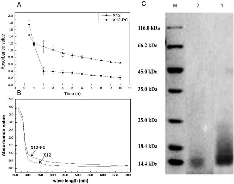
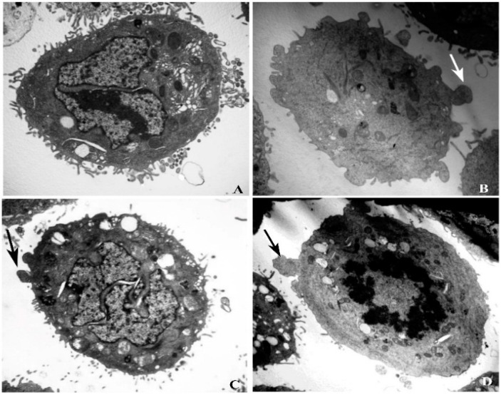
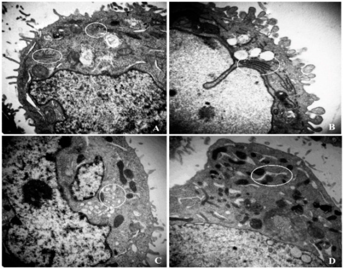
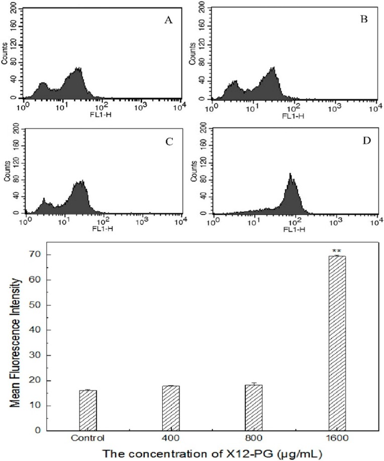
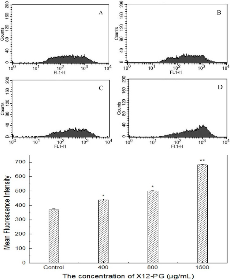
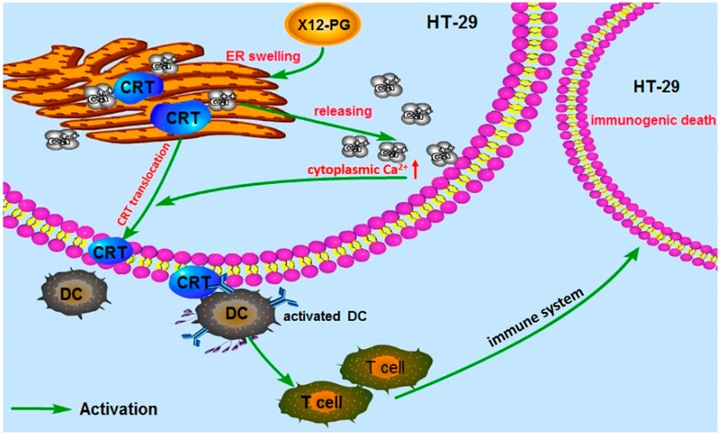
L. paracasei subp. paracasei X12 was previously isolated from a Chinese traditional fermented cheese with anticancer activities and probiotic potential. Herein, the integral peptidoglycan (X12-PG) was extracted by a modified trichloroacetic acid (TCA) method. X12-PG contained the four representative amino acids Asp, Glu, Ala and Lys, and displayed the similar lysozyme sensitivity, UV-visible scanning spectrum and molecular weight as the peptidoglycan standard. X12-PG could induce the production of apoptotic bodies observed by transmission electron microscopy (TEM). X12-PG could significantly induced the translocation of calreticulin (CRT) and the release of high mobility group box 1 protein (HMGB1), the two notable hallmarks of immunogenic cell death (ICD), with the endoplastic reticulum (ER) damaged and subsequently intracellular [Ca(2+)] elevated. Our findings implied that X12-PG could induce the ICD of HT-29 cells through targeting at the ER. The present results may enlighten the prospect of probiotics in the prevention of colon cancer.
Keywords: calcium; calreticulin; immunogenic cell death; peptidoglycan.
Figures
Similar articles
-
Lactobacillus paracasei subsp. paracasei M5L induces cell cycle arrest and calreticulin translocation via the generation of reactive oxygen species in HT-29 cell apoptosis.Food Funct. 2015 Jul;6(7):2257-65. doi: 10.1039/c5fo00248f. Food Funct. 2015. PMID: 26068306
-
Lactobacillus paracasei subsp. paracasei X12 Strain Induces Apoptosis in HT-29 Cells through Activation of the Mitochondrial Pathway.Nutrients. 2023 Apr 28;15(9):2123. doi: 10.3390/nu15092123. Nutrients. 2023. PMID: 37432295 Free PMC article.
-
Whole Peptidoglycan Extracts from the Lactobacillus paracasei subsp. paracasei M5 Strain Exert Anticancer Activity In Vitro.Biomed Res Int. 2018 Feb 7;2018:2871710. doi: 10.1155/2018/2871710. eCollection 2018. Biomed Res Int. 2018. PMID: 29568745 Free PMC article.
-
Immunogenic cell death in anticancer chemotherapy and its impact on clinical studies.Cancer Lett. 2018 Dec 1;438:17-23. doi: 10.1016/j.canlet.2018.08.028. Epub 2018 Sep 11. Cancer Lett. 2018. PMID: 30217563 Review.
-
Multimodal immunogenic cancer cell death as a consequence of anticancer cytotoxic treatments.Cell Death Differ. 2014 Jan;21(1):39-49. doi: 10.1038/cdd.2013.84. Epub 2013 Jul 5. Cell Death Differ. 2014. PMID: 23832118 Free PMC article. Review.
Cited by
-
Modulating Endoplasmic Reticulum Stress in Gastrointestinal Cancers: Insights from Traditional Chinese Medicine.Pharmaceuticals (Basel). 2024 Nov 27;17(12):1599. doi: 10.3390/ph17121599. Pharmaceuticals (Basel). 2024. PMID: 39770441 Free PMC article. Review.
-
Peptidoglycan derived from Lactobacillus rhamnosus MLGA up-regulates the expression of chicken β-defensin 9 without triggering an inflammatory response.Innate Immun. 2020 Nov;26(8):733-745. doi: 10.1177/1753425920949917. Epub 2020 Aug 26. Innate Immun. 2020. PMID: 32847443 Free PMC article.
-
Appraisal of postbiotics in cancer therapy.Front Pharmacol. 2024 Sep 20;15:1436021. doi: 10.3389/fphar.2024.1436021. eCollection 2024. Front Pharmacol. 2024. PMID: 39372197 Free PMC article. Review.
-
Soluble peptidoglycan fragments produced by Limosilactobacillus fermentum with antiproliferative activity are suitable for potential therapeutic development: A preliminary report.Front Mol Biosci. 2023 Feb 15;10:1082526. doi: 10.3389/fmolb.2023.1082526. eCollection 2023. Front Mol Biosci. 2023. PMID: 36876040 Free PMC article.
-
Anti-cancer Substances and Safety of Lactic Acid Bacteria in Clinical Treatment.Front Microbiol. 2021 Oct 12;12:722052. doi: 10.3389/fmicb.2021.722052. eCollection 2021. Front Microbiol. 2021. PMID: 34721321 Free PMC article. Review.
References
-
- McCoy W., Mason J.M., 3rd. Enterococcal endocarditis associated with carcinoma of the sigmoid; report of a case. J. Med. Assoc. State Ala. 1951;21:162–166. - PubMed
Publication types
MeSH terms
Substances
LinkOut - more resources
Full Text Sources
Other Literature Sources
Research Materials
Miscellaneous
