. 2015 Aug 25;10(8):e0135986.
doi: 10.1371/journal.pone.0135986. eCollection 2015.

A Multiplex Assay for Detection of Staphylococcal and Streptococcal Exotoxins

Affiliations

A Multiplex Assay for Detection of Staphylococcal and Streptococcal Exotoxins

Preeti Sharma et al. PLoS One. .

Abstract

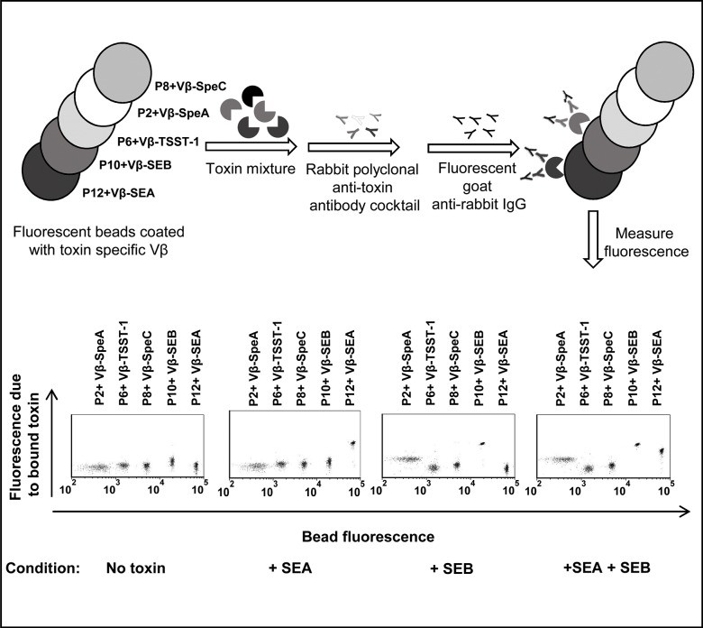
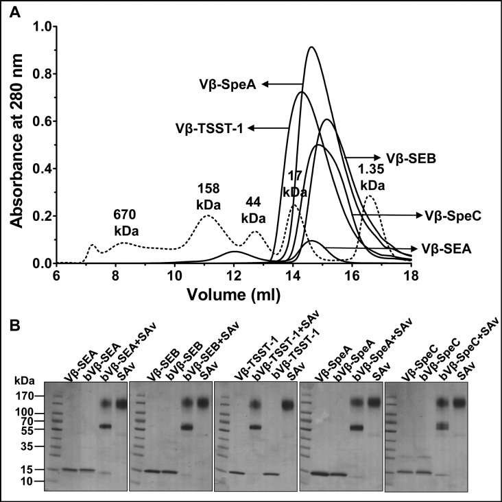
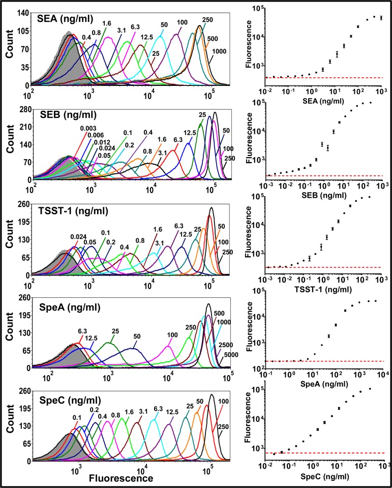
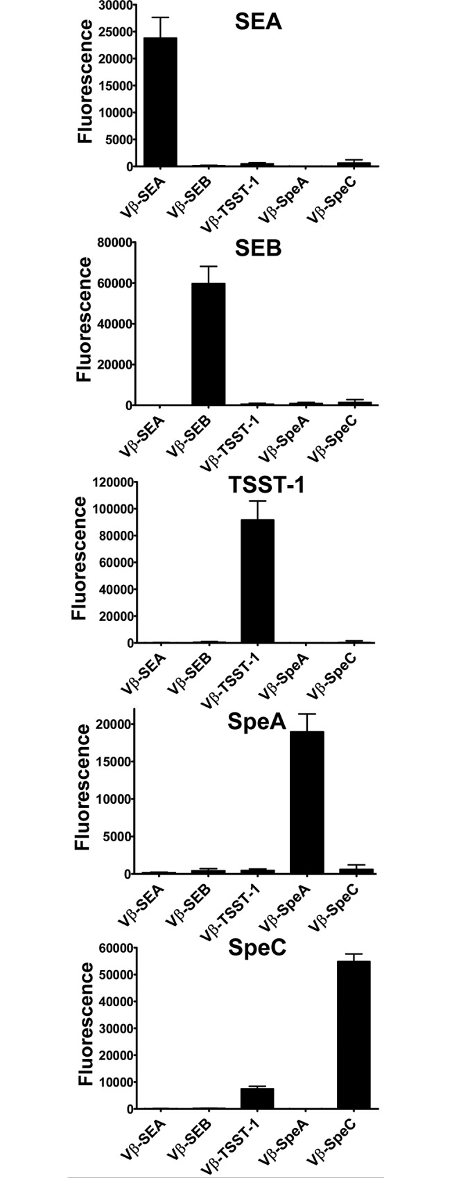
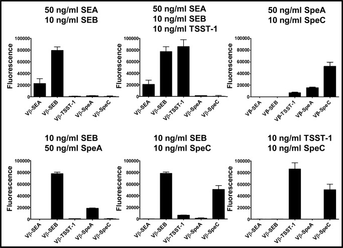
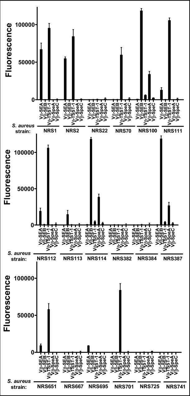
Staphylococcal and streptococcal exotoxins, also known as superantigens, mediate a range of diseases including toxic shock syndrome, and they exacerbate skin, pulmonary and systemic infections caused by these organisms. When present in food sources they can cause enteric effects commonly known as food poisoning. A rapid, sensitive assay for the toxins would enable testing of clinical samples and improve surveillance of food sources. Here we developed a bead-based, two-color flow cytometry assay using single protein domains of the beta chain of T cell receptors engineered for high-affinity for staphylococcal (SEA, SEB and TSST-1) and streptococcal (SpeA and SpeC) toxins. Site-directed biotinylated forms of these high-affinity agents were used together with commercial, polyclonal, anti-toxin reagents to enable specific and sensitive detection with SD50 values of 400 pg/ml (SEA), 3 pg/ml (SEB), 25 pg/ml (TSST-1), 6 ng/ml (SpeA), and 100 pg/ml (SpeC). These sensitivities were in the range of 4- to 80-fold higher than achieved with standard ELISAs using the same reagents. A multiplex format of the assay showed reduced sensitivity due to higher noise associated with the use of multiple polyclonal agents, but the sensitivities were still well within the range necessary for detection in food sources or for rapid detection of toxins in culture supernatants. For example, the assay specifically detected toxins in supernatants derived from cultures of Staphylococcus aureus. Thus, these reagents can be used for simultaneous detection of the toxins in food sources or culture supernatants of potential pathogenic strains of Staphylococcus aureus and Streptococcus pyogenes.


Conflict of interest statement

Competing Interests: David M. Kranz co-founded a company called ImmuVen that acquired rights from the University of Illinois for some of the T cell receptors engineered in his lab. Ningyan Wang, Adam S. Chervin, Cheryl L. Quinn and Jennifer D. Stone were employed by ImmuVen. This does not alter the authors' adherence to PLOS ONE policies on sharing data and materials.

Figures

Fig 1
Fig 1. Schematic diagram and representative flow cytometry data of the multiplex assay.
Biotinylated, high-affinity Vβ proteins (Vβ-SEA, Vβ-SEB, Vβ-TSST-1, Vβ-SpeA and Vβ-SpeC) were immobilized on streptavidin-coated fluorescent beads (P12, P10, P6, P2 and P8 respectively, each having unique fluorescence in the FL-1 channel, λemission = 525 nm). For detecting the presence of one or more toxins (SEA and/or SEB in this example), a mixture of Vβ immobilized-beads was added to an unknown sample. Toxins captured by the Vβ-immobilized beads were detected by rabbit polyclonal anti-toxin antibodies followed by goat-anti rabbit IgG labeled with Alexa fluor 647 (λemission = 688 nm in FL-4 channel). Scatter plots in the absence or presence of toxin(s) are shown in the lower panel. An increase in fluorescence emitted by the beads in the FL-4 channel (Y-axis) indicates the presence of toxin(s) in the unknown sample. Since each class of beads emits distinct fluorescence in FL-1 channel (X-axis), identity of the toxin present is established by identifying the specific, Vβ-immobilized bead(s) undergoing an increase in fluorescence in the FL-4 channel (Y-axis). In the presence of SEA, only the P12 beads (immobilized with Vβ-SEA) exhibited an increase in fluorescence along Y-axis. In the presence of SEB, P10 beads (immobilized with Vβ-SEB) as well as P2 beads (immobilized with Vβ-SpeA) exhibited increases in fluorescence along Y-axis, due to low affinity of Vβ-SpeA toward SEB. Accordingly, in the presence of SEA and SEB, Vβ-immobilized P12, P10 and P2 beads exhibited increases in fluorescence along Y-axis.
Fig 2
Fig 2. Expression and biotinylation of high-affinity Vβ proteins.
(A) Purification of monomeric fractions of refolded, high-affinity Vβ proteins by size-exclusion chromatography. Dashed line indicates the molecular weight standards. (B) Gel-shift assay for monitoring biotinylation of high-affinity Vβ proteins (Vβ). Disappearance of biotinylated-Vβ (bVβ) (~15kDa band) in the presence of streptavidin (SAv) was indicative of biotinylation.
Fig 3
Fig 3. Sensitivity of toxin detection by Vβ-immobilized beads in singleplex assays.
Each biotinylated, high-affinity Vβ protein (Vβ-SEA, Vβ-SEB, Vβ-TSST-1, Vβ-SpeA and Vβ-SpeC) immobilized on individual streptavidin-coated fluorescent beads was subjected to cognate, toxin binding titration (SEA, SEB, TSST-1, SpeA and SpeC respectively). Toxins captured by the Vβ-immobilized beads were detected with rabbit polyclonal anti-toxin antibodies (anti-SEA, anti-SEB, anti-TSST-1, anti-SEB and anti-SpeC respectively) followed by goat-anti rabbit IgG labeled with Alexa fluor 647. Flow cytometry histograms for each binding titration are shown, with fluorescence arising due to toxin binding on X-axis. Fluorescence in the absence of toxin is represented by gray (filled) trace on each histogram. Median fluorescence units from the histograms were used to generate binding curves, shown on the right. Red, dashed line indicates fluorescence in the absence of toxin. Data shown are representative of experiments performed in triplicate.
Fig 4
Fig 4. Multiplex assay in the presence of one toxin.
Solutions containing 50 ng/ml SEA, 10 ng/ml SEB, 10 ng/ml TSST-1, 50 ng/ml SpeA or 10 ng/ml SpeC were tested in multiplex assays. Fluorescence emitted by each Vβ-immobilized bead due to toxin binding, was plotted on bar graphs shown. In all cases, the toxins were detected by the beads immobilized with the high-affinity Vβ engineered for that toxin. The error-bars represent standard deviations from two independent experiments. Similar data (but with higher background) were obtained in other experiments where unoccupied sites on biotin-Vβ immobilized-beads were not blocked with excess biotin (S4 Fig).
Fig 5
Fig 5. Multiplex assay in the presence of a mixture of toxins.
Solutions containing a mixture of two or more toxins were tested in multiplex assays, at the indicated concentrations. Fluorescence emitted by each Vβ-immobilized bead due to toxin binding, was plotted on bar graphs shown. The error-bars represent standard deviations from two independent experiments. Similar data (but with higher background) were obtained in other experiments where unoccupied sites on biotin-Vβ immobilized-beads were not blocked with excess biotin (S5 Fig).
Fig 6
Fig 6. Multiplex assay with supernatants from cultures of various strains of Staphylococcus aureus.
Supernatants (diluted 1:4) from cultures of 18 strains of Staphylococcus aureus obtained from the NARSA repository, were tested in a multiplex assay to determine their toxin expression profile. Fluorescence emitted by each Vβ-immobilized bead due to toxin binding, was plotted on bar graphs shown. Toxin(s) secreted by each strain was determined as an increase in fluorescence signal, compared to the background. The error-bars represent standard deviations from two independent experiments. Similar data (but with higher background) were obtained in other experiments where unoccupied sites on biotin-Vβ immobilized-beads were not blocked with excess biotin (S6 Fig).

References

    1. Salgado-Pabon W, Breshears L, Spaulding AR, Merriman JA, Stach CS, Horswill AR, et al. Superantigens are critical for Staphylococcus aureus Infective endocarditis, sepsis, and acute kidney injury. MBio. 2013;4(4). - PMC - PubMed
    1. Spaulding AR, Salgado-Pabon W, Kohler PL, Horswill AR, Leung DY, Schlievert PM. Staphylococcal and streptococcal superantigen exotoxins. Clin Microbiol Rev. 2013;26(3):422–447. 10.1128/CMR.00104-12 - DOI - PMC - PubMed
    1. McCormick JK, Yarwood JM, Schlievert PM. Toxic shock syndrome and bacterial superantigens: an update. Annu Rev Microbiol. 2001;55:77–104. - PubMed
    1. Asao T, Kumeda Y, Kawai T, Shibata T, Oda H, Haruki K, et al. An extensive outbreak of staphylococcal food poisoning due to low-fat milk in Japan: estimation of enterotoxin A in the incriminated milk and powdered skim milk. Epidemiology and Infection. 2003;130(1):33–40. - PMC - PubMed
    1. Evenson ML, Hinds MW, Bernstein RS, Bergdoll MS. Estimation of human dose of staphylococcal enterotoxin A from a large outbreak of staphylococcal food poisoning involving chocolate milk. Int J Food Microbiol. 1988;7(4):311–316. - PubMed

Publication types

MeSH terms