

The olive leaf extract oleuropein exerts protective effects against oxidant-induced cell death, concurrently displaying pro-oxidant activity in human hepatocarcinoma cells
- PMID: 26305493
- PMCID: PMC6837481
- DOI: 10.1179/1351000215Y.0000000039
The olive leaf extract oleuropein exerts protective effects against oxidant-induced cell death, concurrently displaying pro-oxidant activity in human hepatocarcinoma cells
Abstract
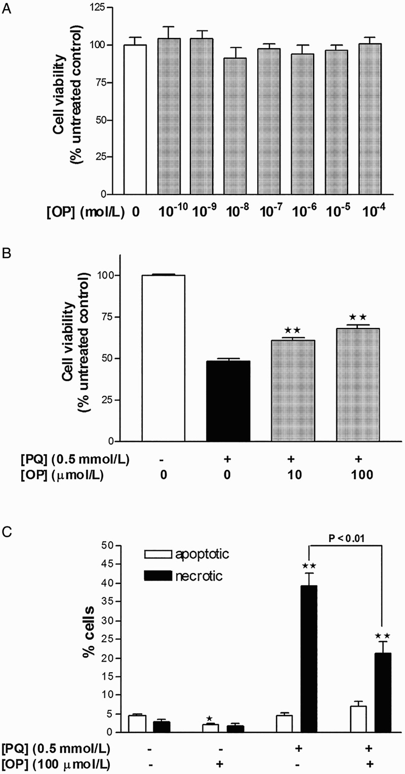
Objectives: Oleuropein (OP), the predominant natural constituent of leaves of the olive tree, exerts anti-inflammatory and antioxidant effects. The purpose of this study was to assess the protective effects of OP under the conditions of paraquat (PQ)-induced oxidative stress in vitro, using the human hepatocarcinoma cell line, HepG2.
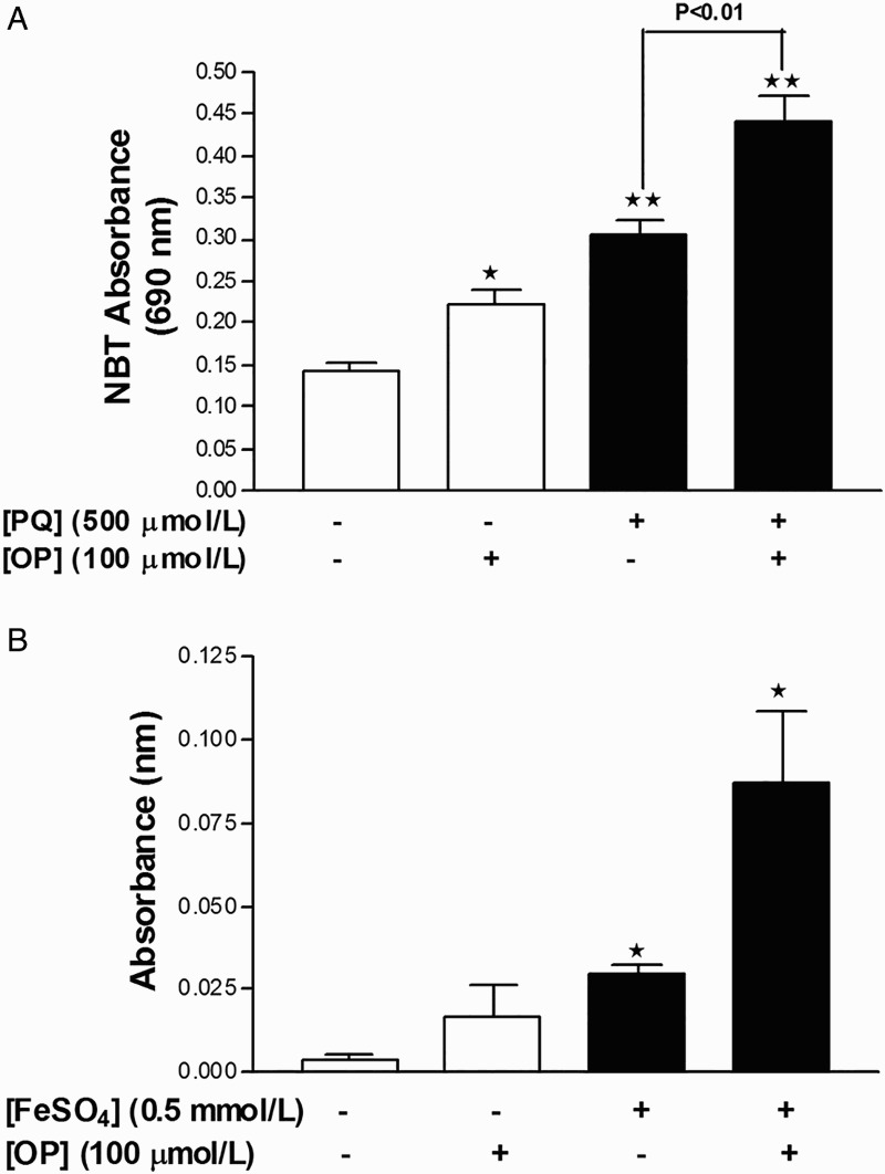
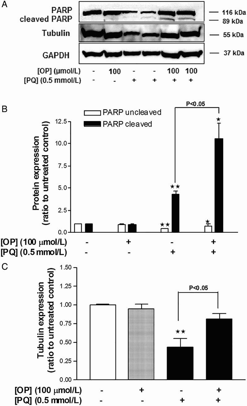
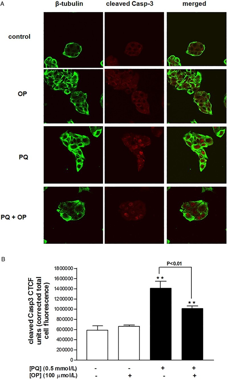
Methods: Cell viability and death were determined using the 3-(4,5-dimethylthiazol-2-yl)-2,5-diphenyltetrazolium bromide assay and 4',6-diamidino-2-phenylindole-propidium iodide staining, respectively. Superoxide anion and lipid peroxidation levels were evaluated using nitroblue tetrazolium and thiobarbituric acid-reactive substances assays, respectively. Apoptosis was assessed by measuring poly(ADP-ribose) polymerase (PARP) and caspase-3 (Casp-3) cleavage via immunoblotting and immunofluorescence analyses.
Results: PQ induced a decrease in cellular viability by promoting necrosis through a mechanism involving superoxide generation and nuclear translocation of cleaved Casp-3. Co-treatment with OP afforded significant protection against the suppressive effects of PQ, as evident from increased cell viability, reduction of Casp-3 immunofluorescence, and normalization of β-tubulin expression levels. Unexpectedly, these OP-mediated protective effects were associated with increased superoxide and malondialdehyde generation and PARP cleavage.
Discussion: OP protects HepG2 cells against PQ-induced necrosis by suppressing Casp-3 cleavage while concomitantly acting as a pro-oxidant agent. This paradoxical mechanism of action of OP requires further investigation.
Keywords: Apoptosis; Hydroxyl radical; Lipid peroxidation; Necrosis; Oleuropein; Oxidative stress; Paraquat; Superoxide.
Figures
Similar articles
-
Polyphenolic extracts from Olea europea L. protect against cytokine-induced β-cell damage through maintenance of redox homeostasis.Rejuvenation Res. 2011 Jun;14(3):325-34. doi: 10.1089/rej.2010.1111. Epub 2011 Jul 11. Rejuvenation Res. 2011. PMID: 21745095
-
Inhibition of 6-hydroxydopamine-induced PC12 cell apoptosis by olive (Olea europaea L.) leaf extract is performed by its main component oleuropein.Rejuvenation Res. 2013 Apr;16(2):134-42. doi: 10.1089/rej.2012.1384. Rejuvenation Res. 2013. PMID: 23394606
-
Olive leaf extracts protect cardiomyocytes against 4-hydroxynonenal-induced toxicity in vitro: comparison with oleuropein, hydroxytyrosol, and quercetin.Planta Med. 2014 Aug;80(12):984-92. doi: 10.1055/s-0034-1382881. Epub 2014 Aug 6. Planta Med. 2014. PMID: 25098929
-
Oleuropein Is Responsible for the Major Anti-Inflammatory Effects of Olive Leaf Extract.J Med Food. 2018 Mar;21(3):302-305. doi: 10.1089/jmf.2017.0070. Epub 2017 Nov 3. J Med Food. 2018. PMID: 29099642
-
Comparative Study on Beneficial Effects of Hydroxytyrosol- and Oleuropein-Rich Olive Leaf Extracts on High-Fat Diet-Induced Lipid Metabolism Disturbance and Liver Injury in Rats.Biomed Res Int. 2020 Jan 8;2020:1315202. doi: 10.1155/2020/1315202. eCollection 2020. Biomed Res Int. 2020. PMID: 31998777 Free PMC article.
Cited by
-
Encapsulation of Oleuropein in Nanostructured Lipid Carriers: Biocompatibility and Antioxidant Efficacy in Lung Epithelial Cells.Pharmaceutics. 2020 May 6;12(5):429. doi: 10.3390/pharmaceutics12050429. Pharmaceutics. 2020. PMID: 32384817 Free PMC article.
-
Extra Virgin Olive Oil Phenolic Extract on Human Hepatic HepG2 and Intestinal Caco-2 Cells: Assessment of the Antioxidant Activity and Intestinal Trans-Epithelial Transport.Antioxidants (Basel). 2021 Jan 15;10(1):118. doi: 10.3390/antiox10010118. Antioxidants (Basel). 2021. PMID: 33467632 Free PMC article.
-
Comparison of the therapeutic effect of the Persian Medicine Protocol with the common treatment of chronic rhinosinusitis: a randomized clinical trial.Electron Physician. 2018 Jul 25;10(7):7017-7027. doi: 10.19082/7017. eCollection 2018 Jul. Electron Physician. 2018. PMID: 30128092 Free PMC article.
-
Virgin Olive Oil Extracts Reduce Oxidative Stress and Modulate Cholesterol Metabolism: Comparison between Oils Obtained with Traditional and Innovative Processes.Antioxidants (Basel). 2020 Aug 27;9(9):798. doi: 10.3390/antiox9090798. Antioxidants (Basel). 2020. PMID: 32867071 Free PMC article.
-
Assessment of the Efficacy of Olive Leaf (Olea europaea L.) Extracts in the Treatment of Colorectal Cancer and Prostate Cancer Using In Vitro Cell Models.Molecules. 2021 Jul 3;26(13):4069. doi: 10.3390/molecules26134069. Molecules. 2021. PMID: 34279409 Free PMC article.
References
-
- Filik L, Ozyilkan O. Olive-oil consumption and cancer risk. Eur J Clin Nutr 2003;57(1):191. - PubMed
-
- Cristina F. Mediterranean diet health benefits may be due to a synergistic combination of phytochemicals and fatty-acids. BMJ 2005;331(7508):E366. - PubMed
-
- de Bock M, Thorstensen EB, Derraik JG, Henderson HV, Hofman PL, Cutfield WS. Human absorption and metabolism of oleuropein and hydroxytyrosol ingested as olive (Olea europaea L.) leaf extract. Mol Nutr Food Res 2013;57(11):2079–85. - PubMed
MeSH terms
Substances
LinkOut - more resources
Full Text Sources
Other Literature Sources
Research Materials
Miscellaneous