. 2015 Sep 29;6(29):28151-63.
doi: 10.18632/oncotarget.4661.

LincRNA-p21 activates endoplasmic reticulum stress and inhibits hepatocellular carcinoma

Affiliations

LincRNA-p21 activates endoplasmic reticulum stress and inhibits hepatocellular carcinoma

Ning Yang et al. Oncotarget. .

Abstract

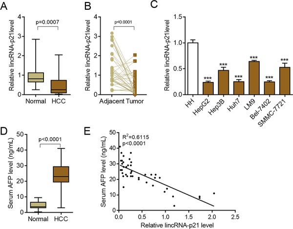
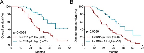
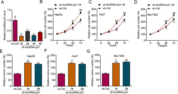
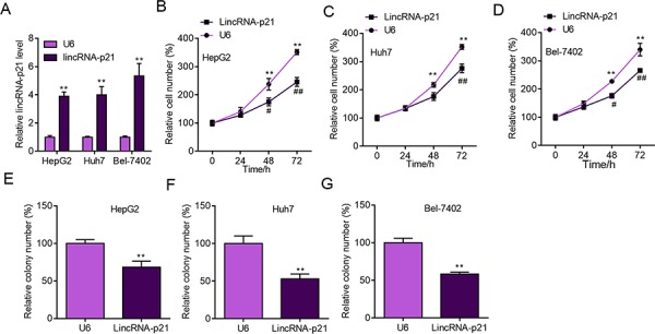
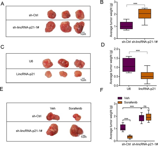
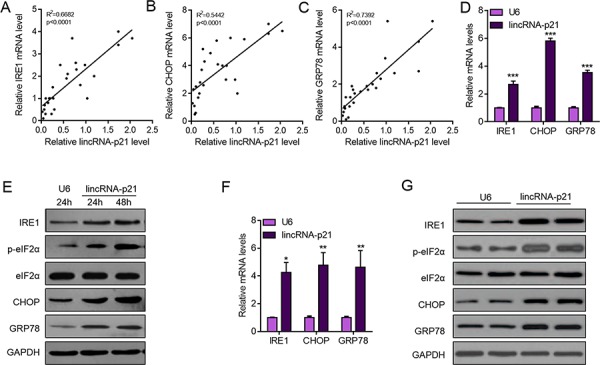
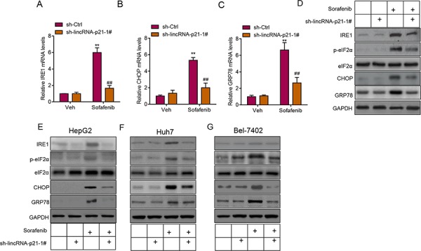
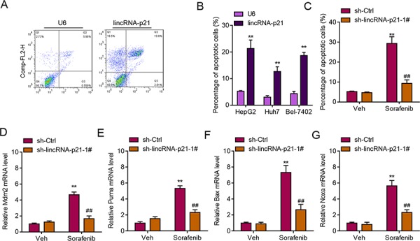
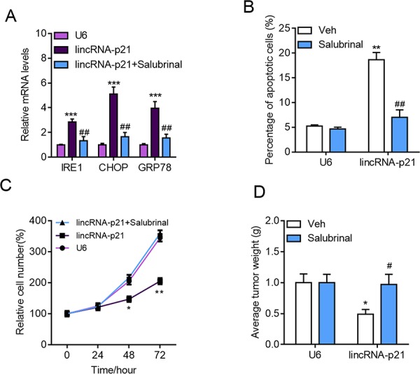
LincRNA-p21 is a downstream long non-coding RNA (lncRNA) transcript of p53. LincRNA-p21 serves as a repressor in p53-dependent transcriptional responses and participates in diverse biological processes, including apoptosis, cell cycle, metabolism and pluripotency. However, the role of lincRNA-p21 in human hepatocellular carcinoma remains to be defined. Here in this work, we demonstrated that lincRNA-p21 acted as a tumor suppressive lncRNA in human hepatocellular carcinoma. We firstly found the downregulation of lincRNA-p21 level in human hepatocellular carcinoma tissues, and showed that low expression of lincRNA-p21 was associated with high disease stage and predicted poor survival. Further we showed that lincRNA-p21 knockdown promoted proliferation and colony formation of HepG2, Huh7 and Bel-7042 cells in vitro, while lincRNA-p21 overexpression obtained oppose results. Using tumor xenograft experiments, we also demonstrated that lincRNA-p21 inhibited HepG2 cell growth in vivo and lincRNA-p21 contributed to sorafenib-induced growth regression of HepG2 cell in vivo. Further mechanism analysis revealed that lincRNA-p21 promoted ER stress both in vitro and in vivo, which facilitated apoptosis of hepatocellular carcinoma cells. Finally, we demonstrated that ER stress accounted for lincRNA-p21 effects on apoptosis, proliferation and in vivo growth of hepatocellular carcinoma. These findings implicate that lincRNA-p21 is a potential prognostic factor and therapeutic target for human hepatocellular carcinoma.

Keywords: ER stress; hepatocellular carcinoma; lincRNA-p21; sorafenib.


Conflict of interest statement

CONFLICTS OF INTEREST

None.

Figures

Figure 1
Figure 1. LincRNA-p21 is down-regulated in hepatocellular carcinoma
A. LincRNA-p21 expression in human hepatocellular carcinoma (HCC, n = 42) or control normal liver tissues (n = 22). B. LincRNA-p21 levels in adjacent normal liver tissues and tumor tissues in HCC. n = 40. Paired Student's t test was performed to analyze the data. C. LincRNA-p21 expression in normal human hepatocytes (HH) and liver cancer cell lines (HepG2, Hep3B, Huh7, LM9, Bel-7042, SMMC-7721). ***p < 0.001 vs. HH D. Serum levels of alpha-fetoprotein (AFP) in HCC patients (n = 42) or normal tissue donors (n = 24). E. Correlation between serum AFP and lincRNA-p21 level (n = 42).
Figure 2
Figure 2. LincRNA-p21 low expression predicts poor survival
The patients were divided to lincRNA-p21 high group and lincRNA-p21 low group by the mean value of total 70 HCC patients. A. Low lincRNA-p21 level predicts poor overall survival in HCC patients. B. Low lincRNA-p21 predicts poor disease-free survival in HCC patients.
Figure 3
Figure 3. LincRNA-p21 knockdown facilitates proliferation and colony formation of liver cancer cells
A. Lentivirus-mediated lincRNA-p21 knockdown by short hairpin RNAs (shRNAs) in HepG2 cells. B–D. LincRNA-p21 knockdown promotes proliferation of HepG2 (B), Huh7 (C), and Bel-7042 (D) cells. **p < 0.01 vs. sh-Ctrl 0 day; #p < 0.05 and ##p < 0.01 vs. sh-Ctrl of the corresponding time points. E–G. LincRNA-p21 knockdown promotes colony formation of HepG2 (E), Huh7 (F), and Bel-7042 (G) cells. **p < 0.01 vs. sh-Ctrl.
Figure 4
Figure 4. LincRNA-p21 overexpression inhibits liver cancer cell proliferation and colony formation
A. Lentivirus-mediated lincRNA-p21 overexpression in HepG2, Huh7 and Bel-7402 cells. The level of lincRNA-p21 was checked 24 hours post lentivirus infection. **p < 0.01 vs. U6. $$p < 0.01 vs. U6. ##p < 0.01 vs. U6. &&p < 0.01 vs. U6. B–D. LincRNA-p21 overexpression inhibits proliferation of HepG2 (B), Huh7 (C), and Bel-7402 (D) cells. *p < 0.05 and **p < 0.01 vs U6 0 day; #p < 0.05 and ##p < 0.01 vs U6 of the corresponding time points. E–G. LincRNA-p21 overexpression inhibits colony formation of HepG2 (E), Huh7 (F) and Bel-7402 (G) cells. **p < 0.01 vs U6.
Figure 5
Figure 5. lincRNA-p21 regulates hepatocarcinoma growth in vivo
A. Representative photograph showing tumor size of sh-Ctrl and sh-LincRNA-p21 groups. HepG2 cells were infected with lentivirus-mediated sh-Ctrl or sh-LincRNA-p21 and tumor xenograft experiment was performed. The mice were sacrificed four weeks after tumor implantation. B. Quantitative data for tumor weight in sh-Ctrl and sh-lincRNA-p21 groups. N = 10 in each group. C. Representative photograph showing tumor size of U6 and lincRNA-p21 groups. HepG2 cells were infected with lentivirus overexpressing U6 or human lincRNA-p21 and tumor xenograft experiment was performed. The mice were sacrificed four weeks after tumor implantation. D. Quantitative data for tumor weight in U6 and lincRNA-p21 groups. N = 10 in each group. E–F. LincRNA-p21 mediates sorafenib-induced inhibition of HepG2 cell growth in vivo. Sorafenib (30 mg/kg/d) was administered in 100 μL by intraperitoneal injection on day three after tumor implantation. N = 10 in each group in (F) ***p < 0.001.
Figure 6
Figure 6. LincRNA-p21 overexpression activates ER stress in hepatocarcinoma
A–C. LincRNA-p21 level is positively correlated with mRNA levels of ER stress markers (IRE1, CHOP, and GRP78) in human hepatocellular carcinoma. N = 26 in each group. D. LincRNA-p21 overexpression promotes the mRNA levels of ER stress markers (IRE1, CHOP, and GRP78) in HepG2 cells. HepG2 cells were infected with lentivirus overexpressing U6 or lincRNA-p21 for 48 hours. ***p < 0.001 vs. U6. E. Western blot showing lincRNA-p21 promotes the activation of ER stress in HepG2 cells. F. LincRNA-p21 overexpression facilitates the mRNA levels of ER stress markers in vivo. Tumor xenograft experiment was performed using HepG2 cells. The mice were sacrificed four weeks after tumor implantation. *p < 0.05 and **p < 0.01 vs. U6. N = 5 in each group. G. Representative western blot showing lincRNA-p21 promotes the activation of ER stress in vivo. Tumor xenograft experiment was performed using HepG2 cells. The mice were sacrificed four weeks after tumor implantation.
Figure 7
Figure 7. LincRNA-p21 contributes to sorafenib-induced ER stress in vivo
A–C. LincRNA-p21 knockdown inhibits sorafenib-induced expression of ER stress markers in vivo. Tumor xenograft experiment was performed using HepG2 cells. The mice were sacrificed four weeks after tumor implantation. Sorafenib (30 mg/kg/d) was administered in 100 μL by intraperitoneal injection on day three after tumor implantation. **p < 0.01 vs. sh-Ctrl + Vehicle (Veh); ##p < 0.01 vs. sh-Ctrl + sorafenib. N = 5 in each group. D. Representative western blot showing lincRNA-p21 knockdown blocks sorafenib-induced activation of ER stress in vivo. Tumor xenograft experiment was performed using HepG2 cells. The mice were sacrificed four weeks after tumor implantation. E–G. Representative western blot showing lincRNA-p21 knockdown blocks sorafenib-induced activation of ER stress in HepG2 (E), Huh7 (F), and Bel-7402 (G) cells. Cells were infected with lentivirus carrying sh-lincRNA-p21 or ctrl shRNA for 24 hours followed by 20 μM sorafenib for 48 hours.
Figure 8
Figure 8. LincRNA-p21 regulates ER stress-related apoptosis
A. Representative FACS results showing lincRNA-p21 overexpression promotes apoptosis of HepG2 cells. B. Quantitative data showing lincRNA-p21 induces apoptosis of HepG2, Huh7, and Bel-7042 cells. Cells were infected with lentivirus overexpressing U6 or lincRNA-p21 for 48 hours. **p < 0.01 vs. U6. C. LincRNA-p21 knockdown blocks sorafenib-induced apoptosis of HepG2 cells. Cells were infected with lentivirus carrying sh-lincRNA-p21 or ctrl shRNA for 24 hours followed by 20 μM sorafenib for 48 hours. **p < 0.01 vs. sh-Ctrl + Vehicle (Veh); ##p < 0.01 vs. sh-Ctrl+sorafenib. D–G. LincRNA-p21 knockdown inhibits expression of sorafenib-induced expression of p53 downstream markers (Mdm2, Puma, Bax and Noxa). Cells were infected with lentivirus carrying sh-lincRNA-p21 or ctrl shRNA for 24 hours followed by 20 μM sorafenib for 48 hours. **p < 0.01 vs. sh-Ctrl +Vehicle (Veh); ##p < 0.01 vs. sh-Ctrl+sorafenib.
Figure 9
Figure 9. Inhibition of ER stress blocks lincRNA-p21 functions in hepatocarcinoma
A–B. The cells were infected with indicated lentivirus for 24 hours followed by salubrinal (10 μM) for another 24 hours. (A) Inhibition of ER stress with salubrinal (10 μM) inhibits lincRNA-p21 induced expression of ER stress markers in HepG2 cells. ***p < 0.001 vs. U6; ##p < 0.01 vs. lincRNA-p21. (B) Salubrinal reduces lincRNA-p21-induced apoptosis in HepG2 cells. **p < 0.01 vs. U6+Vehicle (Veh); ##p < 0.01 vs. lincRNA-p21+Vehicle (Veh). C. Salubrinal inhibition blocks lincRNA-p21-induced repression of HepG2 cell proliferation. The cells were infected with indicated lentivirus with/without salubrinal (10 μM) for indicated times.*p < 0.05 and **p < 0.01 vs. U6 of the same time points. D. Salubrinal (1 mg/kg/d) inhibits lincRNA-p21-induced repression of HepG2 cell growth in vivo. Tumor xenograft experiment was performed using HepG2 cells. The mice were sacrificed four weeks after tumor implantation. *p < 0.05 vs. U6+Vehicle (Veh); #p < 0.05 vs. lincRNA-p21+Vehicle (Veh). N = 10 in each group.

References

    1. Farazi P.A, DePinho R.A. Hepatocellular carcinoma pathogenesis: from genes to environment. Nat. Rev. Cancer. 2006;6:674–687. - PubMed
    1. Poon R.T. Prevention of recurrence after resection of hepatocellular carcinoma: a daunting challenge. Hepatology. 2011;54:757–759. - PubMed
    1. Avila M, Berasain C. Making Sorafenib Irresistible: In Vivo Screening for Mechanisms of Therapy Resistance in Hepatocellular Carcinoma Hits on Mapk14. Hepatology. 2015;61:1755–1757. - PubMed
    1. Moenner M, Pluquet O, Bouchecareilh M, Chevet E. Integrated Endoplasmic Reticulum Stress Responses in Cancer. Cancer Res. 2007;67:10631–10634. - PubMed
    1. Hetz C, Chevet E, Harding H.P. Targeting the unfolded protein response in disease. Nat Rev. Drug Discov. 2013;12:703–719. - PubMed

Publication types

MeSH terms