. 2015 Sep 8;112(36):11371-6.
doi: 10.1073/pnas.1506533112. Epub 2015 Aug 24.

Expanded metabolic versatility of ubiquitous nitrite-oxidizing bacteria from the genus Nitrospira

Affiliations

Expanded metabolic versatility of ubiquitous nitrite-oxidizing bacteria from the genus Nitrospira

Hanna Koch et al. Proc Natl Acad Sci U S A. .

Abstract

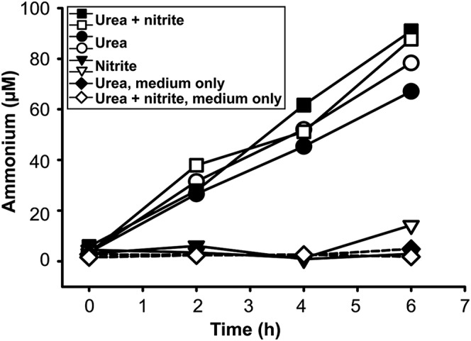
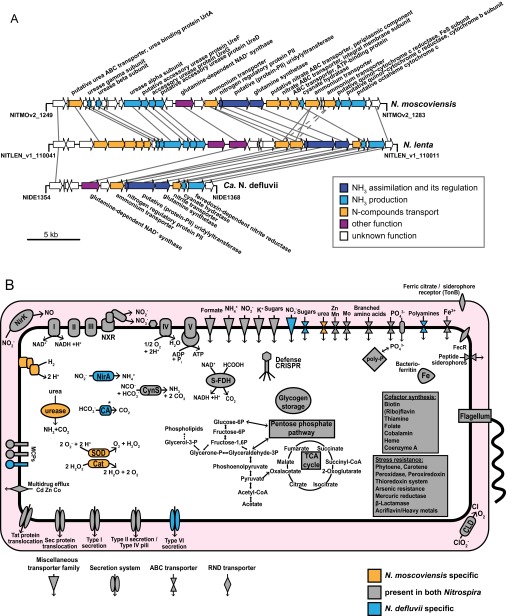
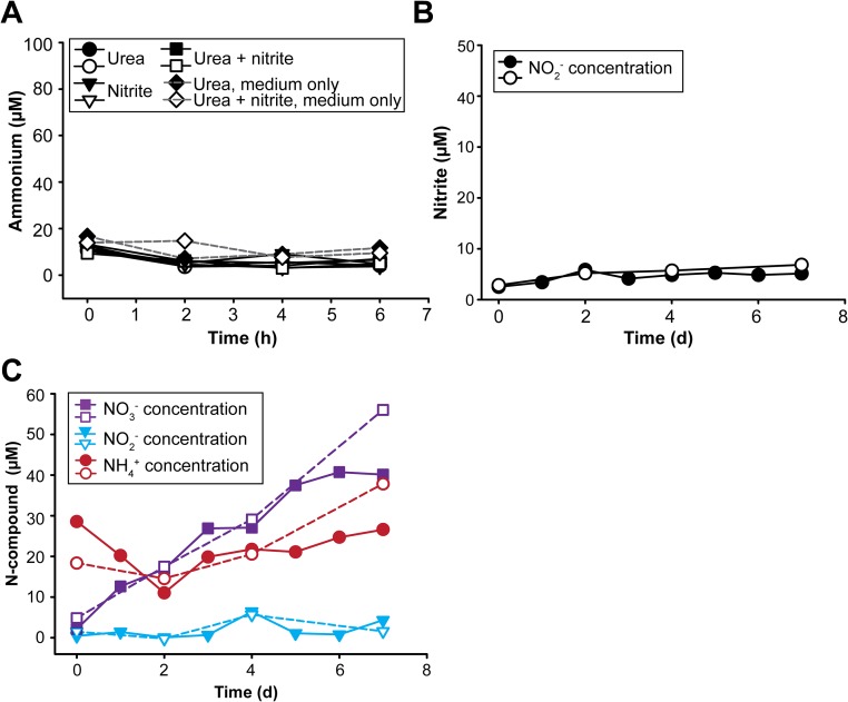
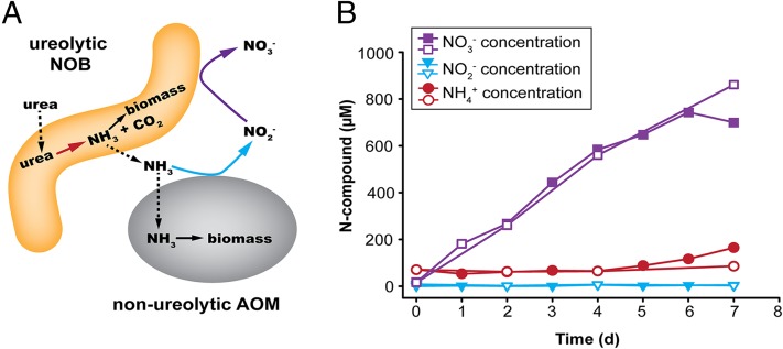
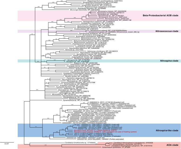
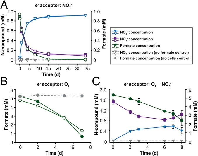
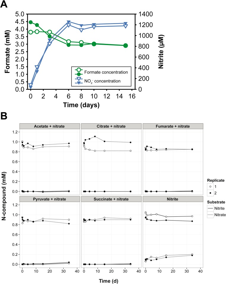
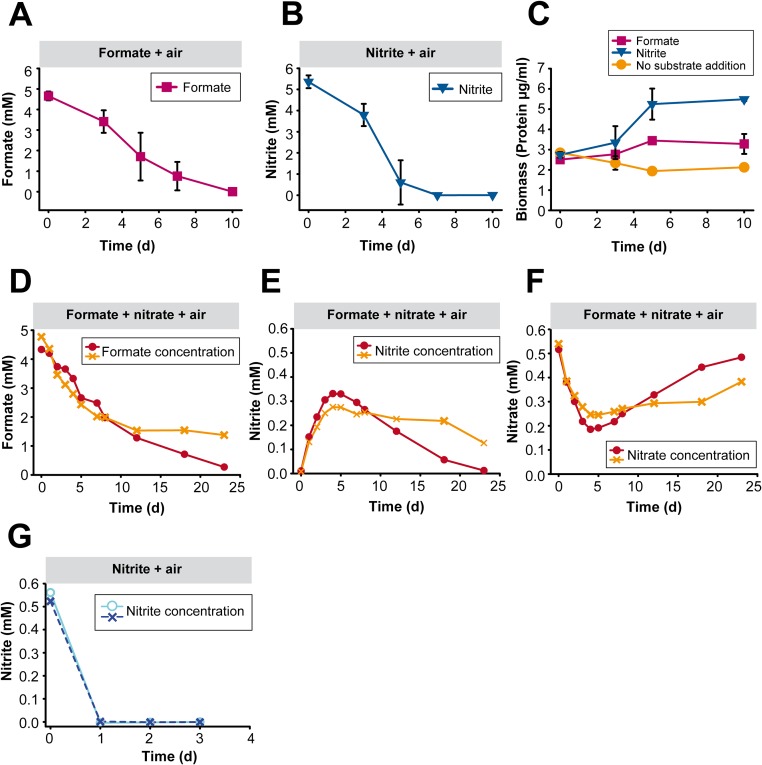
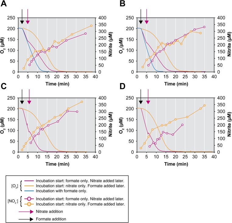
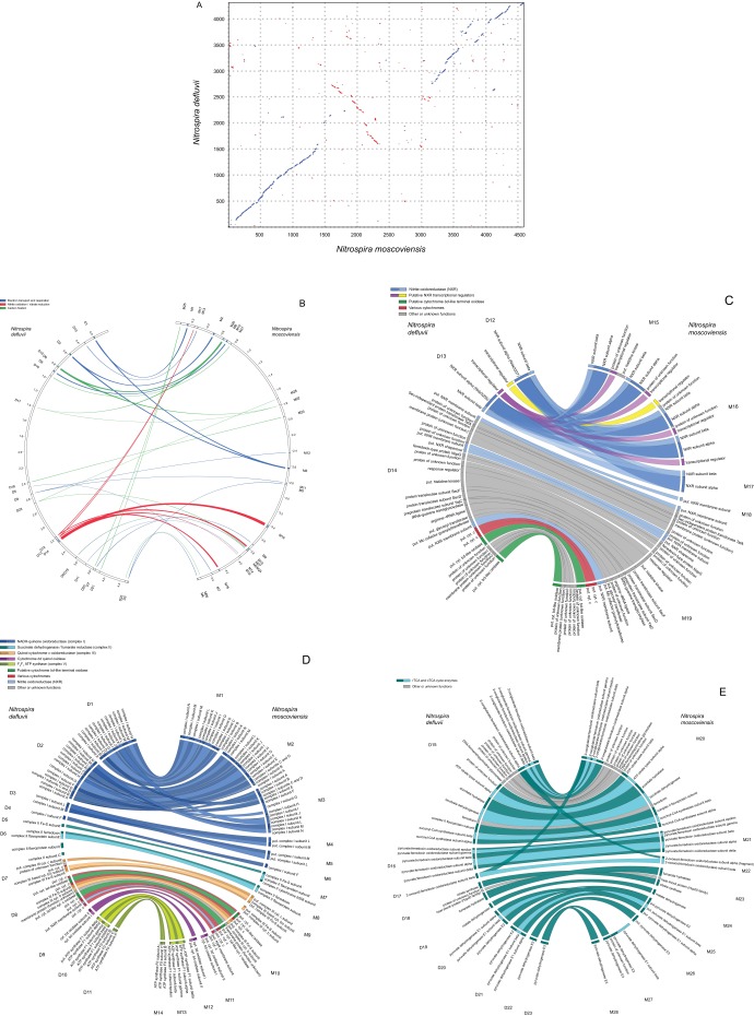
Nitrospira are a diverse group of nitrite-oxidizing bacteria and among the environmentally most widespread nitrifiers. However, they remain scarcely studied and mostly uncultured. Based on genomic and experimental data from Nitrospira moscoviensis representing the ubiquitous Nitrospira lineage II, we identified ecophysiological traits that contribute to the ecological success of Nitrospira. Unexpectedly, N. moscoviensis possesses genes coding for a urease and cleaves urea to ammonia and CO2. Ureolysis was not observed yet in nitrite oxidizers and enables N. moscoviensis to supply ammonia oxidizers lacking urease with ammonia from urea, which is fully nitrified by this consortium through reciprocal feeding. The presence of highly similar urease genes in Nitrospira lenta from activated sludge, in metagenomes from soils and freshwater habitats, and of other ureases in marine nitrite oxidizers, suggests a wide distribution of this extended interaction between ammonia and nitrite oxidizers, which enables nitrite-oxidizing bacteria to indirectly use urea as a source of energy. A soluble formate dehydrogenase lends additional ecophysiological flexibility and allows N. moscoviensis to use formate, with or without concomitant nitrite oxidation, using oxygen, nitrate, or both compounds as terminal electron acceptors. Compared with Nitrospira defluvii from lineage I, N. moscoviensis shares the Nitrospira core metabolism but shows substantial genomic dissimilarity including genes for adaptations to elevated oxygen concentrations. Reciprocal feeding and metabolic versatility, including the participation in different nitrogen cycling processes, likely are key factors for the niche partitioning, the ubiquity, and the high diversity of Nitrospira in natural and engineered ecosystems.

Keywords: Nitrospira; formate; genome; nitrification; urease.


Conflict of interest statement

The authors declare no conflict of interest.

Figures

Fig. 1.
Fig. 1.
Ureolytic activity of a N. moscoviensis pure culture during incubations with urea or both urea and nitrite. Supply of urea led to the accumulation of ammonium in the culture supernatant. Ammonium formation was not detected in the absence of urea (nitrite-only incubations) and in the control experiments without addition of biomass. The results of two biological replicates are shown for each incubation experiment.
Fig. S1.
Fig. S1.
Key genomic and metabolic features of Nitrospira. (A) Schematic representation of the genomic regions in N. moscoviensis and N. lenta that contain the urease genes, the urea ABC transporter, and various other genes involved in the acquisition and metabolism of N compounds. The respective locus in N. defluvii that lacks urease and the urea transporter is shown for comparison. Solid lines connect homologous genes that encode proteins sharing sequence similarities above 50%. Dashed lines connect genes that encode proteins sharing sequence similarities between 30% and 50%. (B) Cell metabolic cartoon constructed from the annotations of the N. moscoviensis and N. defluvii genomes. Core functions, which are shared by both Nitrospira members (gray), and strain-specific features (yellow and blue) are shown. CA, carbonic anhydrase; Cat, catalase; CLD, chlorite dismutase; CRISPR, clustered regularly interspaced short palindromic repeats; CynS, cyanate hydratase (cyanase); MCPs, methyl-accepting chemotaxis proteins; S-FDH, soluble formate dehydrogenase. Enzyme complexes of the electron transport chain are labeled by Roman numerals. The TCA cycle depicts both directions (oxidative and reductive), with the reductive TCA cycle being used by Nitrospira for CO2 fixation. *, N. defluvii possesses a canonical CA, whereas N. moscoviensis has only a putative CA-like protein (NITMOv2_0219) that contains the metal binding sites, but lacks some catalytic residues of canonical CA.
Fig. S2.
Fig. S2.
Full nitrification of urea by reciprocal feeding. (A) Absence of ureolytic activity in N. defluvii. Incubation of N. defluvii cells in medium containing 1 mM urea or 0.5 mM nitrite or both 1 mM urea and 0.5 mM nitrite. No release of free ammonium was observed in any incubation. Control experiments with cell-free medium containing either 1 mM urea or 1 mM urea and 0.5 mM nitrite confirmed that chemical urea degradation did not affect the results. Two biological replicates are shown for all incubations with N. defluvii. (B) Absence of ureolytic activity in N. europaea. Incubation of N. europaea cells in medium containing 1 mM urea as the sole source of ammonia. No ammonia oxidation (production of nitrite) was observed in the two biological replicates. Aliquots of the same N. europaea biomass were used in the coincubation experiment with N. moscoviensis (see Results and Discussion in the main text). (C) Coincubation of N. moscoviensis and urease-negative N. europaea in presence of 50 µM urea as the source of ammonia. The concentrations of free ammonium, nitrite, and nitrate in the culture supernatant during 7 d of incubation are shown. At the start of the incubation, the medium contained some ammonium, most likely due to carryover with the N. europaea inoculum. Full nitrification occurred in each of the two biological replicates.
Fig. 2.
Fig. 2.
Full nitrification by N. moscoviensis and urease-negative AOM through reciprocal feeding. (A) Schematic illustration of the proposed reciprocal feeding interaction between ureolytic NOB such as N. moscoviensis (yellow) and urease-negative AOM such as N. europaea (gray). Solid arrows represent conversions of substrates; dashed arrows the uptake or release of substrates. (B) Concentrations of ammonium, nitrite, and nitrate in a coincubation of N. moscoviensis and urease-negative N. europaea during 7 d of incubation with urea as the sole source of energy and nitrogen. The results of two biological replicates are shown for all incubations.
Fig. S3.
Fig. S3.
Phylogenetic affiliation of the urease alpha subunits (UreC) from Nitrospira, Nitrospina, and other nitrifiers. A Bayesian 80% consensus amino acid tree is shown. The degree of posterior support of a branch is indicated by a single asterisk for >90% posterior probability (PP), a double asterisk for >99% PP or a triple asterisk for >99.9% PP. For metagenomic UreC sequences, the gene ID is followed by the IMG metagenome ID (for UreC received from IMG) and the description of the source habitat. The scale bar shows 7% estimated sequence divergence.
Fig. 3.
Fig. 3.
Formate utilization by a pure culture of N. moscoviensis. (A) Anaerobic consumption of formate with nitrate as terminal electron acceptor. Nitrate was nearly stoichiometrically reduced to nitrite. No nitrite formation from nitrate was observed in the control experiment without formate. The results of two biological replicates are shown for all incubations. (B) Aerobic use of formate with O2 as terminal electron acceptor. For the incubations with N. moscoviensis cells, the results of two biological replicates are shown. Please note that the control experiment with formate but without cells, which confirms the chemical stability of formate, was performed for these incubation conditions only. (C) Aerobic use of formate with both O2 and nitrate as terminal electron acceptors. The oxidation of the formed nitrite became detectable on the seventh day of incubation. Data points show the means of biological replicates (n = 3). Error bars represent SD and are not shown if smaller than symbols. An extended incubation experiment (23 d) confirming the concomitant utilization of formate and nitrite is shown in Fig. S5 DF.
Fig. S4.
Fig. S4.
Incubation experiments of N. moscoviensis with organic substrates in anoxia. (A) Anaerobic consumption of formate (initial concentration 4.5 mM) with nitrate (initial concentration 1.2 mM) as terminal electron acceptor. Nitrate was nearly stoichiometrically reduced to nitrite. The consumption of formate, which was provided in excess, ceased when all nitrate had been reduced. The results of two biological replicates are shown. The divergence of the formate concentrations measured on days 0 and 1 was caused by technical problems with formate measurement. The increase in nitrite indicates that formate was consumed by N. moscoviensis in both replicates during this period. (B) Incubations with various organic compounds under anoxic conditions. Nitrate (1 mM) was provided as terminal electron acceptor in absence of O2. The initial concentration of each organic substrate was 1 mM. The consumption of nitrate and production of nitrite, which would indicate the utilization of the respective organic substrate as electron donor, was not observed in any incubation. The concentrations of the organic substrates at the beginning and end of the incubations were identical (not plotted). A control experiment with nitrite (1 mM) and no organic substrate confirmed the absence of nitrite-oxidizing activity under the anoxic conditions applied. The results of two biological replicates are shown for all incubations.
Fig. S5.
Fig. S5.
Aerobic utilization of formate or nitrite by N. moscoviensis. (A) Aerobic use of formate with O2 as terminal electron acceptor by a pure culture of N. moscoviensis. Data points show the means of biological replicates (n = 3). Error bars represent SD and are not shown if smaller than symbols. This experiment represents an independent replication of the experiment shown in Fig. 3B. (B) Aerobic use of nitrite with O2 as terminal electron acceptor by a pure culture of N. moscoviensis. Data points show the means of biological replicates (n = 3). Error bars represent SD and are not shown if smaller than symbols. This experiment was performed with the same amount of biomass from the same inoculum as the experiment in A. (C) Aerobic growth of N. moscoviensis on formate or nitrite, respectively. Data points show the means of biological replicates (n = 3). Error bars represent SD and are not shown if smaller than symbols. Total biomass protein was measured during the incubations shown in A and B to follow the growth of the cultures in these experiments. In the control experiment, the same amount of N. moscoviensis biomass was incubated in mineral medium without addition of formate or nitrite. Here, total protein decreased likely because of endogenous respiration in the absence of any external electron donor. (D) Long-term incubation of N. moscoviensis with formate and nitrate under oxic conditions. The initial concentrations of formate and nitrate were 5 mM and 0.5 mM, respectively. This graph shows the formate concentration in the culture supernatant. The results of two biological replicates are shown. (E) Nitrite concentrations in the culture supernatant during the incubation experiment shown in D. The initial net increase of the nitrite concentration was caused by nitrate reduction. The following net decrease of nitrite demonstrates the concomitant utilization of nitrite and formate (also see D) from day 6 to the end of the experiment. The results of two biological replicates are shown. (F) Nitrate concentrations in the culture supernatant during the incubation experiment shown in D and E. The initial net decrease and subsequent net increase of the nitrate concentration are consistent with the nitrate-reducing and nitrite-oxidizing activities (D and E) of N. moscoviensis in this experiment. (G) Nitrite oxidation by N. moscoviensis in absence of formate. The rate of nitrite oxidation was considerably higher than in presence of formate (E). Highly similar amounts of N. moscoviensis biomass were used in these incubation experiments (D–G).
Fig. S6.
Fig. S6.
Utilization of O2 and nitrate as terminal electron acceptors by N. moscoviensis. (AD) Microrespirometric measurements of O2 consumption with formate (1 mM initial concentration) as electron donor and in presence or absence of nitrate (5 mM initial concentration) are shown. Curves without symbols depict the O2 concentrations in the supernatant of a N. moscoviensis pure culture. Curves with symbols depict the nitrite concentrations in the supernatant. Each graph represents an independent experiment, and all experiments were performed with highly similar amounts of biomass. Black arrows indicate the addition of formate to the cultures. Purple arrows indicate the addition of nitrate to cultures containing only formate. The reduction in the O2 consumption rates in presence of both electron acceptors, and the production of nitrite, show that electrons from formate were distributed to both O2 and nitrate. Please note that experiments with formate in the total absence of nitrate (blue curves) were carried out only twice (A and B).
Fig. S7.
Fig. S7.
Whole-genome comparison and core metabolism of N. moscoviensis and Nitrospira defluvii. (A) Whole-genome alignment showing the positions of homologous genes in N. moscoviensis and N. defluvii. Sequence matches with the same orientation are plotted blue, whereas inversions are plotted red. (B) Localization of regions encoding the Nitrospira core metabolism for nitrite oxidation, electron transport, and inorganic carbon fixation in the genomes of N. defluvii (Left) and N. moscoviensis (Right). Each semicircle depicts one full genome. Ribbons connect regions containing homologous core metabolism genes. Tags (D1 to D23 for N. defluvii, M1 to M28 for N. moscoviensis) identify the genomic regions shown in C–E. (C) Highly conserved, syntenic gene arrangements within regions encoding nitrite oxidoreductase (NXR) in the genomes of N. defluvii (Left) and N. moscoviensis (right). Regions are separated by spaces and ribbons connect homologous genes. The tags, which identify the regions, are the same as in B. The order of the regions shown here facilitates the synteny comparison and does not reflect the true genomic localization of these regions, which is shown in B. (D) Highly conserved, syntenic gene arrangements within regions encoding the electron transport chains for nitrite oxidation, reverse electron transport, and the utilization of organic substrates in the genomes of N. defluvii (Left) and N. moscoviensis (Right). Regions are separated by spaces and ribbons connect homologous genes. The tags, which identify the regions, are the same as in B. The order of the regions shown here facilitates the synteny comparison and does not reflect the true genomic localization of these regions, which is shown in B. (E) Highly conserved, syntenic gene arrangements within regions encoding the reductive (rTCA) and oxidative (oTCA) tricarboxylic acid cycles in the genomes of N. defluvii (Left) and N. moscoviensis (Right). The rTCA cycle is the CO2 fixation pathway of Nitrospira. Regions are separated by spaces and ribbons connect homologous genes. The tags, which identify the regions, are the same as in B. The order of the regions shown here facilitates the synteny comparison and does not reflect the true genomic localization of these regions, which is shown in B.

References

    1. Lam P, et al. Revising the nitrogen cycle in the Peruvian oxygen minimum zone. Proc Natl Acad Sci USA. 2009;106(12):4752–4757. - PMC - PubMed
    1. Kraft B, et al. Nitrogen cycling. The environmental controls that govern the end product of bacterial nitrate respiration. Science. 2014;345(6197):676–679. - PubMed
    1. Prosser JI. Soil nitrifiers and nitrification. In: Ward BB, Arp DJ, Klotz MG, editors. Nitrification. Am Soc Microbiol; Washington: 2011. pp. 347–383.
    1. Watson SW, Waterbury JB. Characteristics of two marine nitrite oxidizing bacteria, Nitrospina gracilis nov. gen. nov. sp. and Nitrococcus mobilis nov. gen. nov. sp. Arch Mikrobiol. 1971;77:203–230.
    1. Sorokin DY, et al. Nitrification expanded: Discovery, physiology and genomics of a nitrite-oxidizing bacterium from the phylum Chloroflexi. ISME J. 2012;6(12):2245–2256. - PMC - PubMed

Publication types

MeSH terms

Associated data