. 2015 Oct 9;290(41):25103-17.
doi: 10.1074/jbc.M115.658781. Epub 2015 Aug 25.

The Cell Shape-determining Csd6 Protein from Helicobacter pylori Constitutes a New Family of L,D-Carboxypeptidase

Affiliations

The Cell Shape-determining Csd6 Protein from Helicobacter pylori Constitutes a New Family of L,D-Carboxypeptidase

Hyoun Sook Kim et al. J Biol Chem. .

Abstract

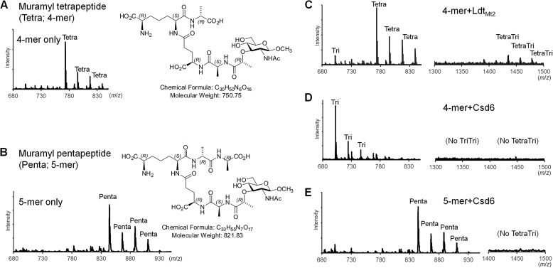
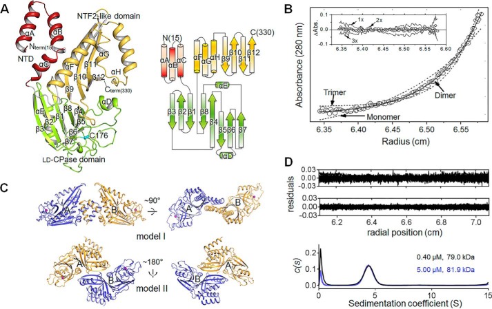
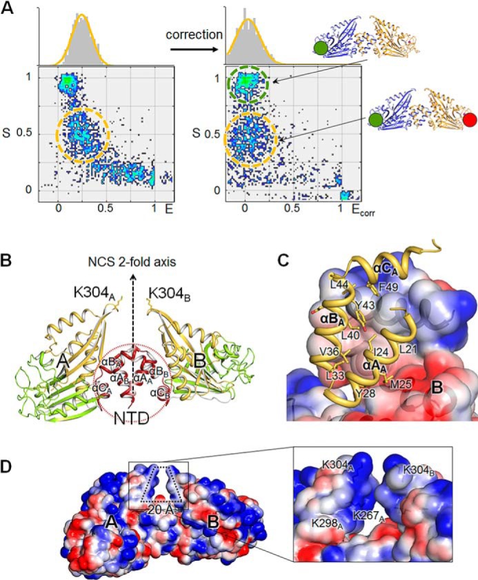
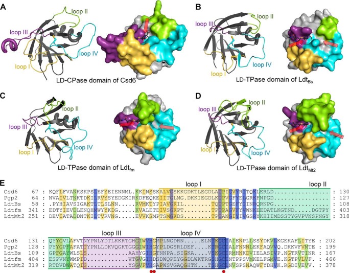
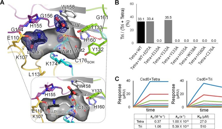
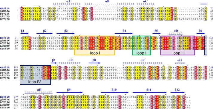
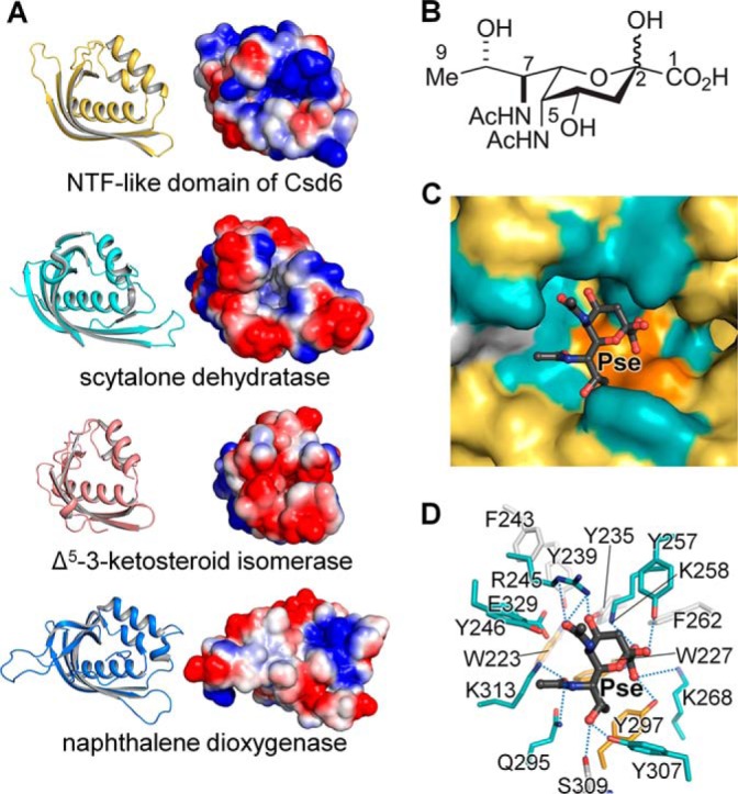
Helicobacter pylori causes gastrointestinal diseases, including gastric cancer. Its high motility in the viscous gastric mucosa facilitates colonization of the human stomach and depends on the helical cell shape and the flagella. In H. pylori, Csd6 is one of the cell shape-determining proteins that play key roles in alteration of cross-linking or by trimming of peptidoglycan muropeptides. Csd6 is also involved in deglycosylation of the flagellar protein FlaA. To better understand its function, biochemical, biophysical, and structural characterizations were carried out. We show that Csd6 has a three-domain architecture and exists as a dimer in solution. The N-terminal domain plays a key role in dimerization. The middle catalytic domain resembles those of l,d-transpeptidases, but its pocket-shaped active site is uniquely defined by the four loops I to IV, among which loops I and III show the most distinct variations from the known l,d-transpeptidases. Mass analyses confirm that Csd6 functions only as an l,d-carboxypeptidase and not as an l,d-transpeptidase. The d-Ala-complexed structure suggests possible binding modes of both the substrate and product to the catalytic domain. The C-terminal nuclear transport factor 2-like domain possesses a deep pocket for possible binding of pseudaminic acid, and in silico docking supports its role in deglycosylation of flagellin. On the basis of these findings, it is proposed that H. pylori Csd6 and its homologs constitute a new family of l,d-carboxypeptidase. This work provides insights into the function of Csd6 in regulating the helical cell shape and motility of H. pylori.

Keywords: Csd6; HP0518; Helicobacter pylori; L,D-carboxypeptidase; cell motility; cell shape; flagellin; peptidoglycan; protein structure; structure-function.


Figures

FIGURE 1.
FIGURE 1.
l,d-CPase activity of Csd6. A and B, mass spectra and structural formulas of the synthesized muramyl tetrapeptide (A) and muramyl pentapeptide (B). The observed peaks correspond to a series of sodium adduct ions of the peptides. C–E, mass spectra of the peptide samples upon incubation with LdtMt2 and Csd6. In the control reaction catalyzed by LdtMt2, the dimeric cross-linked tetra-tripeptide species is produced from the muramyl tetrapeptide (C). In contrast, in the reaction catalyzed by Csd6, the muramyl tetrapeptide is converted to the muramyl tripeptide but not to dimeric cross-linked tetra-tripeptide species (D). Upon incubation with Csd6, the muramyl pentapeptide undergoes no change (E). The observed m/z values for the charged species with bound sodium ions (up to four: [M + Na]+1, [M + 2Na-H]+1, [M + 3Na-2H]+1, and [M + 4Na-3H]+1) agree with the calculated m/z values for muramyl tri-, muramyl tetra-, muramyl penta-, or dimeric cross-linked tetra-tripeptides (average relative mass of neutral species = 679.67, 750.75, 821.83, or 1412.41, respectively).
FIGURE 2.
FIGURE 2.
Overall structure and the oligomeric state of H. pylori Csd6. A, ribbon diagram (upper panel) and topology diagram (bottom panel) of the Csd6-unbound structure. The NTD, l,d-CPase domain, and NTF2-like domain are shown in red, green, and yellow, respectively. The secondary structure elements have been defined by the DSSP program (77). The nucleophile Cys-176 of the l,d-CPase domain is shown in a cyan stick model. B, equilibrium sedimentation data for Csd6 at an ultracentrifugal speed of 16,000 rpm using 3.65 μm protein at 20 °C. The circles are experimental data, and the solid line is a fitting line for an ideal monomer model. The two dotted lines are fitting lines for ideal monomer and trimer models. Distributions of the residuals for monomer, dimer, and trimer models are shown in the inset panel. These data indicate that Csd6 exists as homogeneous dimers in solution. C, two putative models of the Csd6 dimer in the crystal. Dimer model I (upper panel) and model II (bottom panel) are shown in ribbon diagrams. The side chain sulfur atoms of Cys-176 are shown as purple spheres. Model I is favored by the single molecule ALEX-FRET data (shown in Fig. 3A). D, plots of the residuals for dimer species model of Csd6 at 0.40 μm (upper) and 5.00 μm (lower), and the distribution of sedimentation coefficient (c(s) versus s, where s is in Svedberg unit, S) from the sedimentation velocity experiments.
FIGURE 3.
FIGURE 3.
Dimeric structure of H. pylori Csd6. A, determination of the dimeric model of Csd6 by single molecule FRET technique. Single molecule FRET data are presented in a two-dimensional E-S graph, where E is the FRET efficiency and S denotes the Cy3 (donor)/Cy5 (acceptor) molar ratio in a dimer (54). Three clusters correspond to the dimers formed as follows: (i) between two Cy3-Csd6 monomers; (ii) between two Cy5-Csd6 monomers; and (iii) between a Cy3-Csd6 monomer and a Cy5-Csd6 monomer. The cluster appearing at S ∼ 1 (green dotted ellipse) with E ≈ 0 corresponds to Cy3-labeled Csd6 dimers. Csd6 dimers labeled with both Cy3 and Cy5 appear at S ∼ 0.5 (orange dotted ellipse) (55). B, ribbon diagram of the Csd6 homodimer (model I) colored as in Fig. 2A. In this dimer, A and B monomers are related by a noncrystallographic pseudo 2-fold symmetry. C, hydrophobic interactions between the NTDs of A and B monomers, which are shown in the ribbon diagram and the electrostatic surface diagram, respectively. D, electrostatic potential surface diagrams of the Csd6 dimer molecule. A detailed view of the specific hole formed by the dimerization is shown in the black lined box.
FIGURE 4.
FIGURE 4.
Comparison of Csd6 and three l,d-TPases. A–D, the ribbon diagrams (left panels) and surface diagrams (right panels) of the l,d-CPase domain of H. pylori Csd6 (A) and the l,d-TPase domains of B. subtilis LdtBs (B), E. faecium Ldtfm (C), and M. tuberculosis LdtMt2 (D). The four loops I–IV are colored in yellow, green, purple, and light blue, respectively. One or two paths that allow access to the catalytic triad are indicated by red arrows. In the meropenem-complexed LdtMt2 structure (PDB code 4GSU), the paths A and B correspond to left and right arrows, respectively (30). Meropenem was modeled into each domain structure by structural superimposition of the corresponding domains using the meropenem-complexed LdtMt2 (PDB code 4GSU). E, structure-based sequence alignment was performed by PROMALS3D (80), and the alignment was presented by GeneDoc. Structures of the l,d-CPase domain of H. pylori Csd6 (Swiss-Prot accession code O25255; PDB code 4XZZ) and l,d-TPase catalytic domains of B. subtilis LdtBs (O34816; 1Y7M), E. faecium Ldtfm (Q3Y185; 1ZAT), and M. tuberculosis LdtMt2 (O53223; 4GSU) were used in the alignment. In the case of C. jejuni Pgp2 (A1VZP0), no structural information was available, and thus only the sequence is used. Red dots indicate the catalytic triad (His-160, Gly-161, and Cys-176 in H. pylori Csd6). The four loops I–IV are shown in colored boxes as in A.
FIGURE 5.
FIGURE 5.
d-Ala-complexed Csd6 and key residues for the l,d-CPase activity. A, ribbon diagram and the accessible inner surface of the active site in the Csd4-Ala structure. The bound d-Ala molecules (d-Ala′ and d-Ala″) (upper panel) and the mDAP (bottom panel) modeled on the basis of d-Ala′ and Wat1 are shown in stick models. The omit mFoDFc maps for d-Ala′ and d-Ala″ (contoured at 2.0σ) and the side chain oxygen atom of Cys-176 oxidized as the sulfenic acid (contoured at 2.5σ) are colored in blue. The oxyanion hole is asterisked and also shown as blue dotted lines in the bottom panel. Red dotted lines indicate the interactions among residues of the catalytic triad. B, l,d-carboxypeptidation activities of the wild-type Csd6 (with the treatment of EDTA or not) and the mutants (E110A, Y132A, Y133A, H155A, W158A, H160A, and C176A) with the muramyl tetrapeptide. C, SPR experiments with immobilized Csd6 and the muramyl tetrapeptide (or muramyl tripeptide) as an analyte at different concentrations (15.6, 31.3, 62.5, 125, 250, and 500 μm) are shown in traces colored as orange, light blue, purple, green, red, and blue, respectively.
FIGURE 6.
FIGURE 6.
Sequence alignment of Csd6 homologs. Sequence alignment of Csd6 homologs from H. pylori strain 26695 (HP0518; Swiss-Prot accession code O25255), from Wolinella succinogenes (Q7MRJ6), from Arcobacter nitrofigilis (D5V196), from Campylobacter concisus strain 13826 (A7ZCK9), and from Thermovibrio ammonificans (E8T4Z0) was performed and presented by ClustalX (78) and ESPript (79). The secondary structure of Csd6 is presented above the aligned sequences. The four loops I–IV around the active site of the Csd6 l,d-CPase domain are indicated with boxes colored as in Fig. 4E.
FIGURE 7.
FIGURE 7.
NTF-like domain of Csd6. A, ribbon diagrams and electrostatic potential surface diagrams of the NTF-like domains of Csd6, scytalone dehydratase, Δ5-3-ketosteroid isomerase, and naphthalene dioxygenase. B, chemical structure of Pse. C and D, in silico docking of Pse onto the pocket in the NTF-like domain of Csd6. The potential binding pocket is shown in surface representation (C), and the residues around the docked ligand are shown as stick models (D).

Similar articles

Cited by

References

    1. Roesler B. M., Rabelo-Gonçalves E. M., Zeitune J. M. (2014) Virulence factors of Helicobacter pylori: a review. Clin. Med. Insights Gastroenterol. 7, 9–17 - PMC - PubMed
    1. Kusters J. G., van Vliet A. H., Kuipers E. J. (2006) Pathogenesis of Helicobacter pylori infection. Clin. Microbiol. Rev. 19, 449–490 - PMC - PubMed
    1. Roubaud Baudron C., Franceschi F., Salles N., Gasbarrini A. (2013) Extragastric diseases and Helicobacter pylori. Helicobacter 18, 44–51 - PubMed
    1. Malfertheiner P., Selgrad M., Bornschein J. (2012) Helicobacter pylori: clinical management. Curr. Opin. Gastroenterol. 28, 608–614 - PubMed
    1. Ottemann K. M., Lowenthal A. C. (2002) Helicobacter pylori uses motility for initial colonization and to attain robust infection. Infect. Immun. 70, 1984–1990 - PMC - PubMed

Publication types

Substances

LinkOut - more resources