. 2015 Oct 2;290(40):24614-25.
doi: 10.1074/jbc.M115.678193. Epub 2015 Aug 25.

An E2 accessory domain increases affinity for the anaphase-promoting complex and ensures E2 competition

Affiliations

An E2 accessory domain increases affinity for the anaphase-promoting complex and ensures E2 competition

Juliet R Girard et al. J Biol Chem. .

Abstract

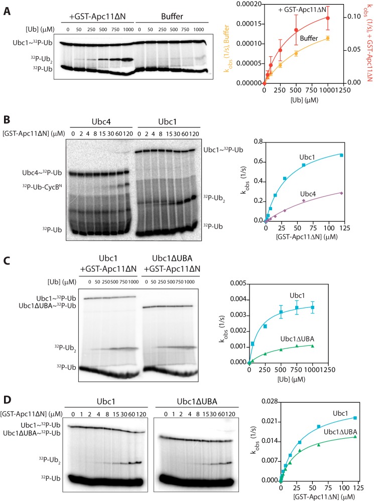
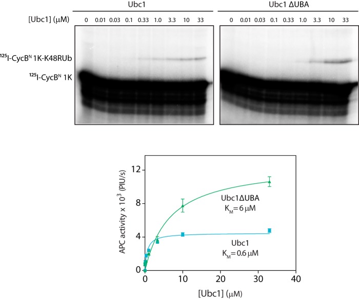
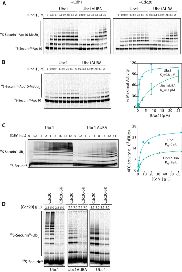
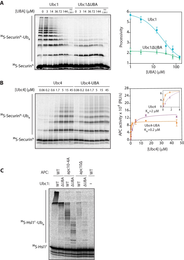
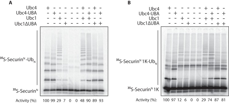
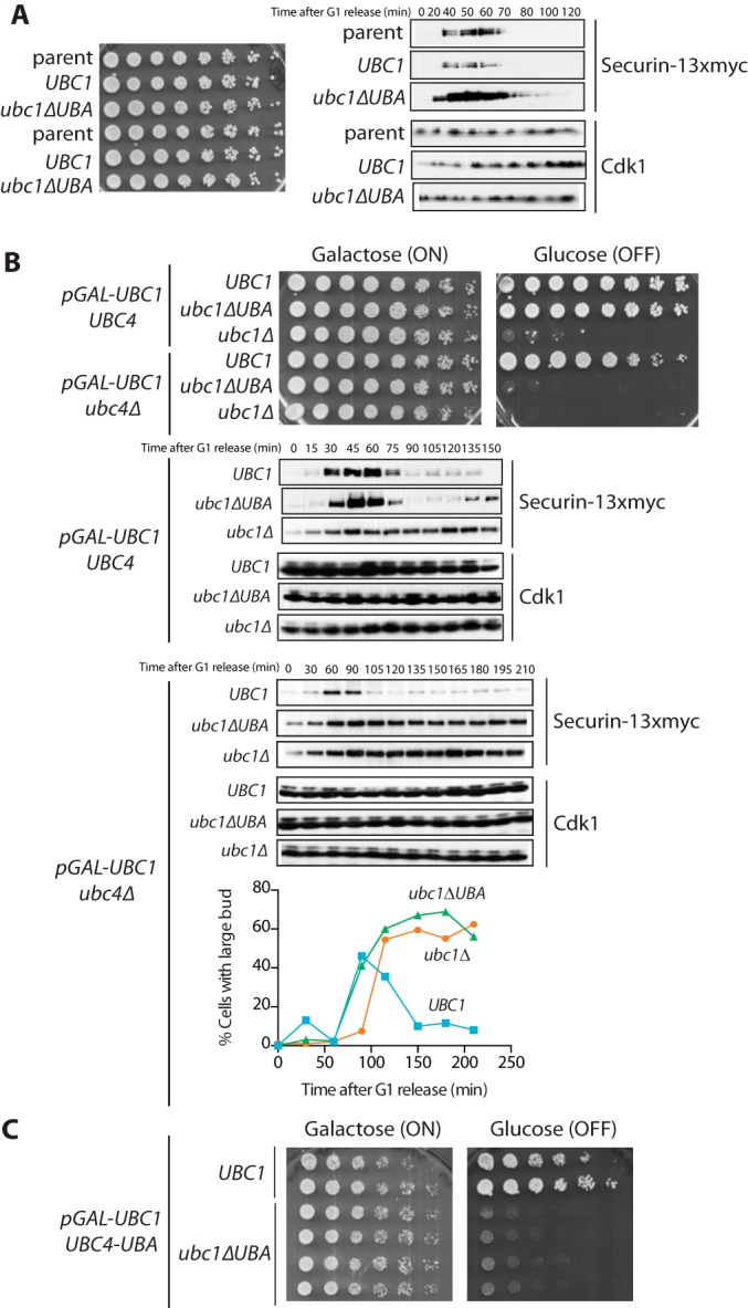
The anaphase-promoting complex/cyclosome (APC/C) is a member of the RING family of E3 ubiquitin ligases, which promote ubiquitin transfer from an E2 ubiquitin-conjugating enzyme to a substrate. In budding yeast, the APC/C collaborates with two E2s, Ubc4 and Ubc1, to promote the initiation and elongation, respectively, of polyubiquitin chains on the substrate. Ubc4 and Ubc1 are thought to compete for the same site on the APC/C, but it is not clear how their affinities are balanced. Here, we demonstrate that a C-terminal ubiquitin-associated (UBA) domain enhances the affinity of Ubc1 for the APC/C. Deletion of the UBA domain reduced apparent APC/C affinity for Ubc1 and decreased polyubiquitin chain length. Surprisingly, the positive effect of the UBA domain was not due to an interaction with the acceptor ubiquitin attached to the APC/C substrate or the donor ubiquitin attached to Ubc1 itself. Instead, our evidence suggests that the UBA domain binds to a site on the APC/C core, thereby increasing Ubc1 affinity and enhancing its ability to compete with Ubc4. The UBA domain is required for normal Ubc1 function and E2 competition in vivo. Thus, the UBA domain of Ubc1 ensures efficient polyubiquitination of substrate by balancing Ubc1 affinity with that of Ubc4.

Keywords: E3 ubiquitin ligase; cell cycle; ubiquitin; ubiquitin-conjugating enzyme (E2 enzyme); yeast.


Figures

FIGURE 1.
FIGURE 1.
The UBA domain does not contribute to acceptor ubiquitin binding or RING enhancement of E2 catalysis. A, Ubc1 (wild type; 0.5 μm) was charged with 32P-radiolabeled K48R ubiquitin and incubated with either GST-Apc11ΔN (80 μm) or buffer. Wild-type ubiquitin was added at the indicated concentrations, and reactions were carried out at 4 °C for 5 s (+GST-Apc11ΔN) or 10 min (buffer alone). Reaction products were analyzed by SDS-PAGE and autoradiography with a phosphorimaging system. The right panel displays the quantification of diubiquitin synthesis assays, showing the dependence of catalytic rate (kobs) on ubiquitin concentration. Autoradiographs were quantified using ImageQuant, and data were fit to the Michaelis-Menten equation in Prism software. The average of three experiments is shown. Error bars represent S.E. B, Ubc1 (wild type) and Ubc4 (each at 0.5 μm) were charged with 32P-radiolabeled K48R ubiquitin and incubated with buffer or increasing concentrations of GST-Apc11ΔN. Unlabeled CycBN (200 μm; Ubc4 reactions) or ubiquitin (100 μm; Ubc1 reactions) was added, and reactions were carried out at 4 °C for 5 s. Reactions were analyzed as in A. Results are representative of three independent experiments. C, Ubc1 (wild type or ΔUBA at 0.5 μm) reactions were carried out as in A except that a subsaturating concentration of GST-Apc11ΔN (6.5 μm) was used. Reactions were carried out at room temperature for 3 (wild-type Ubc1) or 5 min (Ubc1ΔUBA). The average of three experiments is shown. Error bars represent S.E. D, Ubc1 and Ubc1ΔUBA were charged as in A and incubated with buffer or increasing concentrations of GST-Apc11ΔN. Unlabeled wild-type ubiquitin (100 μm) was added, and reactions were carried out, visualized, and quantified as in A. Results are representative of three independent experiments. Ub, ubiquitin.
FIGURE 2.
FIGURE 2.
The UBA domain is independent of ubiquitin on substrate. APC/C purified from yeast cells was mixed with Cdh1 and 125I-CycB 1K. Ubc1 (either wild type or ΔUBA) charged with K48R mutant ubiquitin was added at the indicated concentrations, and reactions were carried out at room temperature for 15 min. Reaction products were analyzed by SDS-PAGE and autoradiography with a phosphorimaging system. The bottom panel displays quantification of results, showing the dependence of APC/C activity on concentration of Ubc1 or Ubc1ΔUBA. Autoradiographs were quantified using ImageQuant, and data were fit to the Michaelis-Menten equation in Prism software. The average of four experiments is shown. Error bars represent S.E. PIU/s, phosphorimaging units/s.
FIGURE 3.
FIGURE 3.
UBA domain does not bind to APC/C activator. A, [35S]SecurinN-Apc10, Cdh1, and Cdc20 were generated by in vitro translation in rabbit reticulocyte lysate. [35S]SecurinN-Apc10 was bound to immunoprecipitated APC/C (apc10Δ cdh1Δ), and unbound substrate was washed away. Cdh1 and Cdc20 were purified from reticulocyte lysate and mixed with fusion substrate-bound APC/C. Ubc1 (either wild type or ΔUBA) charged with methylated ubiquitin (MeUb) was added in increasing concentrations, and reactions were carried out at room temperature for 15 min. Reaction products were analyzed by SDS-PAGE and autoradiography with a phosphorimaging system. Results are representative of three independent experiments. B, APC/C assays were performed as in A except that no activator subunit was added. The graph at right shows mean values ±S.E. normalized to maximal Ubc1 activity of three independent experiments. C, [35S]SecurinN and Cdh1 were generated by in vitro translation in rabbit reticulocyte lysate and purified. Immunoprecipitated APC/C was mixed with [35S]SecurinN and increasing amounts of Cdh1. Ubc1 or Ubc1ΔUBA (10 μm) was charged with wild-type ubiquitin (Ub), and reactions were carried out at room temperature for 15 min. Reaction products were analyzed as in A. The graph at right shows quantification of APC/C activity as a function of Cdh1 concentration for the experiment at left. Results are representative of three independent experiments. PIU/s, phosphorimaging units/s. D, [35S]SecurinN, Cdc20, and Cdc20-5K were generated as in C and mixed with purified APC/C. E2s (each at 5 μm) charged with wild-type ubiquitin were added, and reactions were carried out at room temperature for 30 min. Reaction products were analyzed as in A. Results are representative of three independent experiments. Error bars represent S.E.
FIGURE 4.
FIGURE 4.
The UBA domain promotes APC/C binding. A, [35S]SecurinN was translated in vitro in rabbit reticulocyte lysate, purified, and mixed with purified APC/C, Cdh1, and varying concentrations of recombinant UBA domain. Ubc1 (either wild type or ΔUBA; 10 μm) charged with wild-type ubiquitin (Ub) was added, and reactions were carried out at room temperature for 15 min. Reaction products were analyzed by SDS-PAGE and autoradiography with a phosphorimaging system. Autoradiographs were quantified using ImageQuant. The right panel displays quantification of processivity of APC/CUbc1 and APC/CUbc1ΔUBA as a function of the concentration of free UBA domain. Processivity was calculated by quantifying individual ubiquitinated products, multiplying the amount of product by the number of ubiquitins in the product, and dividing by the total amount of modified products. Data were fit to the log(inhibitor) versus response equation in Prism software. The average of three experiments is shown. Error bars represent S.E. B, purified [35S]SecurinN, APC/C, and Cdh1 were combined as in A. Ubc4 or Ubc4-UBA charged with wild-type ubiquitin was added at the indicated concentrations, and reactions were carried out at room temperature for 15 min. Reaction products were analyzed as in A. The right panel displays quantification of the dependence of APC/C activity on concentration of Ubc4 or Ubc4-UBA. Data were fit to the Michaelis-Menten equation in Prism software. The average of three experiments is shown. Error bars represent S.E. The inset shows a close-up of the graph at lower E2 concentrations. PIU/s, phosphorimaging units/s. C, [35S]Hsl1F was translated in vitro in rabbit reticulocyte lysate, purified, and mixed with Cdh1 and APC/C immunoprecipitated from wild-type, apc10Δ mutant, or apc10-4A mutant yeast. E2s (all at 5 μm final concentration) were charged with wild-type ubiquitin and added, and reactions were carried out at room temperature for 15 min. Reaction products were analyzed as in A.
FIGURE 5.
FIGURE 5.
The UBA domain is important for E2 competition. A, [35S]SecurinN was translated in vitro in rabbit reticulocyte lysate, purified, and mixed with purified APC/C and Cdh1. The indicated E2s (each at 3 μm final concentration) were charged with wild-type ubiquitin (Ub) and added. Reactions were carried out at room temperature for 20 min. Reaction products were analyzed by SDS-PAGE and autoradiography with a phosphorimaging system. The numbers below show quantification of APC/C activity (i.e. total modified substrate) in each lane with Ubc4 activity normalized to 100. Results are representative of three independent experiments. B, APC/C assays were performed as in A except that [35S]SecurinN 1K was used as the substrate. Results are representative of three independent experiments.
FIGURE 6.
FIGURE 6.
The UBA domain is important for Ubc1 function in vivo. A, left, strains were grown to midlog phase at 30 °C in medium containing 2% glucose, diluted to an A600 of 0.1, plated as serial dilutions on 2% glucose, and grown for 2 days at 30 °C. Right, asynchronous cultures (A600 = 0.2) were arrested in G1 with α factor (1 μg/ml) for 3 h and released from G1 arrest by washing away α factor (zero time point). Cell samples were taken at the indicated times, lysed, and analyzed by Western blotting against the indicated proteins. Results are representative of three independent experiments. B, top, strains were grown to midlog phase at 30 °C in medium containing 2% galactose and raffinose, diluted to an A600 of 0.1, plated as serial dilutions on 2% galactose and raffinose or 2% glucose, and grown for 2 days at 30 °C. Results are representative of three independent experiments. Bottom, asynchronous cultures were arrested in G1 with α factor (1 μg/ml) for 5 h. During the last 2 h of α factor treatment, cultures were incubated with 2% glucose. Cells were released from G1 by washing away α factor, and resuspended in medium containing 2% glucose (zero time point). Cell samples were taken at the indicated times, lysed, and analyzed by Western blotting against the indicated proteins. Parallel samples were taken, and a budding index was counted by microscopy. Results are representative of three independent experiments. C, the indicated strains were grown and plated as in B.

Similar articles

Cited by

References

    1. Primorac I., Musacchio A. (2013) Panta rhei: the APC/C at steady state. J. Cell Biol. 201, 177–189 - PMC - PubMed
    1. Chang L., Barford D. (2014) Insights into the anaphase-promoting complex: a molecular machine that regulates mitosis. Curr. Opin. Struct. Biol. 29, 1–9 - PubMed
    1. Rodrigo-Brenni M. C., Foster S. A., Morgan D. O. (2010) Catalysis of lysine 48-specific ubiquitin chain assembly by residues in E2 and ubiquitin. Mol. Cell 39, 548–559 - PMC - PubMed
    1. Rodrigo-Brenni M. C., Morgan D. O. (2007) Sequential E2s drive polyubiquitin chain assembly on APC targets. Cell 130, 127–139 - PubMed
    1. Williamson A., Wickliffe K. E., Mellone B. G., Song L., Karpen G. H., Rape M. (2009) Identification of a physiological E2 module for the human anaphase-promoting complex. Proc. Natl. Acad. Sci. U.S.A. 106, 18213–18218 - PMC - PubMed

Publication types