

Myeloid Sarcoma: An Unusual Case of Mediastinal Mass and Malignant Pleural Effusion with Review of Literature
- PMID: 26306072
- PMCID: PMC4542764
- DOI: 10.1007/s12288-015-0536-z
Myeloid Sarcoma: An Unusual Case of Mediastinal Mass and Malignant Pleural Effusion with Review of Literature
Abstract
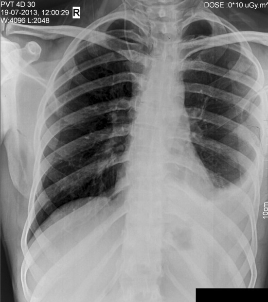
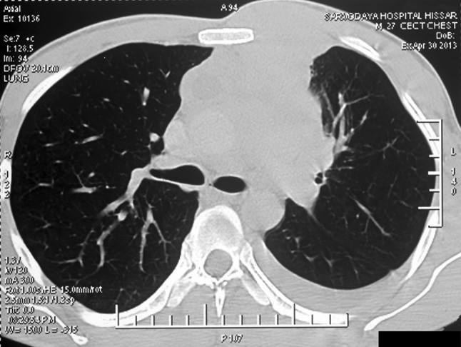
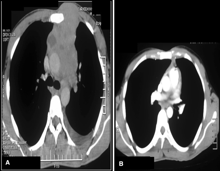
Myeloid sarcoma is an extramedullary tumor seen most commonly in patients with acute myeloid leukemia and less frequently in chronic myeloid leukemia, myelodysplastic syndrome and rarely, in an isolated form without any other underlying malignancy. Malignant pleural effusion in hematological malignancies is rare when compared with solid tumors. We present an unusual case of myeloid sarcoma in which a mediastinal mass with pleural effusion was the initial presentation. A 27 year old gentleman presented with complaints of fever, chest pain and swelling in the anterior chest wall for 6 months. Examination revealed a lump measuring 5 × 5 cm on the left side of the chest wall. Hematological evaluation showed hemoglobin-14.2 g/dL, platelet count-233 × 10(9)/L, TLC-117 × 10(6)/L with normal differential counts. Contrast enhanced computerised tomography (CECT) confirmed the presence of a soft tissue mass in the superior mediastinum abutting against the chest wall. Core biopsy was suggestive of myeloid sarcoma and immunohistochemistry was positive for myeloperoxidase and negative for CD3, CD 20 and CD 23. Pleural fluid analysis showed the presence of malignant cells. Bone marrow examination did not show an excess of blasts. A final diagnosis of extramedullary myeloid sarcoma with malignant pleural effusion was made. The patient was given induction chemotherapy (3 + 7 regimen) with daunorubicin and cytosine arabinoside. Repeat CECT done on day 28 showed complete resolution of pleural effusion and significant reduction in the size of mediastinal mass. The patient has successfully completed three cycles of consolidation therapy following which there has been complete resolution of the mass. He remains asymptomatic on close follow up.
Keywords: Acute myeloid leukemia; Malignant cytology; Myeloid sarcoma.
Figures


Similar articles
-
[Isolated myeloid sarcoma involving the mediastinum].Acta Med Croatica. 2011 Sep;65 Suppl 1:133-8. Acta Med Croatica. 2011. PMID: 23126041 Croatian.
-
Polyserositis as a Unique Presentation of Isolated Myeloid Sarcoma: A Case Report.Anticancer Res. 2022 Jul;42(7):3595-3599. doi: 10.21873/anticanres.15846. Anticancer Res. 2022. PMID: 35790247
-
Myeloid sarcoma diagnosed on pleural effusion cytology: A case report and literature review.Diagn Cytopathol. 2021 Aug;49(8):E316-E319. doi: 10.1002/dc.24739. Epub 2021 Mar 22. Diagn Cytopathol. 2021. PMID: 33751858 Review.
-
Isolated myeloid sarcoma with pericardial and pleural effusions as first manifestation: A case report.Medicine (Baltimore). 2022 Oct 21;101(42):e31026. doi: 10.1097/MD.0000000000031026. Medicine (Baltimore). 2022. PMID: 36281103 Free PMC article.
-
[Pleural effusion as the first manifestation of pleural isolated myeloid sarcoma: a case report and literature review].Zhonghua Jie He He Hu Xi Za Zhi. 2020 Oct 12;43(10):839-843. doi: 10.3760/cma.j.cn112147-20200526-00639. Zhonghua Jie He He Hu Xi Za Zhi. 2020. PMID: 32992437 Review. Chinese.
Cited by
-
Uterine Mass and Menorrhagia: A Rare Presentation of Acute Myeloid Leukemia with Arduous Clinical Course.Balkan Med J. 2018 May 29;35(3):282-284. doi: 10.4274/balkanmedj.2017.0941. Epub 2017 Dec 8. Balkan Med J. 2018. PMID: 29219111 Free PMC article. No abstract available.
-
Myeloid Sarcoma Of Vulva: A Short Update.Indian J Hematol Blood Transfus. 2016 Jun;32(Suppl 1):69-71. doi: 10.1007/s12288-016-0662-2. Epub 2016 Mar 1. Indian J Hematol Blood Transfus. 2016. PMID: 27408359 Free PMC article. No abstract available.
-
Cardiac Myeloid Sarcoma: Review of Literature.J Clin Diagn Res. 2017 Mar;11(3):XE01-XE04. doi: 10.7860/JCDR/2017/23241.9499. Epub 2017 Mar 1. J Clin Diagn Res. 2017. PMID: 28511492 Free PMC article. Review.
-
A Case of a Constricted Vessel: The Impact of Acute Myeloid Leukemia on the Superior Vena Cava.Cureus. 2023 Nov 28;15(11):e49616. doi: 10.7759/cureus.49616. eCollection 2023 Nov. Cureus. 2023. PMID: 38161934 Free PMC article.
-
Isolated cardiac involvement of a primary myeloid sarcoma: a case report of an unusual cause of pulmonary oedema.Eur Heart J Case Rep. 2023 Feb 18;7(3):ytad088. doi: 10.1093/ehjcr/ytad088. eCollection 2023 Mar. Eur Heart J Case Rep. 2023. PMID: 36895307 Free PMC article.
References
Publication types
LinkOut - more resources
Full Text Sources
Other Literature Sources
Research Materials