

doi: 10.1055/s-0035-1556851.
Physical Evaluation of the Asian Blepharoplasty Patient
Affiliations
- PMID: 26306081
- PMCID: PMC4536068
- DOI: 10.1055/s-0035-1556851
Item in Clipboard
Physical Evaluation of the Asian Blepharoplasty Patient
Semin Plast Surg.
2015 Aug.
Abstract
In contrast with Caucasian blepharoplasty, which is usually done for rejuvenation purposes, eyelid surgery for Asians is mainly done for cosmetic reasons. Here the authors present preoperative evaluation methods of Asian eyelids to overcome anatomical variations and determine the right aesthetic options. Proper selection of the most suitable operative methods guarantees optimal surgical results.
Keywords: Asian blepharoplasty; Asian double crease; Asian eyelid; Asian eyelid anatomy; Asian eyelid morphology; Asian oculoplastic surgery; blepharoptosis; epicanthoplasty; eyelid anatomy; eyelids.
Figures

(A) A young Asian woman without brow elevation. (B) Hyperactive frontalis due to hidden blepharoptosis.

(A) Without brow elevation. (B) Correction of ptosis improves brow position and frontalis hyperactivity.

(A) Preoperative primary gaze: margin reflex distance 1 (MRD1) = 2 mm. (B) Postoperative brow elevation. (C) Brow descends when postoperative primary gaze is MRD1 = 5.5 mm.

Characteristics of the Asian eyelid: Puffiness, no fold, deep set, epicanthus, canthal tilt: up-slanting, vertically narrow.
Removal of hypertrophied fat is almost routinely done during Asian blepharoplasty. Pretarsal fullness is also observed.

Lacrimal gland prolapse also exaggerates the lateral bulginess.

Floppy eyelid syndrome in a patient who has highly extensible eyelid skin.

Margin reflex distance 1 is the same, but eyes look bigger with a crease line.

(A) Multiple shallow creases are usually located above the natural crease line. (B) After making a single deep crease line, multiple lines disappear and the brow is moved to its original position.

Crease shape: The relationship between the epicanthal fold and the height of the crease.

Criteria for fold height.

(A) Shallow crease: Weakening of crease line causes eyelash inversion and skin laxity, which gives a tired look. (B) Deep crease: Deepening of the crease line helps to evert eyelash direction to give a more vivid look.

Upturned cilia with exposure of palpebral conjunctiva after ptosis surgery.

Criteria for cosmetic surgery of the Asian eyelid.
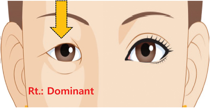
Right (Rt.) dominant eye and right ptosis. The patient feels discomfort because the dominant eye is affected. There is compensatory hyperfunction of the left levator (left eye looks normal) with the possibility of postoperative ptosis of the left eye due to Hering's law.
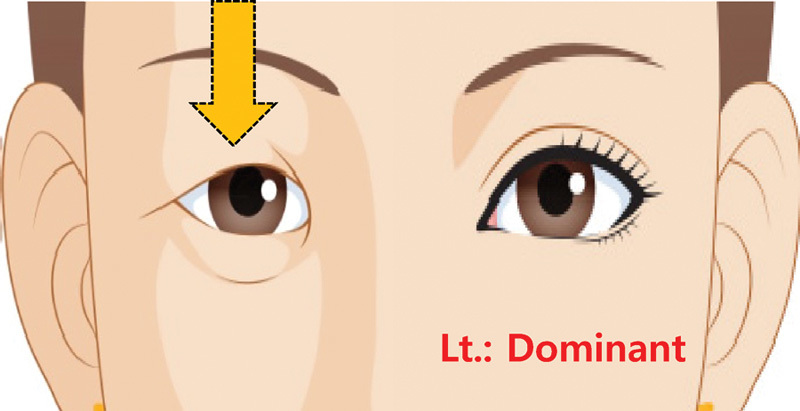
Left (Lt.) dominant eye and right ptosis. The patient feels less discomfort because the dominant eye is not affected. There is no compensatory hyperfunction of the right levator (the right eye looks ptotic) with less possibility of postoperative ptosis of the left eye.

Right (Rt.) dominant eye and left ptosis. The patient feels less discomfort because the dominant eye is not affected. There is no compensatory hyperfunction of the left levator (the left eye looks ptotic) with less possibility of postoperative ptosis of the right eye.

Left (Lt.) dominant eye and left ptosis. The patient feels discomfort because the dominant eye is affected. There is compensatory hyperfunction of the right levator (the right eye looks normal) with the possibility of postoperative ptosis of the right eye due to Hering's law.

Miles test: Dominant eye.

Degree of upward palpebral fissure slanting of the Asian eyelid.
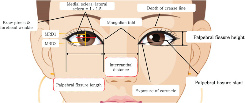
Criteria to be used in the evaluation of Asian eyelids.

Slant reduction surgery (lateral canthal lengthening and vertical enlargement). (A) Preoperative. (B) Postoperative 2 weeks. (C) Postoperative 4 weeks.
Preoperative evaluation for vertical enlargement of the palpebral aperture.

Slant reduction surgery (lateral canthal lengthening and vertical enlargement).

Lateral canthal lengthening procedure.

Classification of medial epicanthal fold. Source: Arch Aesthetic Plast Surg 2014 Feb;20(1):15-19. Available at: http://dx.doi.org/10.14730/aaps.2014.20.1.15
Medial canthal stretching test: Stretching of the medial canthus to find the proper point to lengthen the fissure.

Evaluating the amount to be removed: Medial canthal stretching test.

Medial canthal stretching test with tapes.

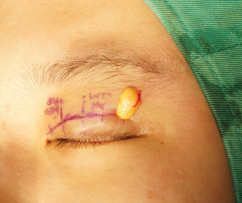
Preoperative measurement of epicanthoplasty and double fold making.

Measure the length of canthal web.

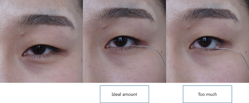
Lateral canthal stretching test.

(A) Lateral canthal stretching test: Tight canthus patient with tight canthal web displays limited exposure of conjunctiva. (B) Lateral canthal stretching test: Loose canthus patient with loose canthal web displays exposure of bulbar and palpebral conjunctiva.

Slant reduction surgery for reverse ptosis: Vertical enlargement procedure. (A) Preoperative. (B) Postoperative 2 weeks.

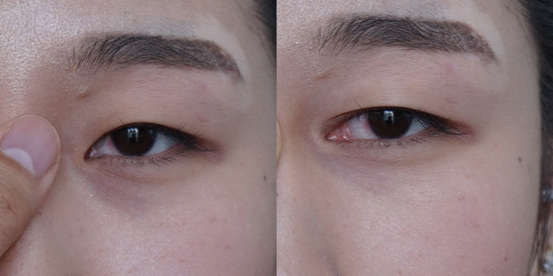
Evaluating lower lid tone. (A) Snap back test. (B) Distraction test.

Pretarsal augmentation (love band). (A) Preoperative: Looks normal and natural. (B) Postoperative: Slightly overexaggerated, but the patient is satisfied with the results. Beauty is in the eyes of the beholder.

Tear trough ligament: Orbicularis retaining ligament complex. OOM, orbicularis oculi muscle; ORL, orbicularis retaining ligament.

Part of the periosteum where tear trough ligament arises is more densely adherent to the orbital rim than that of the orbicularis retaining ligament.
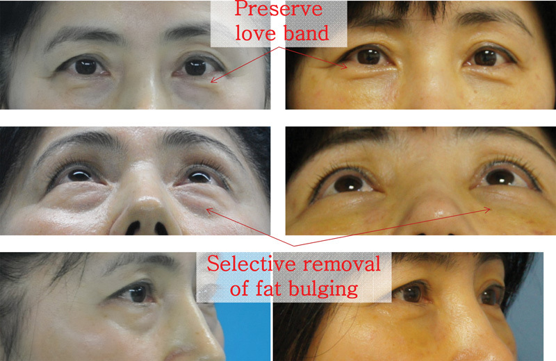
Selective removal of fat bulging while maintaining love band.
References
-
- Lee E I, Kim N H, Park R H, Park J B, Ahn T J. The relationship between eyebrow elevation and height of the palpebral fissure: should postoperative brow descent be taken into consideration when determining the amount of blepharoptosis correction? Arch Aesthetic Plast Surg. 2014;20(1):20–25.
-
- Miles W R. Ocular dominance demonstrated by unconscious sighting. J Exp Psychol. 1929;12(2):113–126.
-
- Glat P M Jelks G W Jelks E B Wood M Gadangi P Longaker M T Evolution of the lateral canthoplasty: techniques and indications Plast Reconstr Surg 199710061396–1405., discussion 1406–1408 - PubMed
-
- Park J I. Z-epicanthoplasty in Asian eyelids. Plast Reconstr Surg. 1996;98(4):602–609. - PubMed
-
- Wong C H, Hsieh M K, Mendelson B. The tear trough ligament: anatomical basis for the tear trough deformity. Plast Reconstr Surg. 2012;129(6):1392–1402. - PubMed
LinkOut - more resources
Full Text Sources
Other Literature Sources
