. 2015 Jul 8;48(Pt 4):1102-1113.
doi: 10.1107/S1600576715010420. eCollection 2015 Aug 1.

Methods for analysis of size-exclusion chromatography-small-angle X-ray scattering and reconstruction of protein scattering

Affiliations

Methods for analysis of size-exclusion chromatography-small-angle X-ray scattering and reconstruction of protein scattering

Andrew W Malaby et al. J Appl Crystallogr. .

Abstract

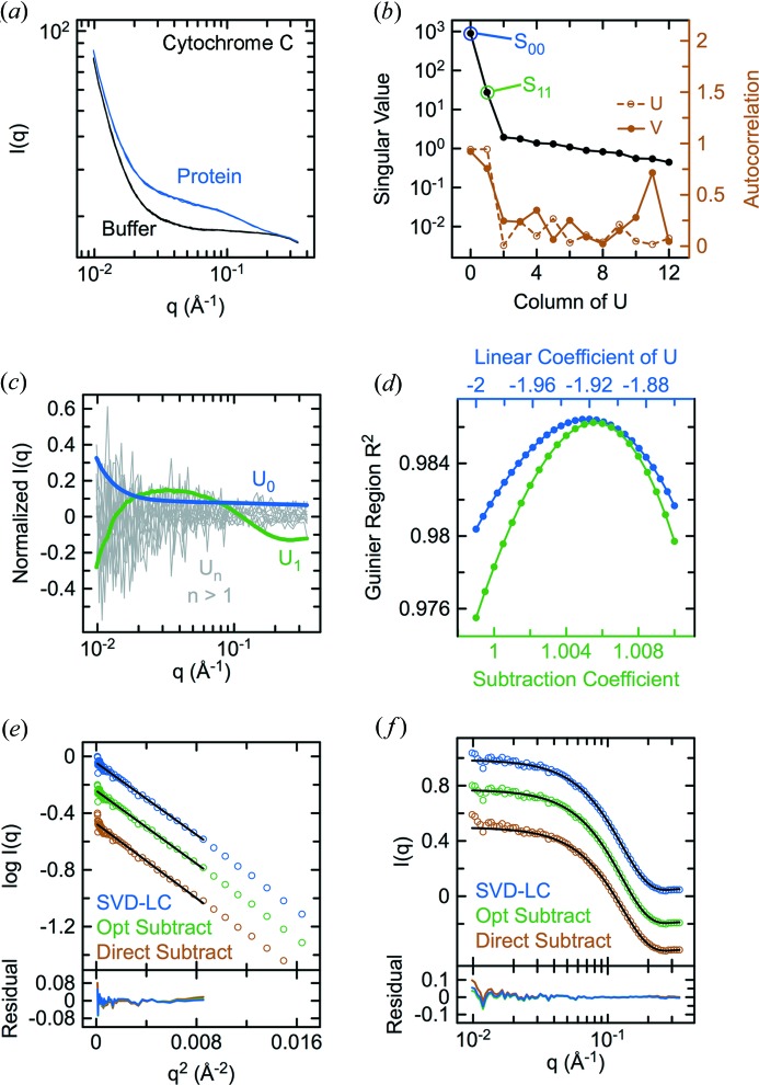
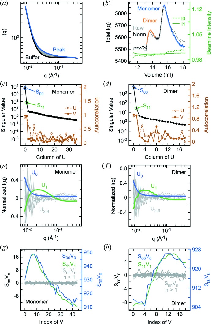
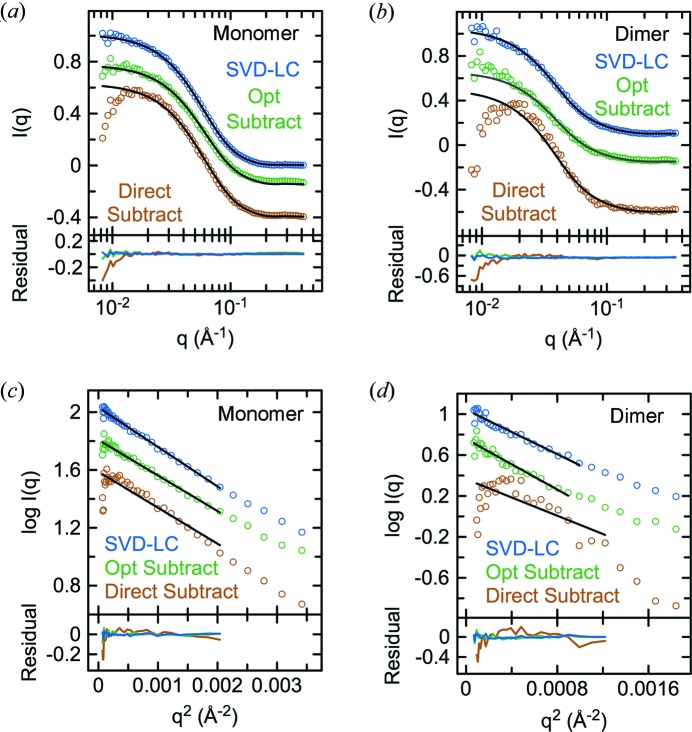
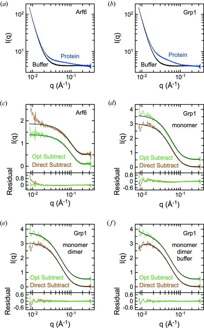
Size-exclusion chromatography in line with small-angle X-ray scattering (SEC-SAXS) has emerged as an important method for investigation of heterogeneous and self-associating systems, but presents specific challenges for data processing including buffer subtraction and analysis of overlapping peaks. This paper presents novel methods based on singular value decomposition (SVD) and Guinier-optimized linear combination (LC) to facilitate analysis of SEC-SAXS data sets and high-quality reconstruction of protein scattering directly from peak regions. It is shown that Guinier-optimized buffer subtraction can reduce common subtraction artifacts and that Guinier-optimized linear combination of significant SVD basis components improves signal-to-noise and allows reconstruction of protein scattering, even in the absence of matching buffer regions. In test cases with conventional SAXS data sets for cytochrome c and SEC-SAXS data sets for the small GTPase Arf6 and the Arf GTPase exchange factors Grp1 and cytohesin-1, SVD-LC consistently provided higher quality reconstruction of protein scattering than either direct or Guinier-optimized buffer subtraction. These methods have been implemented in the context of a Python-extensible Mac OS X application known as Data Evaluation and Likelihood Analysis (DELA), which provides convenient tools for data-set selection, beam intensity normalization, SVD, and other relevant processing and analytical procedures, as well as automated Python scripts for common SAXS analyses and Guinier-optimized reconstruction of protein scattering.

Keywords: Guinier optimization; linear combination; singular value decomposition; size-exclusion chromatography; small-angle X-ray scattering.


Figures

Figure 1
Figure 1
Guinier optimization of buffer subtraction for cyt c. (a) Raw scattering data sets with buffer or 4 mg ml−1 horse heart cyt c. (b) Singular values and autocorrelation of the columns of U and V after SVD of the data in (a). (c) Columns of the orthonormal U matrix corresponding to the rank-ordered singular values in (b). (d) R 2 values from automated Guinier optimization of the U 0 coefficient for linear combination of the two most significant SVD basis components or the scaling constant for buffer subtraction. Guinier (e) and CRYSOL (f) fits of the theoretical scattering for the crystal structure (PDB entry 1hrc; Bushnell et al., 1990 ▸) to the protein scattering curves obtained by SVD–LC, optimized buffer subtraction or direct subtraction.
Figure 2
Figure 2
SVD analysis and reconstruction of protein scattering from an SEC–SAXS experiment for Arf6. (a) Raw scattering data sets for Arf6NΔ13Q67L acquired in-line with chromatography on a 24 ml Superdex-200 column. (b) Raw and normalized total intensity elution profiles calculated by summing the intensities for each data set in (a) over the entire q range. Also shown are the incident (I 0) and transmitted (I 1) beam intensity profiles. (c) Singular values and autocorrelation of the columns of U and V after SVD of the data in (a). (d) Columns of the orthonormal U matrix corresponding to the rank-ordered singular values in (c). (e) Columns of the orthonormal V matrix multiplied by the corresponding rank-ordered singular values in (c). (f) CRYSOL fits of the theoretical scattering for the Arf6NΔ13Q67L-GTPγS crystal structure [PDB entry 2j5x (Pasqualato et al., 2001 ▸), with or without addition of a His6 tag from chain A in 2r09] to the protein scattering reconstructed by SVD–LC, Guinier-optimized subtraction or direct subtraction.
Figure 3
Figure 3
SVD analysis of an SEC–SAXS experiment for Grp163–399. (a) Raw scattering data sets for Grp163–399 acquired in-line with chromatography on a 24 ml Superdex-200 column. (b) Raw and normalized total intensity elution profiles calculated by summing the intensities for each data set in (a) over the entire q range. Also shown are the incident (I 0) and transmitted (I 1) beam intensity profiles. Singular values and autocorrelations of the columns of U and V after SVD of the monomer (c) and dimer (d) data sets corresponding to the peak regions indicated in (b). Columns of U corresponding to the rank-ordered singular values in (c) and (d) for the monomer (e) and dimer (f) regions. Columns of V multiplied by the corresponding rank-ordered singular values in (c) and (d) for the monomer (g) and dimer (h) regions.
Figure 4
Figure 4
Evaluation of methods for reconstructing the monomer and dimer scattering for the Grp163–399 SEC–SAXS experiment. CRYSOL fits of the monomer (a) and dimer (b) scattering reconstructed by SVD–LC, Guinier-optimized subtraction or direct subtraction with the theoretical scattering for either chain A or chains A and B (alternative definition of asymmetric unit) of the His6-Grp163–399 crystal structure (PDB entry 2r09). Guinier fits of the monomer (c) or dimer (d) scattering reconstructed by SVD–LC, Guinier-optimized subtraction, or direct subtraction.
Figure 5
Figure 5
Alternative analysis of the Grp163–399 SEC–SAXS data. (a) Transposed scattering data fitted with a sum of exponentially modified Gaussians as described in the text. (b) Individual components from a fit of the total scattering profile [equivalent to a summation of the transposed data in (a)] with a sum of exponentially modified Gaussians, where b is a baseline constant and eg1, eg2 and eg3 are the individual amplitude-weighted exponential Gaussian terms. (c) Comparison of the fitted baseline constant b(q) and the raw scattering data. Note that b(q) strongly resembles the expected buffer scattering. (d) R 2 values for Guinier optimization of the scaling constant for buffer subtraction using b(q) as the buffer. Comparison of protein scattering curves (e) and weighted Guinier fits (f) for direct and Guinier-optimized buffer subtraction using b(q) as buffer with the protein scattering curves and Guinier fits for Guinier-optimized reconstruction following SVD. For comparison, the curves were scaled by linear least squares.
Figure 6
Figure 6
Conventional SAXS experiments for Arf6 and Grp1 after SEC. SAXS data sets for Arf6NΔ13Q67L (a) and Grp163–399 (b) acquired after chromatography on a 3 ml Superdex-200 Increase column. Individual curves are from consecutive acquisitions under flow. (c) Fit of the His6-Arf6NΔ13Q67L CRYSOL model to the direct and Guinier-optimized buffer subtractions after averaging the data in (a). (d) Fit of the His6-Grp163–399 chain A monomer CRYSOL model to the direct and Guinier-optimized buffer subtractions after averaging the data in (b). Fits of the subtractions in (d) to a linear combination of CRYSOL models for the His6-Grp163–399 chain A monomer and chain A/B dimer either without (e) or including (f) a buffer scattering component. Black lines represent fitted models. All fits were performed by general linear least squares in DELA using theoretical models from CRYSOL.

Similar articles

Cited by

References

    1. Biou, V., Aizel, K., Roblin, P., Thureau, A., Jacquet, E., Hansson, S., Guibert, B., Guittet, E., van Heijenoort, C., Zeghouf, M., Perez, J. & Cherfils, J. (2010). J. Mol. Biol. 402, 696–707. - PubMed
    1. Brookes, E., Pérez, J., Cardinali, B., Profumo, A., Vachette, P. & Rocco, M. (2013). J. Appl. Cryst. 46, 1823–1833. - PMC - PubMed
    1. Bushnell, G. W., Louie, G. V. & Brayer, G. D. (1990). J. Mol. Biol. 214, 585–595. - PubMed
    1. Chen, B., Zuo, X., Wang, Y. X. & Dayie, T. K. (2012). Nucleic Acids Res. 40, 3117–3130. - PMC - PubMed
    1. David, G. & Pérez, J. (2009). J. Appl. Cryst. 42, 892–900.