Review
. 2015 Oct;12(4):896-909.
doi: 10.1007/s13311-015-0385-3.

Neuroinflammation: Ways in Which the Immune System Affects the Brain

Affiliations
Review

Neuroinflammation: Ways in Which the Immune System Affects the Brain

Richard M Ransohoff et al. Neurotherapeutics. 2015 Oct.

Abstract

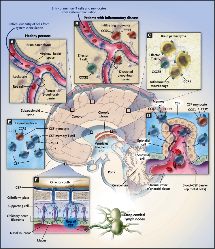
Neuroinflammation is the response of the central nervous system (CNS) to disturbed homeostasis and typifies all neurological diseases. The main reactive components of the CNS include microglial cells and infiltrating myeloid cells, astrocytes, oligodendrocytes, and the blood-brain barrier, cytokines, and cytokine signaling. Neuroinflammatory responses may be helpful or harmful, as mechanisms associated with neuroinflammation are involved in normal brain development, as well as in neuropathological processes. This review examines the roles of various cell types that contribute to the immune dysregulation associated with neuroinflammation. Microglia enter the CNS very early in embryonic development and, as such, play an essential role in both the healthy and diseased brain. B-cell diversity contributes to CNS disease through both antibody-dependent and antibody-independent mechanisms. The influences of these B-cell mechanisms on other cell types, including myeloid cells and T cells, are reviewed in relationship to antibody-mediated CNS disorders, paraneoplastic neurological diseases, and multiple sclerosis. New insights into neuroinflammation offer exciting opportunities to investigate potential therapeutic targets for debilitating CNS diseases.

Keywords: B cells; Central nervous system; immunology; inflammation; microglial cells; multiple sclerosis.; paraneoplastic syndromes.


Figures

Fig. 1
Fig. 1
Central memory T cells of cerebral spinal fluid (CSF) are mediators of central nervous system immune surveillance. From The New England Journal of Medicine, Israel F. Charo and Richard M. Ransohoff, The many roles of chemokines and chemokine receptors in inflammation, 354, pages 610–621. Copyright © 2006. Massachusetts Medical Society. Reprinted with permission from Massachusetts Medical Society [6].
Fig. 2
Fig. 2
Microglia-mediated engulfment of retinal ganglion cell (RGC) inputs is developmentally regulated. (A) Schematic of retinogeniculate pruning and strategy used for assessing engulfment: contralateral (red) and ipsilateral (blue) inputs overlap at early postnatal ages [postnatal day 5 (P5)]. Inputs from both eyes prune throughout the dorsal lateral geniculate nucleus (dLGN) during the first postnatal week, which is largely complete by postnatal day 9/10 (P9/10). Engulfment was analyzed throughout the dLGN. (B) Engulfment of RGC inputs is significantly increased during peak pruning in the dLGN (P5). *p < 0.001 by 1-way analysis of variance, n = 3 mice/age. (C) Engulfment in P5 dLGN occurs most significantly in synapse-enriched (contralateral and ipsilateral dLGN) versus nonsynaptic (optic tract) regions. *p < 0.01 by Student’s t test, n = 3 P5 mice. All error bars represent SEM. From Dorothy P Schafer, et al. Microglia sculpt postnatal neural circuits in an activity and complement-dependent manner, Neuron, 74(4), pages 691–705, 2012, Elsevier Inc., doi:10.1016/j.neuron.2012.03.026 [29].
Fig. 3
Fig. 3
N-methyl-D-aspartate receptor (NMDAR) antibodies in a 2-year-old male following herpes simplex virus encephalitis (HSVE). An unusual case presenting with leukoencephalopathic magnetic resonance imaging changes arising post-HSVE. (A–C) Serial axial T2 fluid attenuation inversion recovery images show localized cortical changes in the left thalamus and occipital lobe resulting from HSVE (not shown). However, after improvement from HSVE the patient relapsed with worsening of cognition and behavior and motor regression. This was associated with bilateral white matter signal changes (leuckencephalopathy) evident in (A) and (B). (C) Following treatment with intravenous immunoglobulin (IVIG), neuroimaging 2 months later demonstrated substantial resolution of the white matter change. (D) Neurologic relapse correlated with raised NMDAR antibodies in both serum and cerebrospinal fluid (CSF), and demonstrated a clinical response to immunotherapy with reduction of antibody levels. Ab = antibody. From N-methyl-D-aspartate receptor antibodies in post-Herpes simplex virus encephalitis neurological relapse, Yael Hacohen, et al., Mov Disord. 29(1). Copyright (c) 2014 [Movement Disorder Society] [69].
Fig. 4
Fig. 4
Central nervous system (CNS) autoimmunity demonstrated in N2-LacZ and wild-type (WT) mice. (A) T cells from transgenic mice (CD8-BG1 and CD4-BG2) reject tumor but do not cause autoimmune brain disease. Experimental design: 5 × 106 million CD8+ T cells with and without 5 × 106 CD4+ T cells from BG1, BG2, or WT mice were transferred into WT or N2-LacZ hosts and challenged with β-galactosidase (βgal)-expressing WP4 cells. After 30 days, mice were secondarily challenged with AdV-β-gal and pertussis toxin (PTx). Tumor growth in N2-LacZ or WT mice was assessed every 2–3 days. (B) T and B cells collaborate to generate neuronal targeting. Brain sections from a neurologically ill mouse were stained and arrows indicate dying neurons in the dentate gyrus. Arrowheads indicate normal reference neurons. H&E = hematoxylin and eosin; TUNEL = terminal deoxynucleotidyl transferase dUTP nick end labeling. From T cells targeting a neuronal paraneoplastic antigen mediate tumor rejection and trigger CNS autoimmunity with humoral activation. Nathalie E. Blachère, et al., Eur J Immunol. 44. Copyright (c) 2014 [WILEY-VCH Verlag GmbH & Co. KGaA, Weinheim] [78].
Fig. 5
Fig. 5
Direct implication of interleukin (IL)-6 from B cells mediating a proinflammatory T-cell response. (A) IL-6 production by B cells isolated from patients with multiple sclerosis (MS) is increased compared with healthy controls (HC) after in vitro stimulation. *p < 0.05. (B) IL-6 production from B cells from patients with MS before and after rituximab treatment (Rtx: 1000 mg  × 2 infusions, 2 weeks apart). *p < 0.05; NS = not significant (p > 0.05). (C) Mice with a B-cell IL-6 deficiency (B-IL-6−/−) develop an attenuated form of experimental autoimmune encephalomyelitis (EAE), implying that B cells drive disease exacerbation through the production of IL-6. EAE progression was monitored for 32 days after immunization with myelin oligodendrocyte glycoprotein (MOG) in B-WT (blue circles) and B-IL-6−/− mice (pink circles). (D) In the EAE model, IL-17 and interferon (IFN)-γ secretion by CD4 splenic T cells from B-WT (blue circles) and B-IL-6−/− mice (pink circles) shows impaired T helper 17 cell responses. Error bars represent SEM. ©Tom A. Barr, et al. 2009. Originally published in the Journal of Experimental Medicine. doi:10.1084/jem.20111675 [94].

References

    1. Alliot F, Lecain E, Grima B, Pessac B. Microglial progenitors with a high proliferative potential in the embryonic and adult mouse brain. Proc Natl Acad Sci U S A. 1991;88:1541–1545. doi: 10.1073/pnas.88.4.1541. - DOI - PMC - PubMed
    1. Alliot F, Godin I, Pessac B. Microglia derive from progenitors, originating from the yolk sac, and which proliferate in the brain. Brain Res Dev Brain Res. 1999;117:145–152. doi: 10.1016/S0165-3806(99)00113-3. - DOI - PubMed
    1. Bilimoria PM, Stevens B. Microglia function during brain development: New insights from animal models. Brain Res 2014. - PubMed
    1. Matyszak MK. Inflammation in the CNS: balance between immunological privilege and immune responses. Prog Neurobiol. 1998;56:19–35. doi: 10.1016/S0301-0082(98)00014-8. - DOI - PubMed
    1. Galea I, Bechmann I, Perry VH. What is immune privilege (not)? Trends Immunol. 2007;28:12–18. doi: 10.1016/j.it.2006.11.004. - DOI - PubMed