. 2015 Aug 26:6:8146.
doi: 10.1038/ncomms9146.

Chamber identity programs drive early functional partitioning of the heart

Affiliations

Chamber identity programs drive early functional partitioning of the heart

Christian Mosimann et al. Nat Commun. .

Abstract

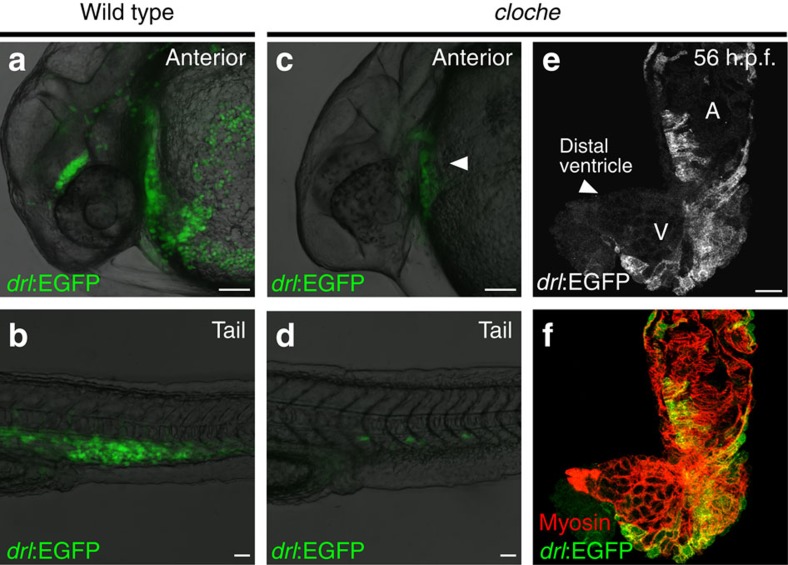
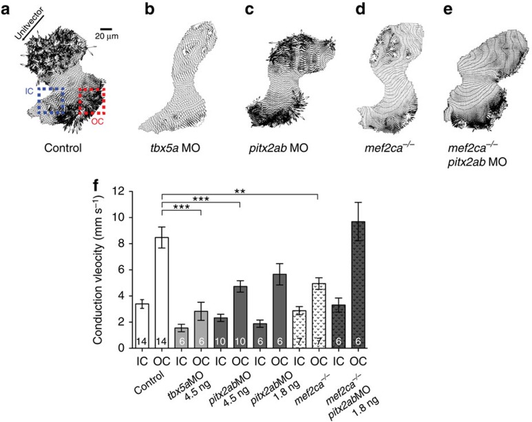
The vertebrate heart muscle (myocardium) develops from the first heart field (FHF) and expands by adding second heart field (SHF) cells. While both lineages exist already in teleosts, the primordial contributions of FHF and SHF to heart structure and function remain incompletely understood. Here we delineate the functional contribution of the FHF and SHF to the zebrafish heart using the cis-regulatory elements of the draculin (drl) gene. The drl reporters initially delineate the lateral plate mesoderm, including heart progenitors. Subsequent myocardial drl reporter expression restricts to FHF descendants. We harnessed this unique feature to uncover that loss of tbx5a and pitx2 affect relative FHF versus SHF contributions to the heart. High-resolution physiology reveals distinctive electrical properties of each heart field territory that define a functional boundary within the single zebrafish ventricle. Our data establish that the transcriptional program driving cardiac septation regulates physiologic ventricle partitioning, which successively provides mechanical advantages of sequential contraction.


Conflict of interest statement

L.I.Z. is a founder and stock holder of Fate, and a scientific advisor for Stemgent. The remaining authors declare no competing financial interests.

Figures

Figure 1
Figure 1. drl transgene reporter expression in the early lateral plate mesoderm and cardiovascular development.
(a) The zebrafish draculin (drl) locus; blue depicts coding exons (with ATG and Stop codons), yellow are untranslated regions, a DANA retro-transposon is located in the upstream region, arrows and red bar indicate PCR product used for cloning the regulatory sequence. (b) drl:EGFP reporter expression during gastrulation (80% epiboly, inset shows the embryo in bright-field view), dorsal to the right; scale bar, 100 μm; note the scattered band of cells that form the precursors of the lateral plate mesoderm. (c,d) Bud stage embryos probed for endogenous drl (c, mRNA in situ hybridization, ISH) versus drl:EGFP (d, composite bright-field and EGFP confocal stack image) depicting the continuous lateral plate mesoderm; anterior to the top, posterior to the bottom and dorsal to the right; scale bar, 100 μm. (e) EGFP fluorescence from homozygous drl:EGFP transgenic zebrafish; 5–7 somite stage view of the converging lateral plate mesoderm; scale bar, 100 μm. (fl) Genetic lineage tracing using drl:creERT2 × ubi:Switch (schematic on top), 4-OHT-induced at shield stage, labels the lateral plate mesoderm descendants at 36 h.p.f., including cardiac (outlined arrowhead in f; scale bar, 250 μm), pectoral fin mesoderm (solid arrowhead in f,g; scale bar, 50 μm), red blood cells (RBCs, h; scale bar, 50 μm), kidney (asterisks in i,j; scale bar, 50 μm), plus vasculature and scattered superficial trunk muscle cells (solid arrowheads in i). (k,l) 4-OHT induction at 12 somite stage and imaged at 36 h.p.f. refines tracing exclusively to the circulatory system and blood, note absence of kidney tracing (asterisks in k); scale bar, 50 μm.
Figure 2
Figure 2. drl transgenic reporters track cardiac development and distinguish FHF from SHF myocardium.
(a,b) Confocal projection of the anterior lateral plate mesoderm (ALPM) with hatching gland most anteriorly at 16 somite stage (ss) marked by drl:EGFP, dorsal view (a, green in b) and lmo2:dsRED2 (endothelial and haematopoietic precursors in red, b); asterisks in b indicate migrating myeloid precursors, solid arrowhead indicates merging endocardium precursors, and open arrowheads indicate positions of presumptive myocardium precursors; anterior to the top and posterior to the bottom; scale bar, 100 μm. (cf) Confocal projections of the linear heart tube at 26 h.p.f., anterior to the left, arterial pole (AP) and venous pole (VP) indicated, (c,d) top-down 2-μm projection, (e,f) transversal optical 2-μm section through heart tube. drl expression (drl:EGFP counter-stained with anti-GFP) in both the central endocardium and the myocardium (counter stained with anti-myosin/MF20 in red, and 4,6-diamidino-2-phenylindole (DAPI) in blue for nuclei in d,f; scale bar, 10 μm. (g,h) 56 h.p.f. two-chambered hearts transgenic for drl:EGFP top-down 2-μm confocal projection counter stained for anti-GFP (g,h), anti-myosin/MF20 in red, DAPI in blue for nuclei in h. EGFP is expressed in outer ventricular curvature and part of atria, with dotted lines indicating the margins of expression domains, the solid arrowhead point to drl:EGFP-negative regions at sinoatrial node (SA) and distal ventricle including outflow tract in g; scale bar, 10 μm. (ik) Ventral view of a two-chambered heart from a drl:EGFP; ltbp3:TagRFP2Acre double-transgenic embryo at 72 h.p.f.; at this stage, drl:EGFP-positive FHF-derived cardiac cells (i, green in k) comprise central portion of the cardiac tube, while the SHF-derived distal ventricular cardiomyocytes population (arrowhead in i) is labelled by the ltbp3:TagRFP2Acre transgene (j, red in k); scale bar, 50 μm. (l,m) drl:creERT2 × myl7:loxP-Amcyan-loxP-ZsYellow (schematic) induced at shield stage labels all myocardial descendants at 72 h.p.f. including the outflow tract cardiomyocytes in the distal ventricle (arrowhead, l); scale bar, 50 μm. Atrium (A) and ventricle (V) labelled in g,i,l.
Figure 3
Figure 3. Lack of endocardial signals does not affect integration of FHF with SHF.
(a,b) Anterior (a) and tail region (b) expression of drl:EGFP in wild-type zebrafish at 24–28 h.p.f., scale bar, 50 μm; note the circulating red blood cells. (c,d) Anterior (c) and tail region (d) drl:EGFP expression at 24–28 h.p.f. in the recessive zebrafish mutant cloche, which lacks all endothelial and haematopoietic lineages; scale bar, 50 μm; note the formation of linear heart tube in cloche (solid arrowhead in c and residual angioblasts in the tail (d). (e,f) Top-down 2-μm confocal projection of 56 h.p.f. cloche mutant heart, drl:EGFP expression counter stained with anti-GFP (e, green in f) and cardiomyocytes stained with myosin/MF20 (red in f); scale bar, 50 μm; note how despite missing endocardium the heart features both demarcated FHF and SHF myocardium, solid arrowhead in e indicates the distal ventricle region. Atrium (A) and ventricle (V) labelled in e.
Figure 4
Figure 4. tbx5a and pitx2ab affect the relative contribution of FHF versus SHF to the heart tube.
(aj) Top-down 2-μm confocal sections through zebrafish 54–56 h.p.f. hearts; scale bar, 20 μm. (ad) Hearts mutant for cloche (clo) to enable imaging of myocardium without endocardial and erythrocyte signal in drl:EGFP transgenics, stained for EGFP expression from drl:EGFP (green, monochrome channel image in right column) and myosin/MF20 (red); (a,b) cloche-only control, (c,d) morpholino knockdown of tbx5a leads to expansion of FHF. (e,f) Loss of pitx2ab results in diminishing of FHF and expansion of SHF. (gj) Genetic interplay of mef2ca with pitx2ab on FHF versus SHF contribution in wild-type hearts. (g,h) Loss of mef2ca leads to only mild reduction of FHF contribution. (i,j) Concomitant loss of mef2ca and pitx2ab reverts the pitx2ab morpholino (MO)-mediated impact on FHF formation. (k) Graph displaying the ratio of FHF area labelled by drl:EGFP expression to the whole ventricular area determined by anti-myosin staining in hearts from experiments represented in af. (l) Graph showing percentage of newly added cardiomyocytes to the ventricle after photoconversion of myl7:nlsKikGR at 27 h.p.f. and imaged at 56 h.p.f. (see also Supplementary Fig. 6). (m) Graph showing the restoration of the FHF area labelled by drl:EGFP expression to the whole ventricular area determined by anti-myosin staining in hearts with combined loss of pitx2ab and mef2ca comparing with the single morphant or control hearts. Error bars in km=s.d., asterisks indicate significance, significance tested by two-tailed unpaired t-test, P<0.05. (n) At 54–56 hpf, whole-embryo microarray comparison of tbx5a versus pitx2ab knockdown reveals cell adhesion genes significantly deregulated (red dots); note the upregulation of the SHF regulator mef2ca on pitx2ab knockdown (orange dot). (o) mRNA in situ hybridization of acta2a as representative deregulated gene at 52–56 h.p.f.; closeups of heart regions, anterior to the left, in indicated conditions, dotted outlines indicate heart tube; scale bar, 100 μm (see also Supplementary Fig. 8 for more examples).
Figure 5
Figure 5. Myocardial conduction velocity gradient depends on proper integration of FHF with SHF.
(ae) Vector field maps of control (a) hearts revealing the difference in cardiomyocyte connectivity of outer curvature (OC, red square ROI), presumptive FHF and inner curvature (IC, blue square ROI), presumptive SHF. Hearts of tbx5a (b), pitx2ab (c) morphants and mef2ca mutants (d) show the marked slowing and change from complex to more uniform conduction in OC; partial recovery of these polarities occur in combined mef2ca mutant with loss of pitx2ab (e); unit vector shows the principal direction of propagation. (f) Mean estimated conduction velocities reveal marked deceleration of conduction velocities in tbx5a, pitx2ab morphant and mef2ca mutant hearts. Note the complete loss of the velocity coupling gradient in tbx5a morphant hearts and partial loss in pitx2ab morphant hearts and mef2ca mutants compared with control hearts. The reduction of pitx2ab in mef2ca mutants completely rescued the conduction velocity coupling gradient with marked acceleration of conduction velocities in OC; error bar=s.d., asterisks indicate significance, statistical significance tested with one-way analysis of variance, with Tukey's post test, P<0.05. All experiments performed at 54 h.p.f.
Figure 6
Figure 6. Myocardial connectivity depends on proper integration of FHF with SHF.
(ae) Vector polar plots, in which each point represents a vector of the vector field map plotted in respect to its magnitude (velocity) and direction (angle from unit vector), red points show the frequency of angle distribution at 22.5° intervals. (a) Wild type as control reference. Loss of tbx5a (b) results in almost uniform conduction, while loss of pitx2ab (c) and mef2ca (d) partially impairs the coupling gradients as compared to the control (a) hearts. Combined loss of pitx2ab and mef2ca (e) show marked recovery of large velocity vectors. Note the discrete absence of all angles between +160° and+200° in these hearts. (fj) Frequency angle polar plots averaged from ROIs depicted in (Fig. 5a); majority of the vectors in control (f) hearts of OC/FHF point away from OFT revealing orthogonal mean coupling directions between OC/FHF and IC/SHF. Reduction of tbx5a (g) and pitx2ab (h) levels leads to complete loss of the orthogonal coupling and impairment in mef2ca mutants (i). Combined mef2ca mutants with loss of pitx2ab (j) partially rescues the orthogonal coupling direction. (k) Histogram of angle variability between inner and outer ventricular curvatures plotted as the angle s.d. in degrees. The angle variability is higher in outer curvature of control hearts, but this difference is diminished in the absence of tbx5a, pitx2ab, mef2ca, and combined pitx2ab morphant and mef2ca mutant hearts. Asterisks indicates significance, statistical significance tested with one-way analysis of varinace, with Tukey's post test, P<0.05. All experiments performed at 54 h.p.f.

References

    1. Ishitobi H. et al. Molecular basis for Flk1 expression in hemato-cardiovascular progenitors in the mouse. Development 138, 5357–5368 (2011). - PMC - PubMed
    1. Van Handel B. et al. Scl represses cardiomyogenesis in prospective hemogenic endothelium and endocardium. Cell 150, 590–605 (2012). - PMC - PubMed
    1. Wang Q., Lan Y., Cho E. S., Maltby K. M. & Jiang R. Odd-skipped related 1 (Odd 1) is an essential regulator of heart and urogenital development. Dev. Biol. 288, 582–594 (2005). - PMC - PubMed
    1. Garrity D. M., Childs S. & Fishman M. C. The heartstrings mutation in zebrafish causes heart/fin Tbx5 deficiency syndrome. Development 129, 4635–4645 (2002). - PubMed
    1. Black B. L. Transcriptional pathways in second heart field development. Semin. Cell Dev. Biol. 18, 67–76 (2007). - PMC - PubMed

Publication types

MeSH terms

Associated data