. 2015 Oct;46(10):2926-34.
doi: 10.1161/STROKEAHA.115.010592. Epub 2015 Aug 25.

PD-L1 Monoclonal Antibody Treats Ischemic Stroke by Controlling Central Nervous System Inflammation

Affiliations

PD-L1 Monoclonal Antibody Treats Ischemic Stroke by Controlling Central Nervous System Inflammation

Sheetal Bodhankar et al. Stroke. 2015 Oct.

Abstract

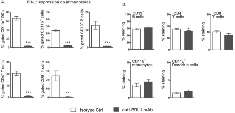
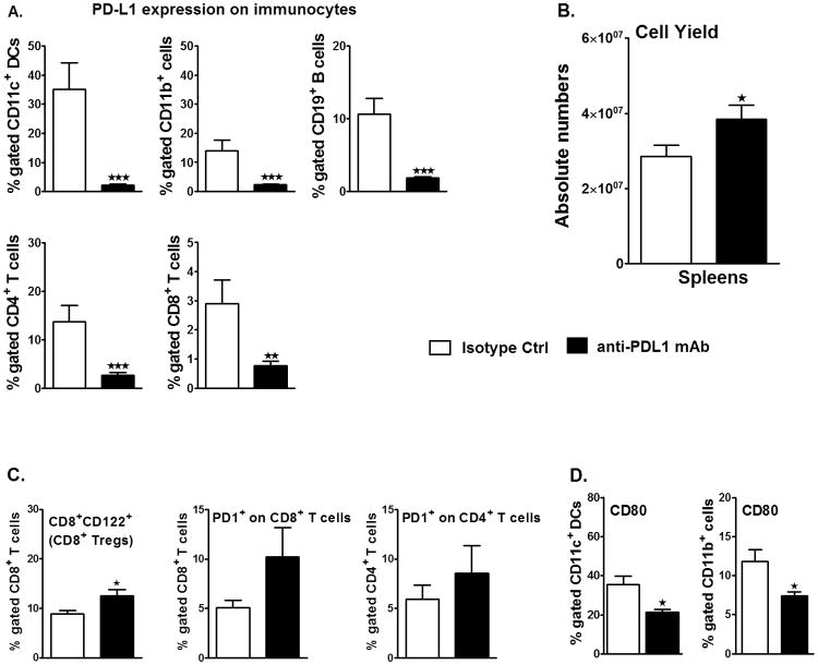
Background and purpose: Both pathogenic and regulatory immune processes are involved in the middle cerebral artery occlusion (MCAO) model of experimental stroke, including interactions involving the programmed death 1 (PD-1) receptor and its 2 ligands, PD-L1 and PD-L2. Although PD-1 reduced stroke severity, PD-L1 and PD-L2 appeared to play pathogenic roles, suggesting the use of anti-PD-L monoclonal antibody therapy for MCAO.

Methods: Male C57BL/6 mice were treated with a single dose of anti-PD-L1 monoclonal antibody 4 hours after MCAO and evaluated for clinical, histological and immunologic changes after 96 hours of reperfusion.

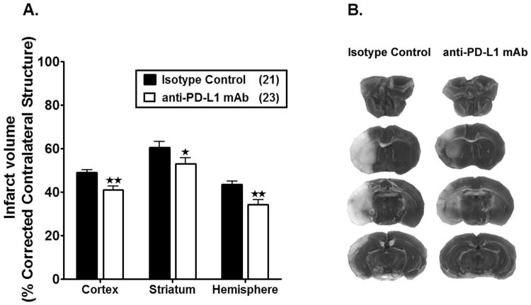
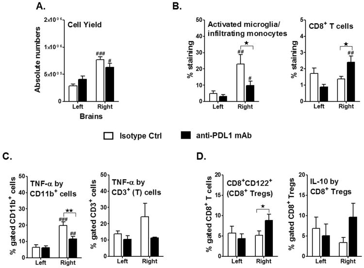
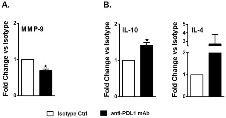
Results: Blockade of the PD-L1 checkpoint using a single injection of 200 μg anti-PD-L1 monoclonal antibody given intravenously 4 hours after occlusion significantly reduced MCAO infarct volumes and improved neurological outcomes after 96 hours of reperfusion. Treatment partially reversed splenic atrophy and decreased central nervous system infiltrating immune cells concomitant with enhanced appearance of CD8(+) regulatory T cells in the lesioned central nervous system hemisphere.

Conclusions: This study demonstrates for the first time the beneficial therapeutic effects of PD-L1 checkpoint blockade on MCAO, thus validating proposed mechanisms obtained in our previous studies using PD-1- and PD-L-deficient mice. These results provide strong support for the use of available humanized anti-PD-L1 antibodies for treatment of human stroke subjects.

Keywords: anti-PD-L1 antibody therapy; interleukin-10; middle cerebral artery occlusion; reperfusion; stroke.


Figures

Figure 1
Figure 1. A single dose of anti-PD-L1 mAb depletes PD-L1 expression without affecting the cell composition in naive male WT mice
A. Percentage of PD-L1 expression on CD11c+ dendritic cells, CD11b+ monocytes, CD19+ B-cells and CD4+ and CD8+ T-cells in spleens of anti-PD-L1 vs. isotype control mAb treated adult naïve C57BL/6J WT male mice as determined after 96 h by flow cytometry. B. Frequency of CD19+ B-cells, CD4+ and CD8+ T-cells, CD11b+ monocytes and CD11c+ dendritic cells in spleens of anti-PD-L1 vs. isotype control mAb treated naïve WT male mice as determined by flow cytometry. Values represent mean of 4 mice for the control group and 5 mice for the anti-PD-L1 mAb treated group. Statistical analysis was performed with Student's t test. Significant differences between control and anti-PD-L1 mAb-treated groups are indicated as *p≤0.05, **p≤ 0.01 and ***p≤ 0.001.
Figure 2
Figure 2. Treatment with anti-PD-L1 mAb, 4 h after MCAO, reduces infarct volume in male WT mice
Infarct volume (% corrected contralateral structure) in cortex, striatum and hemisphere were determined by 2,3,5-triphenyltetrazolium chloride staining in isotype control-treated vs. anti-PD-L1 mAb-treated adult male WT mice. All mice underwent 1 hour of middle cerebral artery occlusion (MCAO), with treatments at 4 h after MCAO followed by 96 hours of reperfusion. A. Infarct volumes in anti-PD-L1 Ab- treated (n=23) male WT mice were significantly reduced compared to the isotype control-treated (n=21) mice. Values represent mean ± SEM. *p<0.05, **p<0.01. B. Representative cerebral sections showing localization of the ischemic lesions in anti-PD-L1 vs. isotype control mAb treated mice. Note-Three mice from the anti-PD-L1 mAb treated group were excluded due to hemorrhagic transformation.
Figure 3
Figure 3. Treatment with anti-PD-L1 mAb, 4 h after MCAO, reduces inflammatory responses but leads to accumulation of CD8 Tregs in the ischemic brain
Mononuclear cells were isolated, following 96 h reperfusion, from brains of WT male mice, that received either isotype control or anti-PD-L1 treatment, 4 h after MCAO and were analyzed for: A. Total cell count via hemocytometer. Values represent mean numbers (±SEM) of indicated cell subsets from 13-14 mice per group, from at least 4 separate experiments. B. CD11b+CD45high activated microglia (MG)/monocytes and CD8+ T-cells were obtained from the non-ischemic (left) and ischemic (right) hemispheres of the isotype or anti-PD-L1 mAb-treated mice. Values represent mean numbers (±SEM) of indicated cell subsets, from 12-13 mice per group, from at least 4 separate experiments. Determinations of: C. TNF-α production by CD11b+ cells, likely activated microglia/monocytes, and TNF-α+CD3+ T-cells in the non-ischemic (left) and ischemic (right) hemispheres of the isotype or anti-PD-L1 mAb-treated mice, 96 hours after MCAO. Values represent mean numbers (±SEM) of indicated cell subsets from 10-12 mice of each group, from at least 4 separate experiments. D. CD8+CD122+ T-cells and IL-10 production by gated CD8+CD122+ T-cells. Data are representative of 4 independent experiments with brains processed from 13-14 individual mice (mean ± SEM). Statistical analysis was performed with Student's t test with Welch's correction. Significant differences between sample means are indicated as #p≤0.05; ##p≤ 0.01; ###p≤ 0.001 as compared to their respective left hemisphere and *p≤0.05; **p≤ 0.01 compared to the ischemic right hemisphere of isotype-treated WT mice, post-MCAO.
Figure 4
Figure 4. Treatment with anti-PD-L1 mAb, 4 h after MCAO, not only reduces the overall pro-inflammatory status but induces an anti-inflammatory milieu in the ischemic brain hemisphere
Brains were collected from isotype and anti-PD-L1 mAb-treated mice, 96 hours after occlusion, and mRNA prepared from ipsilateral (right) hemispheres of brain tissues for RT-PCR analysis. Fold change of mRNA levels is presented for A. inflammatory factor Mmp-9 and B. anti-inflammatory cytokines, Il-10 & Il-4. Values for A. and B. represent mean (±SEM) values for fold differences over the Ct values for isotype-treated ischemic brains. Significant differences between the right ischemic hemispheres of the isotype-treated and anti-PD-L1 mAb-treated groups were determined using Student's t-test. Significant differences between sample means are indicated *p≤0.05 compared to the ischemic right hemisphere of isotype-treated WT mice, post-MCAO.
Figure 5
Figure 5. Treatment with anti-PD-L1 mAb, 4 h after MCAO, rescues splenic atrophy, increases the expression of regulatory molecules on T-cells and decreases the expression of CD80 on APCs in spleens, post-MCAO
Ninety six hours after MCAO, mononuclear cells were isolated from spleens of isotype-treated or anti-PD-L1 mAb-treated mice and were analyzed for: A. Percentage of PD-L1 expression on CD11c+ dendritic cells, CD11b+ monocytes, CD19+ B-cells, CD4+ and CD8+ T-cells by flow cytometry. Values represent mean numbers (±SEM) of indicated cell subsets from 8-20 mice in each group, from at least 5 separate experiments B. Total cell count via hemocytometer. Values represent mean numbers (±SEM) of indicated cell subsets from 24-30 mice in each group, from at least 6 separate experiments; C. For CD8+CD122+ T-cells, data are representative of 4-5 independent experiments with spleens processed from 17-19 individual mice (mean ± SEM). For PD-1 expression on gated CD4+ T-cells and CD8+ T-cells, data are representative of 2 independent experiments with spleens processed from 4-5 individual mice (mean ± SEM). D. CD80 expression on gated CD11c+ and CD11b+ cells in the spleens. Values represent mean numbers (±SEM) of indicated cell subsets, from 8-12 individual mice from 4 separate experiments. Statistical analysis was performed with Student's t test with Welch's correction. Significant differences between isotype-treated and anti-PD-L1 mAb-treated groups, post-MCAO, are indicated as *p≤0.05, **p≤ 0.01 and ***p≤ 0.001.

References

    1. Dirnagl U, Iadecola C, Moskowitz MA. Pathobiology of ischaemic stroke: An integrated view. Trends Neurosci. 1999;22:391–397. - PubMed
    1. Lakhan S, Kirchgessner A, Hofer M. Inflammatory mechanisms in ischemic stroke: Therapeutic approaches. J Transl Med. 2009;7:97. - PMC - PubMed
    1. Schaller B, Graf R. Cerebral ischemia and reperfusion: The pathophysiologic concept as a basis for clinical therapy. J Cereb Blood Flow Metab. 2004;24:351–371. - PubMed
    1. Iadecola C, Anrather J. The immunology of stroke: From mechanisms to translation. Nat Med. 2011;17:796–808. - PMC - PubMed
    1. Macrez R, Ali C, Toutirais O, LeMauff B, Defer G, Dirnagle U, et al. Stroke and the immune system: From pathophysiology to new therapeutic strategies. Lancet Neurol. 2011;10:471–480. - PubMed

Publication types