. 2015 Sep;27(9):2600-15.
doi: 10.1105/tpc.15.00234. Epub 2015 Aug 25.

HIGH CHLOROPHYLL FLUORESCENCE145 Binds to and Stabilizes the psaA 5' UTR via a Newly Defined Repeat Motif in Embryophyta

Affiliations

HIGH CHLOROPHYLL FLUORESCENCE145 Binds to and Stabilizes the psaA 5' UTR via a Newly Defined Repeat Motif in Embryophyta

Nikolay Manavski et al. Plant Cell. 2015 Sep.

Abstract

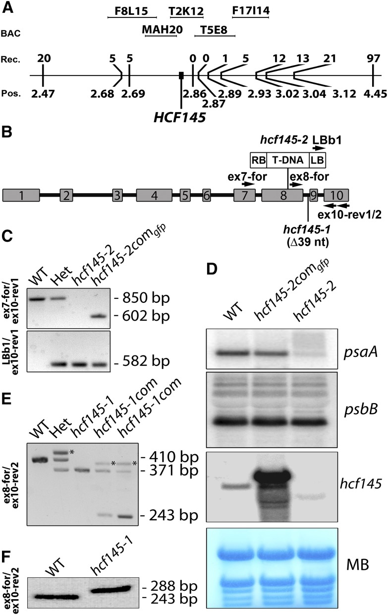
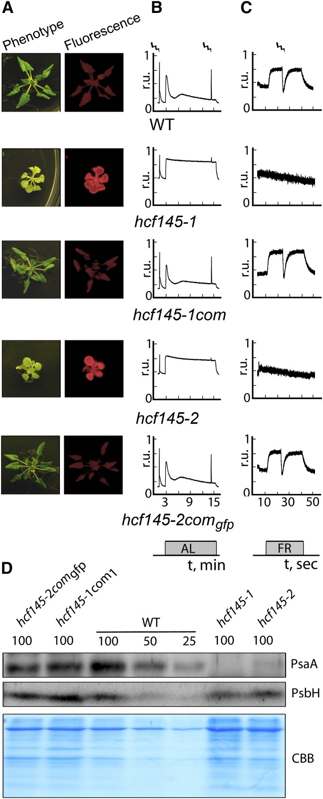
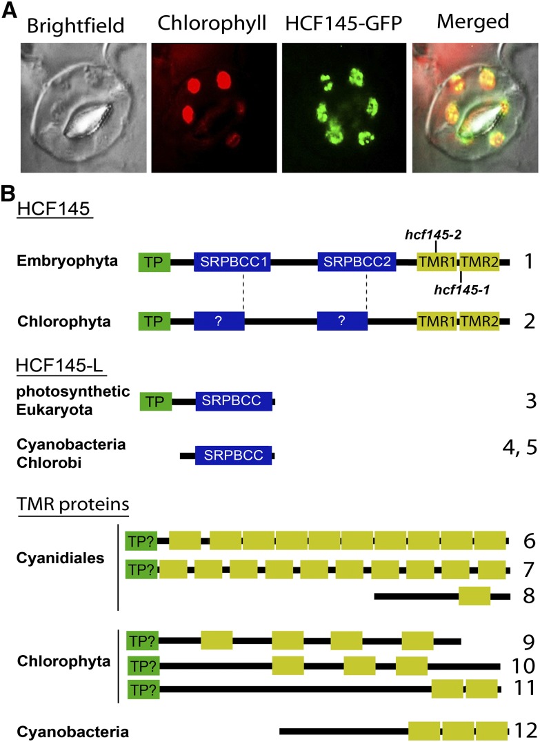
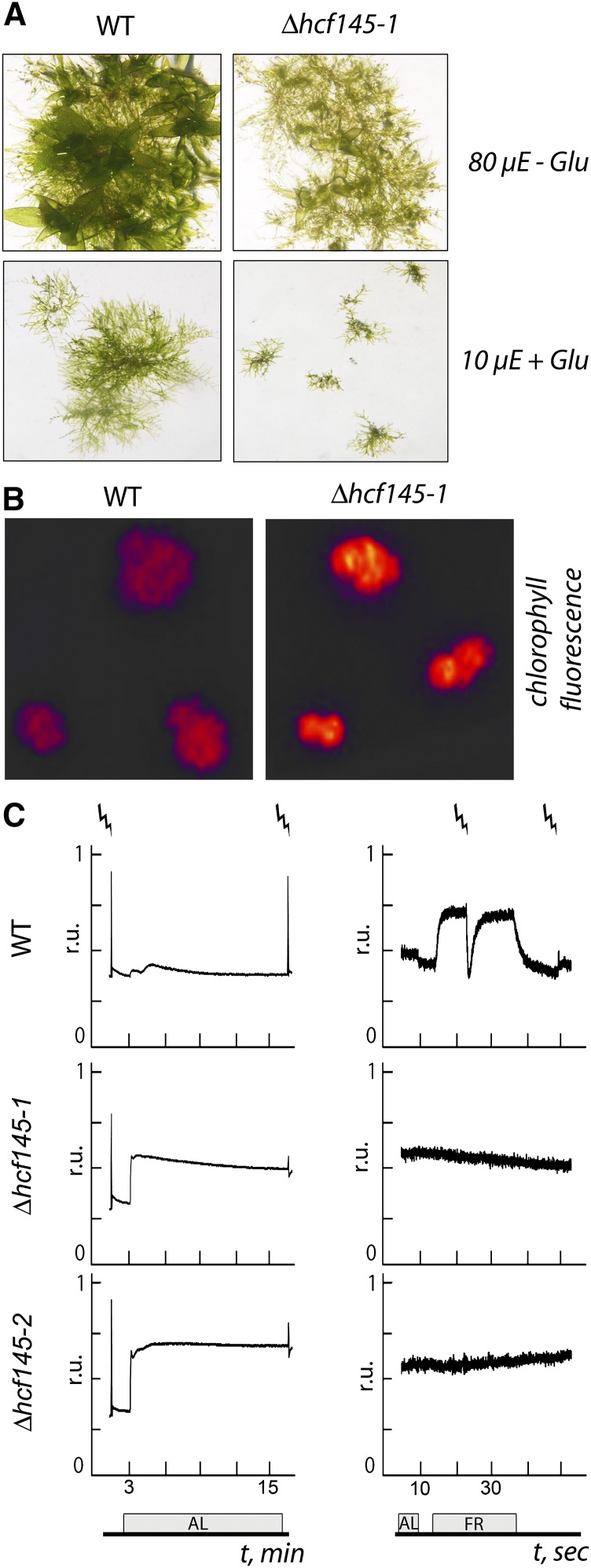
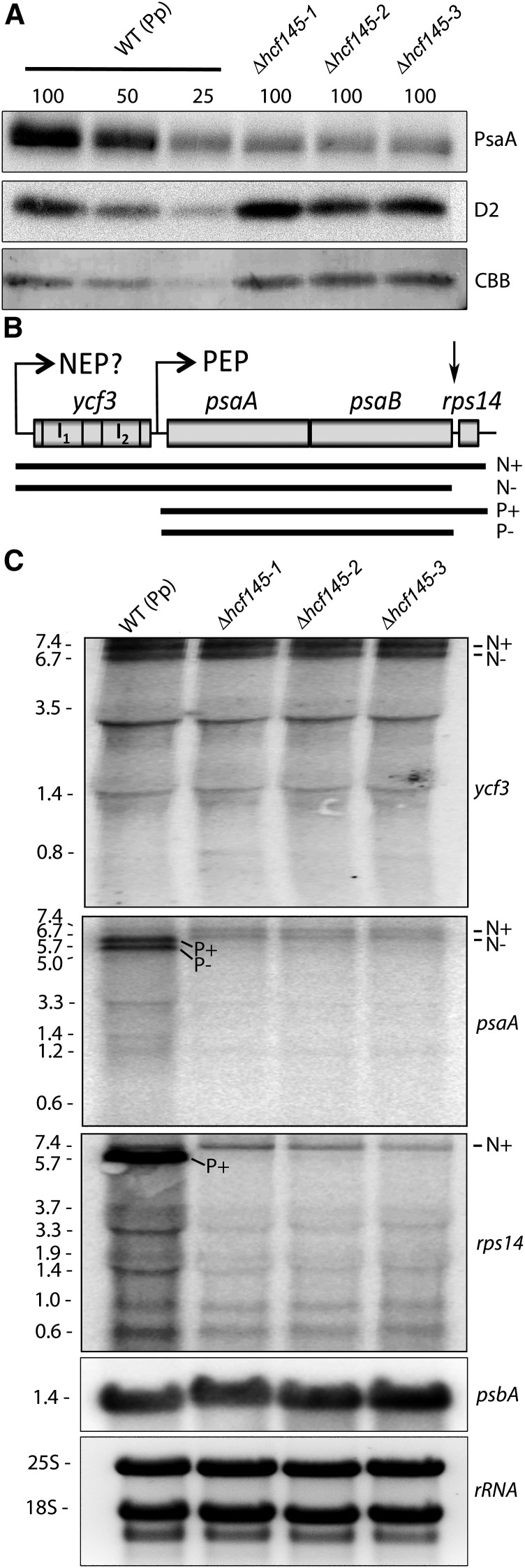
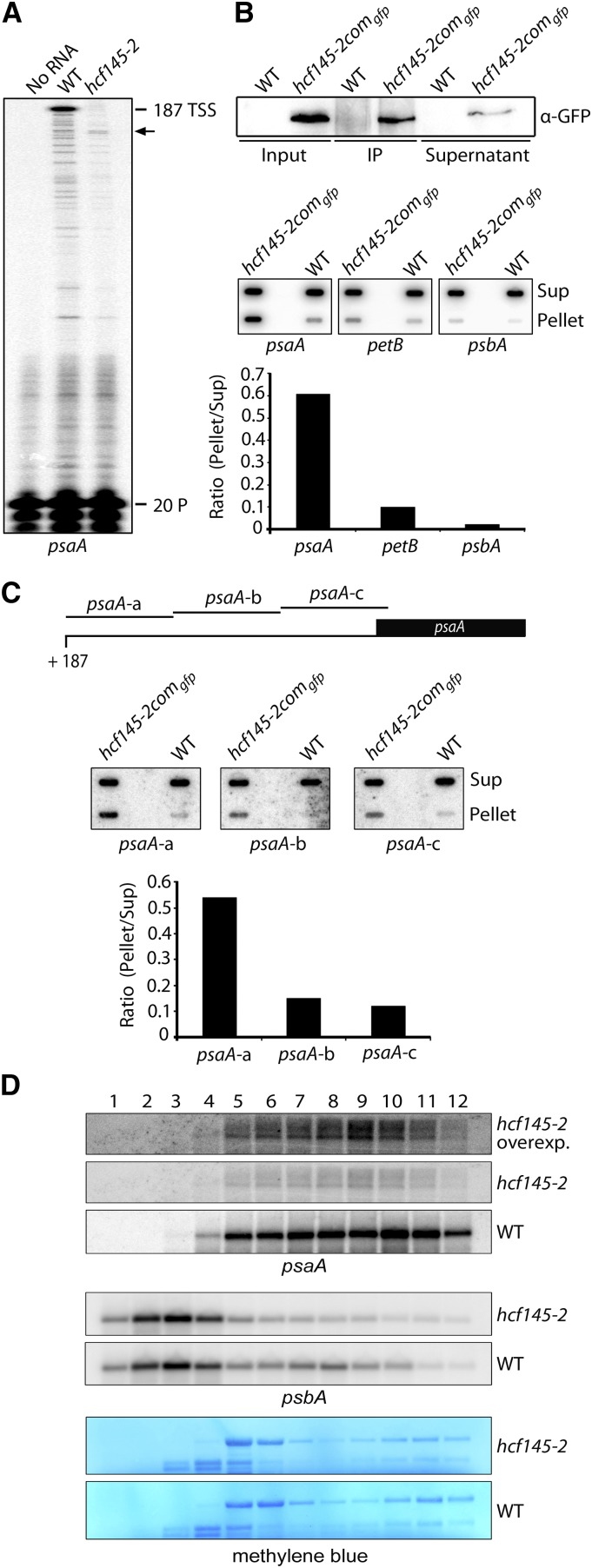
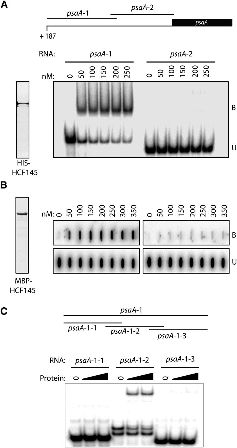
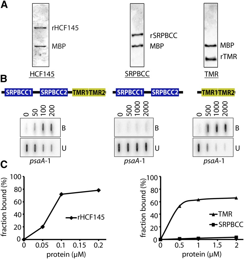
The seedling-lethal Arabidopsis thaliana high chlorophyll fluorescence145 (hcf145) mutation leads to reduced stability of the plastid tricistronic psaA-psaB-rps14 mRNA and photosystem I (PSI) deficiency. Here, we genetically mapped the HCF145 gene, which encodes a plant-specific, chloroplast-localized, modular protein containing two homologous domains related to the polyketide cyclase family comprising 37 annotated Arabidopsis proteins of unknown function. Two further highly conserved and previously uncharacterized tandem repeat motifs at the C terminus, herein designated the transcript binding motif repeat (TMR) domains, confer sequence-specific RNA binding capability to HCF145. Homologous TMR motifs are often found as multiple repeats in quite diverse proteins of green and red algae and in the cyanobacterium Microcoleus sp PCC 7113 with unknown function. HCF145 represents the only TMR protein found in vascular plants. Detailed analysis of hcf145 mutants in Arabidopsis and Physcomitrella patens as well as in vivo and in vitro RNA binding assays indicate that HCF145 has been recruited in embryophyta for the stabilization of the psaA-psaB-rps14 mRNA via specific binding to its 5' untranslated region. The polyketide cyclase-related motifs support association of the TMRs to the psaA RNA, presumably pointing to a regulatory role in adjusting PSI levels according to the requirements of the plant cell.


Figures

Figure 1.
Figure 1.
Mapping, PCR, and RNA Gel Blot Analysis of the Arabidopsis hcf145 Mutants. (A) Genetic mapping of the HCF145 mutation localized at map position (Pos.) 2.83 Mb on chromosome 5. Backcrosses to the Arabidopsis accession Landsberg erecta using 1281 F2 mutants obtained 0 recombinants (Rec.) in the region covered by the BACs T2K12 and T5E8. (B) Schematic representation of the HCF145 gene, indicating the 39-nucleotide deletion (∆39 nt) in hcf145-1, the T-DNA insertion in hcf145-2, and the location of the oligonucleotides used for PCR analysis. (C) Genotyping of wild-type, heterozygous, hcf145-2, and complemented hcf145-2comgfp lines by PCR analysis. Genomic and T-DNA primers used are shown on the left and in (B). The data demonstrate homozygosity of hcf145-2, successful complementation (upper panel), and the presence of the T-DNA in hcf145-2 (lower panel). (D) Expression of psaA and hcf145 in wild-type, mutant, and complemented lines. RNA gel blot analysis was performed using 10 µg total leaf RNA with strand-specific 80-mer oligonucleotides (psaA) and a full-length HCF145 cDNA (hcf145) as hybridization probes. MB, methylene blue. (E) Selection of the homozygous and complemented hcf145-1 mutants by PCR analysis using the primers ex8-for and ex10-rev2. A 410-bp fragment was amplified in the wild type and plants heterozygous for hcf145-1 (Het). A smaller fragment of 371 bp was amplified in hcf145-1, hcf145-1com, and Het lines. A 243-bp fragment, corresponding to the cDNA, was amplified in the hcf145-1com lines. The asterisks mark mismatching products of the amplified fragments in the heterozygous and hcf145-1com lines, migrating at higher molecular weight. (F) RT-PCR analysis of the wild type and hcf145-1. Exon-specific primers were used to amplify the cDNA of mutant and wild-type plants. The sizes of the products are indicated.
Figure 2.
Figure 2.
Spectroscopic and Immunological Analyses of the Arabidopsis hcf145 Mutants. (A) Photographs and chlorophyll a fluorescence images of 3-week-old plants grown on sucrose-supplemented medium at 10 µmol photons m−2 s−1. (B) Chlorophyll a fluorescence measurements. Strokes indicate saturating light pulses, and bars indicate the duration of actinic light (AL) application. r.u., relative units. (C) P700 redox kinetics. Strokes indicate saturating light pulses, and bars indicate the duration of far-red light (FR) application. r.u., relative units. (D) Immunoblot analyses using specific antisera for the large PSI core subunit PsaA and the PSII subunit PsbH. Protein loading corresponds to 4, 2, and 1 µg chlorophyll (100, 50, and 25, respectively). CBB, Coomassie blue.
Figure 3.
Figure 3.
Subcellular Localization of HCF145 and Its Motifs in HCF145, HCF145-L, and TMR Proteins. (A) Fluorescence micrographs demonstrating presence of the HCF145-GFP fusion in the chloroplasts of the hcf145-2comgfp guard cells. (B) Schematic representation and motif organization of HCF145, HCF145-L, and representative TMR proteins in photosynthetic organisms. N-terminal chloroplast transit peptide (TP; green boxes), SRPBCC motifs in HCF145 and HCF145-L proteins (blue boxes), and repeated TMR domains (yellow boxes) are shown. The positions of the mutations found in the hcf145-1 and hcf145-2 lines are indicated. (For multiple sequence alignments of the SRPBCC motifs in HCF145 and HCF145-L, and the TMR proteins, see Supplemental Figures 4, 5, and 7). The accession numbers correspond to the proteins numbered from 1 to 12: 1, Arabidopsis HCF145 (At5g08720); 2, C. subellipsoidea (XM_005645014); 3. Arabidopsis (At4g01650); 4, C. tepidum (AE006470); 5, Synechococcus sp CC9902 (CP000097); 6, C. merolae (XM_005538687); 7, G. sulphuraria (XM_005705295); 8, C. merolae (XM_005538035); 9, C. variabilis (XM_005846292); 10, C. subellipsoidea (C-169 XP_005645828); 11, Ostreococcus lucimarinus (XM_001415510); 12, Microcoleus sp PCC 7113 (AFZ22266).
Figure 4.
Figure 4.
Phenotype of the Moss Δhcf145 Mutants. (A) Wild type and Δhcf145-1 grown for 3 weeks in the absence (-Glu) and presence of glucose (+ Glu) at 80 and 10 μmol photons m−2 s−1, respectively. The phenotype of Δhcf145-1 is in all respects representative for all three knockout lines. (B) Chlorophyll fluorescence imaging of the wild type and Δhcf145-1, which is representative for all three knockout lines. (C) Chlorophyll a fluorescence (left) and P700 redox kinetics (right) in the wild type, Δhcf145-1, and Δhcf145-2. Strokes indicate saturating light pulses, and bars the duration of actinic light (AL) or far-red light (FR) application.
Figure 5.
Figure 5.
Immunological and RNA Gel Blot Analysis of Δhcf145 Mutants. (A) Immunoblot analysis with PsaA- and D2-specific antibodies. Protein loading corresponds to 4, 2, and 1 µg chlorophyll (100, 50, and 25, respectively). CBB, Coomassie blue. (B) Organization of the ycf3-psaA-psaB-rps14 gene cluster in P. patens. I1 and I2, intron 1 and intron 2, respectively. Primary transcripts generated by the NEP and the PEP and proposed processing products are shown below. N+ and N-, proposed NEP transcripts with and without rps14, respectively; P+ and P-, PEP transcripts with and without rps14, respectively. The ycf3, psaA, psbA, and rps14 probes were generated by PCR using primer pairs ppYcf3_f and -r, ppPsaA_f and -r, ppPsba_f and -r, and ppRps14_f and -r. For the amplification of an intron-less ycf3 probe, we used a cDNA as template. (C) RNA gel blot analysis using 10 µg total RNA from the wild-type and Δhcf145 lines and hybridization probes specific for ycf3, psaA, rps14, and psbB. The band sizes are given in kilobases on the left.
Figure 6.
Figure 6.
Determination of the psaA 5′ Termini, RNA Immunoprecipitation, and psaA Polysome Loading of hcf145-2. (A) Primer extension analysis with radiolabeled primer (20 P) indicates that the 5′ UTR of psaA is truncated in the Arabidopsis hcf145-2 mutant as indicated by arrows. TSS, transcription start site. (B) Detection of RNA coimmunoprecipitating with HCF145. Upper panel: Immunoprecipitation of the HCF145-GFP fusions from chloroplast extracts of hcf145-2comgfp using GFP antibodies. HCF145-GFP expression was driven by the 35S promoter. IP, immunoprecipitate. Lower panel: Coprecipitated RNAs of the supernatant (Sup) and the pellet were applied to slot blots. Filters were hybridized with the psaA 5′ UTR, petB ORF-, and psbA 5′-region-specific probes. The signals were quantified using ImageQuant software and the ratio of bound versus unbound RNA is indicated in the bar graph. (C) Fine-mapping of the psaA 5′ UTR RNAs that coimmunoprecipitate with HCF145. Slot-blot hybridization (as in [B]) with three probes covering the entire psaA 5′ UTR. The ratio of bound versus unbound RNA is indicated in the bar graph. (D) RNA gel blot analysis of polysome fractions 1 to 12 taken from the sucrose gradient of the wild-type and hcf145-2 plants. The probes used are indicated. rRNA was stained with methylene blue. Due to the low expression of psaA in the hcf145-2 mutant, the filter was overexposed (overexp.) for better comparison.
Figure 7.
Figure 7.
Recombinant HCF145 Binds with High Specificity to the 5′ End of the psaA 5′ UTR. (A) Generation of two overlapping transcripts covering the entire psaA 5′ UTR for binding assays (upper panel). EMSA using the two psaA 5′ UTR probes (lower panel). The binding reaction contained the indicated concentrations of recombinant HCF145 (rHCF145). Purified protein was stained with Coomassie blue (left panel). (B) Filter binding assay performed with the purified MBP-HCF145 fusion. Purified protein was visualized with Coomassie blue (left panel). psaA 5′ UTR probes and amounts of rHCF145 are indicated. B, bound RNA; U, unbound RNA. (C) EMSA using rHCF145 (see [B]) and three overlapping probes covering the psaA-1 probe in the 5′ region of the 5′ UTR (see [A]). A schematic representation of the probes is shown in the upper panel and the sequences of the probes are indicated in Supplemental Figure 2.
Figure 8.
Figure 8.
Purification and Filter Binding Assays of Defined Motifs of rHCF145 Protein. (A) Release of the entire recombinant HCF145 protein, a fragment containing the SRPBCC domains, as well as the tandem repeated TMR motifs from the MBP fusion. (B) Filter binding assays of purified proteins shown in (A) using increasing concentrations of protein up to 200 nM for the full-length protein and up to 2000 nM for the truncated versions. B, bound RNA; U, unbound RNA. (C) Quantitative determination of the RNA fraction bound to HCF145 to evaluate the dissociation constant (Kd) value.
Figure 9.
Figure 9.
HCF145 Is Predominantly Expressed in Dark-Grown Seedlings. Immunological analysis of HCF145, PsbP, and CP47 accumulation in Arabidopsis seedlings etiolated for 8 d and subsequently illuminated for 4, 8, 24, and 96 h at 50 μmol photons m−2 s−1. Seedlings grown for 8 d in the light were used for comparison.

References

    1. Amann K., Lezhneva L., Wanner G., Herrmann R.G., Meurer J. (2004). ACCUMULATION OF PHOTOSYSTEM ONE1, a member of a novel gene family, is required for accumulation of [4Fe-4S] cluster-containing chloroplast complexes and antenna proteins. Plant Cell 16: 3084–3097. - PMC - PubMed
    1. Barkan A. (1993). Nuclear mutants of maize with defects in chloroplast polysome assembly have altered chloroplast RNA metabolism. Plant Cell 5: 389–402. - PMC - PubMed
    1. Barkan A. (2011). Studying the structure and processing of chloroplast transcripts. Methods Mol. Biol. 774: 183–197. - PubMed
    1. Barkan A., Klipcan L., Ostersetzer O., Kawamura T., Asakura Y., Watkins K.P. (2007). The CRM domain: an RNA binding module derived from an ancient ribosome-associated protein. RNA 13: 55–64. - PMC - PubMed
    1. Barkan A., Small I. (2014). Pentatricopeptide repeat proteins in plants. Annu. Rev. Plant Biol. 65: 415–442. - PubMed

Publication types

MeSH terms

LinkOut - more resources