. 2016 Feb;24(1):17-25.
doi: 10.1038/mt.2015.150. Epub 2015 Aug 26.

Gene Therapy Restores Hair Cell Stereocilia Morphology in Inner Ears of Deaf Whirler Mice

Affiliations

Gene Therapy Restores Hair Cell Stereocilia Morphology in Inner Ears of Deaf Whirler Mice

Wade W Chien et al. Mol Ther. 2016 Feb.

Abstract

Hereditary deafness is one of the most common disabilities affecting newborns. Many forms of hereditary deafness are caused by morphological defects of the stereocilia bundles on the apical surfaces of inner ear hair cells, which are responsible for sound detection. We explored the effectiveness of gene therapy in restoring the hair cell stereocilia architecture in the whirlin mouse model of human deafness, which is deaf due to dysmorphic, short stereocilia. Wild-type whirlin cDNA was delivered via adeno-associated virus (AAV8) by injection through the round window of the cochleas in neonatal whirler mice. Subsequently, whirlin expression was detected in infected hair cells (IHCs), and normal stereocilia length and bundle architecture were restored. Whirlin gene therapy also increased inner hair cell survival in the treated ears compared to the contralateral nontreated ears. These results indicate that a form of inherited deafness due to structural defects in cochlear hair cells is amenable to restoration through gene therapy.


Figures

Figure 1
Figure 1
The inner ear and whirlin gene therapy. Sound is transmitted through the outer and middle ear into the inner ear, where the cochlea resides. The cochlea contains hair cells, which are mechanosensory cells that transduce sound energy into neural input to the brain. The organ of Corti contains three rows of outer hair cells and a single row of inner hair cells. The inner hair cells synapse with the primary afferent auditory nerve fibers, which send axons via the VIIIth cranial nerve to the auditory brainstem. Many forms of hereditary hearing loss are caused by genetic mutations that lead to abnormalities of the hair cells. In this study, we explore whether cochlear gene therapy can be used to restore hair cell stereocilia morphology and function in whirler mutant mice, which are a model for human DFNB31 nonsyndromic deafness.
Figure 2
Figure 2
AAV8-whirlin gene therapy restores whirlin expression in infected IHCs. (a) Inner hair cells in control mice (whrn+/wi) show whirlin (green) localized at the tips of the stereocilia (red). Examples of whirlin expression in stereocilia of IHCs are indicated by thin white arrows. (b) Hair cells in whirler (whrnwi/wi) mice lack whirlin expression at the stereocilia tips of IHCs. In addition, whrnwi/wi inner hair cells have aberrantly short stereocilia and supernumerary rows of stereocilia (white arrowhead). (c) AAV8-whirlin gene therapy delivered to cochleas of adult whrnwi/wi mice restored whirlin expression at the stereocilia tips in infected IHCs (white arrow). A neighboring noninfected IHC is also shown (white arrowhead). (d) The contralateral (nonsurgery) ear of the same mouse in (c) showed absence of whirlin expression in all IHCs. (e) AAV8-whirlin gene therapy delivered to cochleas of neonatal whrnwi/wi mice restored whirlin expression in infected IHCs. Infected IHCs show whirlin immunolocalization at the stereocilia tips (white arrow), while adjacent noninfected IHCs do not show whirlin expression (white arrowhead). Infected IHCs appear to have longer stereocilia than adjacent noninfected IHCs. (f) IHCs in the control (contralateral, nonsurgery) ear (same mouse as in e) show no whirlin expression. F-actin is labeled red with phalloidin. (g) A whrnwi/wi mouse cochlea that received AAV8-whirlin at P2 and was examined at P30. An infected IHC and a neighboring non-infected IHC are indicated by white arrow and white arrowhead, respectively. (h) A whrnwi/wi mouse cochlea that received AAV8-whirlin gene therapy at P2 and was examined at P90. An infected IHC and a neighboring non-infected IHC are indicated by white arrow and white arrowhead, respectively. Whirlin expression at the tips of the stereocilia in infected IHCs persisted until at least P90. (i) The percentage of IHCs infected (IHCs demonstrating whirlin expression at stereocilia tips) in whrnwi/wi ears that underwent AAV8-whirlin gene therapy during the neonatal period. Error bars represent standard errors. All images were taken from the apical turn of the cochlea and were acquired at 63×. Scale bar = 10 μm.
Figure 3
Figure 3
AAV8-whirlin gene therapy restores stereocilia length and reduces supernumerary stereocilia rows. (a) Measurements of the length of IHC stereocilia in the tallest row. The number of IHCs counted and the number of animals used for each group in each cochlear region are shown in the table. The “n” in figure legend indicates the total number of animals used for each group. (b) Stereocilia length distributions in all four groups. The average stereocilia lengths are sorted into 1 μm bins. The percentage of measured IHCs whose stereocilia lengths fall into each bin is shown. The labels are the same as in panel A. (c) IHC stereocilia row numbers. The labels are the same as in panel a. Error bars represent standard error. Asterisks indicate statistical significance (P < 0.05).
Figure 4
Figure 4
AAV8-GFP does not alter stereocilia morphology in whrnwiwi IHCs. (a) Injection of the control virus AAV8-GFP into the whirler cochlea did not alter stereocilia morphology or bundle architecture in infected IHCs. Infected IHCs (arrow) showed GFP expression that colabeled with myosin 7a. GFP was not localized to the stereocilia tips. The neighboring noninfected IHCs (arrowheads) showed no GFP expression. Both GFP-infected and noninfected IHCs had short stereocilia and supernumerary rows. Images were acquired at 63×. Scale bar represents 10 μm. (b) Quantification of stereocilia lengths in IHCs infected with the control virus AAV8-GFP (GFP) versus neighboring noninfected IHCs (control). The stereocilia lengths are similar between the two groups (1.14 versus 1.18 μm, respectively). The differences are not statistically significant (P > 0.5). n = the number of animals. Error bars represent standard error.
Figure 5
Figure 5
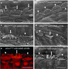
Correlative scanning electron microscopy (SEM) and confocal microscopy of inner hair cells after AAV8-whirlin gene therapy. (a) A row of IHCs from a normal control (whrn+/wi) cochlea (middle turn) showing stereocilia arranged in staircase pattern with two to three rows (white arrows). (b) A row of IHCs from a whrnwi/wi cochlea (no AAV8-whirlin gene therapy, basal turn) showing all whrnwi/wi IHCs have short stereocilia bundles arranged in supernumerary rows (white arrowheads). (c) and (d) Examples of whrnwi/wi cochleas that received AAV8-whirlin gene therapy (middle and basal turns, respectively). Some IHCs have longer stereocilia (white arrow), while the adjacent IHC has short stereocilia (white arrowhead) that are reminiscent of the whrnwi/wi cochlea without gene therapy (compare to a and b). (e) and (f) Correlative confocal and SEM images of a single whrnwi/wi specimen after AAV8-whirlin gene therapy (from apical turn). The middle IHC (white arrow) was infected with AAV8-whirlin and has elongated stereocilia arranged in fewer rows (resembling the wild-type cochlea), while the adjacent two IHCs (white arrowheads) were not infected with AAV8-whirlin and have shorter stereocilia arranged in supernumerary rows (resembling bundles from whrnwi/wi mice). This confirms that the whrnwi/wi IHCs with elongated stereocilia bundles were infected with AAV8-whirlin and expressed whirlin at the stereocilia tips. Scale bars represent 5 μm.
Figure 6
Figure 6
AAV8-whirlin gene therapy increases IHC survival. (a) Confocal image of the contralateral cochlea from a whrnwi/wi mouse at P30 (base) that did not undergo AAV8-whirlin gene therapy. Significant IHC degeneration has begun with no surviving IHC in this image. (b) Confocal image of a whrnwi/wi cochlea (base) after AAV8-whirlin gene therapy. Myosin 7a, which labels the hair cells, is shown in magenta, and phalloidin, which labels F-actin in the stereocilia, is shown in red. Whirlin is shown in green. Even though IHC degeneration has begun, many IHCs are still intact (thin white arrows). There is one IHC with abundant whirlin expression in the cytoplasm (thick white arrow). Images were taken at 63×. Scale bar represents 10 μm. (c) A normal control cochlea is shown for comparison. (d) IHC survival in whrnwi/wi ears injected with AAV8-whirlin gene therapy (whrnwi/wi with AAV8-whirlin) vs. the contralateral noninjected ears (Contralateral). The whrnwi/wi ears that underwent AAV8-whirlin gene therapy had increased IHC survival relative to the contralateral (no AAV8-whirlin) ears. This difference was significant at the cochlear base (P < 0.001). Error bars represent standard errors. n = the number of animals.
Figure 7
Figure 7
AAV8-whirlin did not restore hearing in whirler mice. In both the whrnwi/wi ears injected with AAV8-whirlin (whrnwi/wi with AAV8-whirlin) and untreated whrnwi/wi ears (whrnwi/wi no surgery), no auditory brainstem responses (NR) were detected even at 90 dB sound pressure level. Control mice that underwent AAV8-whirlin gene therapy or sham surgery had a small elevation in ABR thresholds in all three frequencies tested. n = the number of animals. All ABR testing was done at age P30 to P90.

References

    1. Morton, NE (1991). Genetic epidemiology of hearing impairment. Ann N Y Acad Sci 630: 16–31. - PubMed
    1. Lenz, DR and Avraham, KB (2011). Hereditary hearing loss: from human mutation to mechanism. Hear Res 281: 3–10. - PubMed
    1. Drummond, MC, Belyantseva, IA, Friderici, KH and Friedman, TB (2012). Actin in hair cells and hearing loss. Hear Res 288: 89–99. - PMC - PubMed
    1. Richardson, GP, de Monvel, JB and Petit, C (2011). How the genetics of deafness illuminates auditory physiology. Annu Rev Physiol 73: 311–334. - PubMed
    1. Belyantseva, IA, Labay, V, Boger, ET, Griffith, AJ and Friedman, TB (2003). Stereocilia: the long and the short of it. Trends Mol Med 9: 458–461. - PubMed

Publication types

MeSH terms